. 2025 Feb;31(2):478-489.
doi: 10.1038/s41591-024-03420-w. Epub 2025 Jan 17.

Genomic reanalysis of a pan-European rare-disease resource yields new diagnoses

Steven Laurie #  1   2 Wouter Steyaert #  3   4 Elke de Boer #  3   5 Kiran Polavarapu #  6 Nika Schuermans #  7   8   9 Anna K Sommer #  10 German Demidov  11 Kornelia Ellwanger  11 Ida Paramonov  1   2 Coline Thomas  12 Stefan Aretz  10   13 Jonathan Baets  14   15   16 Elisa Benetti  17   18 Gemma Bullich  1   2 Patrick F Chinnery  19   20 Jill Clayton-Smith  21   22 Enzo Cohen  23 Daniel Danis  24 Jean-Madeleine de Sainte Agathe  25 Anne-Sophie Denommé-Pichon  26   27 Jordi Diaz-Manera  28 Stephanie Efthymiou  29 Laurence Faivre  26   30   31   32   33 Marcos Fernandez-Callejo  1   2 Mallory Freeberg  12 José Garcia-Pelaez  34   35   36 Lena Guillot-Noel  37 Tobias B Haack  11 Mike Hanna  38 Holger Hengel  39   40 Rita Horvath  19 Henry Houlden  29 Adam Jackson  21   22 Lennart Johansson  41 Mridul Johari  42 Erik-Jan Kamsteeg  3 Melanie Kellner  39   40 Tjitske Kleefstra  3   5   43   44 Didier Lacombe  45   46 Hanns Lochmüller  1   6   47   48   49 Estrella López-Martín  50 Alfons Macaya  51 Anna Marcé-Grau  51 Aleš Maver  52 Heba Morsy  29   53 Francesco Muntoni  54   55 Francesco Musacchia  56   57 Isabelle Nelson  23 Vincenzo Nigro  56   57 Catarina Olimpio  19   58 Carla Oliveira  35   36   37 Jaroslava Paulasová Schwabová  59 Martje G Pauly  60   61   62 Borut Peterlin  52 Sophia Peters  10 Rolph Pfundt  3   5 Giulio Piluso  56 Davide Piscia  1   2 Manuel Posada  50 Selina Reich  39   40 Alessandra Renieri  17   18   63 Lukas Ryba  64 Karolis Šablauskas  3   65 Marco Savarese  42 Ludger Schöls  39   40 Leon Schütz  11 Verena Steinke-Lange  66   67 Giovanni Stevanin  37 Volker Straub  28 Marc Sturm  11 Morris A Swertz  41 Marco Tartaglia  68 Iris B A W Te Paske  3   4 Rachel Thompson  6 Annalaura Torella  56   57 Christina Trainor  28 Bjarne Udd  42   69   70 Liedewei Van de Vondel  14   15   71 Bart van de Warrenburg  5   72 Jeroen van Reeuwijk  3   5 Jana Vandrovcova  29 Antonio Vitobello  26   27 Janet Vos  3   4 Emílie Vyhnálková  64 Robin Wijngaard  3   4 Carlo Wilke  39   40 Doreen William  73   74 Jishu Xu  11   39   40 Burcu Yaldiz  3 Luca Zalatnai  1   2 Birte Zurek  11 Solve-RD DITF-GENTURISSolve-RD DITF-ITHACASolve-RD DITF-EURO-NMDSolve-RD DITF-RNDSolve-RD consortiumAnthony J Brookes  75 Teresinha Evangelista  23 Christian Gilissen  3   4 Holm Graessner  11   76 Nicoline Hoogerbrugge  3   4 Stephan Ossowski  11   77 Olaf Riess  11   76 Rebecca Schüle  39   40 Matthis Synofzik  39   40 Alain Verloes  78   79 Leslie Matalonga  1   2 Han G Brunner  3   5   80 Katja Lohmann  61 Richarda M de Voer  3   4 Ana Töpf  28 Lisenka E L M Vissers  3   5 Sergi Beltran  1   81 Alexander Hoischen  3   4   82
Collaborators, Affiliations

Genomic reanalysis of a pan-European rare-disease resource yields new diagnoses

Steven Laurie et al. Nat Med. 2025 Feb.

Erratum in

  • Publisher Correction: Genomic reanalysis of a pan-European rare-disease resource yields new diagnoses.
    Laurie S, Steyaert W, de Boer E, Polavarapu K, Schuermans N, Sommer AK, Demidov G, Ellwanger K, Paramonov I, Thomas C, Aretz S, Baets J, Benetti E, Bullich G, Chinnery PF, Clayton-Smith J, Cohen E, Danis D, de Sainte Agathe JM, Denommé-Pichon AS, Diaz-Manera J, Efthymiou S, Faivre L, Fernandez-Callejo M, Freeberg M, Garcia-Pelaez J, Guillot-Noel L, Haack TB, Hanna M, Hengel H, Horvath R, Houlden H, Jackson A, Johansson L, Johari M, Kamsteeg EJ, Kellner M, Kleefstra T, Lacombe D, Lochmüller H, López-Martín E, Macaya A, Marcé-Grau A, Maver A, Morsy H, Muntoni F, Musacchia F, Nelson I, Nigro V, Olimpio C, Oliveira C, Paulasová Schwabová J, Pauly MG, Peterlin B, Peters S, Pfundt R, Piluso G, Piscia D, Posada M, Reich S, Renieri A, Ryba L, Šablauskas K, Savarese M, Schöls L, Schütz L, Steinke-Lange V, Stevanin G, Straub V, Sturm M, Swertz MA, Tartaglia M, Te Paske IBAW, Thompson R, Torella A, Trainor C, Udd B, Van de Vondel L, van de Warrenburg B, van Reeuwijk J, Vandrovcova J, Vitobello A, Vos J, Vyhnálková E, Wijngaard R, Wilke C, William D, Xu J, Yaldiz B, Zalatnai L, Zurek B; Solve-RD DITF-GENTURIS; Solve-RD DITF-ITHACA; Solve-RD DITF-EURO-NMD; Solve-RD DITF-RND; Solve-RD consortiu… See abstract for full author list ➔ Laurie S, et al. Nat Med. 2025 Aug;31(8):2819-2820. doi: 10.1038/s41591-025-03754-z. Nat Med. 2025. PMID: 40537530 Free PMC article. No abstract available.

Abstract

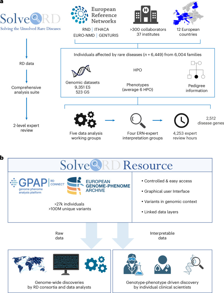
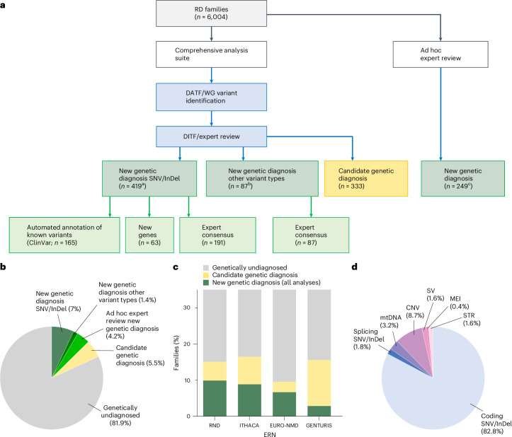
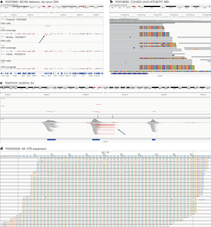
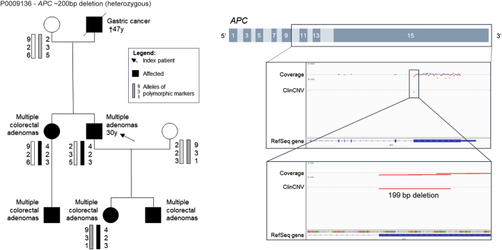
Genetic diagnosis of rare diseases requires accurate identification and interpretation of genomic variants. Clinical and molecular scientists from 37 expert centers across Europe created the Solve-Rare Diseases Consortium (Solve-RD) resource, encompassing clinical, pedigree and genomic rare-disease data (94.5% exomes, 5.5% genomes), and performed systematic reanalysis for 6,447 individuals (3,592 male, 2,855 female) with previously undiagnosed rare diseases from 6,004 families. We established a collaborative, two-level expert review infrastructure that allowed a genetic diagnosis in 506 (8.4%) families. Of 552 disease-causing variants identified, 464 (84.1%) were single-nucleotide variants or short insertions/deletions. These variants were either located in recently published novel disease genes (n = 67), recently reclassified in ClinVar (n = 187) or reclassified by consensus expert decision within Solve-RD (n = 210). Bespoke bioinformatics analyses identified the remaining 15.9% of causative variants (n = 88). Ad hoc expert review, parallel to the systematic reanalysis, diagnosed 249 (4.1%) additional families for an overall diagnostic yield of 12.6%. The infrastructure and collaborative networks set up by Solve-RD can serve as a blueprint for future further scalable international efforts. The resource is open to the global rare-disease community, allowing phenotype, variant and gene queries, as well as genome-wide discoveries.


Conflict of interest statement

Competing interests: M. Synofzik has received consultancy honoraria from Janssen, Ionis, Orphazyme, Servier, Reata, GenOrph and AviadoBio, all unrelated to the present manuscript. B.v.d.W. has received consultancy honoraria from, and/or has served on advisory boards for, Servier, Biohaven Pharmaceuticals, Vico Therapeutics and Biogen, all unrelated to the present manuscript. The other authors declare no competing interests.

Figures

Fig. 1
Fig. 1. Overview of the Solve-RD analysis and interpretation framework and community resource established.
a, Solve-RD brought together rare-disease data and expertise. Central to Solve-RD are four core ERNs relating to rare diseases; via these expert disease networks, patients with rare diseases were recruited from 43 research groups from 37 institutes in 12 European countries (Belgium, Czech Republic, Finland, France, Germany, Hungary, Italy, the Netherlands, Portugal, Slovenia, Spain and the United Kingdom) and Canada. The work involved >300 collaborators in the submission, analysis and interpretation of rare-disease data. The RD-REAL framework allows sharing of data and expertise on a continental scale, consisting of (1) expert curated data, (2) a comprehensive analysis suite and (3) a two-level (that is, molecular and clinical) expert review. The complete dataset comprises 9,645 individuals from 6,004 families and includes phenotypes in Phenopacket format (average of six HPO terms per affected individual), pedigrees and genomic data (genomes and exomes). b, Illustration of the utility of this resource to the global rare-disease community. In total, RD-REAL data of >23,000 individuals with >100 million unique genomic variants are available via RD-Connect GPAP and EGA. This represents a growing resource containing data that have been submitted since the start of Solve-RD. Interpretable data (genetic variants, phenotypes and pedigrees) are standardized and annotated, and are made available for querying, analysis and interpretation in RD-Connect GPAP for authorized users. In addition, all raw and processed data are available for download at EGA under a controlled-access model. All icons, except logos of services (GPAP; EGA) and consortia/networks (Solve-RD; European Reference Networks) that are contributors of this publication, created with Biorender.com.
Fig. 2
Fig. 2. Systematic reanalysis of genomic datasets for the genetic diagnosis of rare diseases.
a, Flowgram of systematic analysis of 6,004 families. Yield per analysis type (genetic diagnoses by SNV/InDel and other variant types; candidate genetic diagnoses and genetic diagnoses by ad hoc expert review) are shown. For SNV/InDels, we evaluated why the 464 variants previously identified in 419 families had not been classified as disease causing. b, Chart summarizing diagnostic yield across 6,004 families in Solve-RD. c, Chart summarizing yield per disease category (ERN); the denominator is 6,004 families. d, Chart summarizing the different variant types that led to a molecular diagnosis in 506 of 6,004 families as part of the systematic reanalysis effort of Solve-RD. aDisease-causing SNVs or short insertions/deletions were identified in 419 families. bDisease-causing non-SNV variants identified in 87 families, including three cases of compound heterozygosity involving an SNV and a CNV/SV, identified through the ‘other variant type’ analyses, and are counted only under ‘New genetic diagnosis other variant types’. cIn 114 of 147 cases where we could confirm the variant identified in the ad hoc analysis, we established that it would also have been found by the standard analysis. RD, rare disease; splicing SNV/InDel, noncanonical splicing sites; WG, work group.
Fig. 3
Fig. 3. Examples of ‘beyond standard’ variant types by Solve-RD.
ad, Illustrative examples of previously unsolved rare-disease probands for which a new variant other than a coding SNV/InDel resulted in a new diagnosis. a, De novo CNV affecting BICRA (P0012861). b, MEI variant in COL6A2 (P0014682). c, SV in SCN11A (P0011371). d, STR expansion affecting AR (P0002409).
Fig. 4
Fig. 4. Example of a new discovery by Solve-RD.
a,b, An example of discoveries enabled by the Solve-RD resource. a, RAB14 de novo variants in two cases from this project contribute to the establishment of a new genotype–phenotype relationship. The first individual (P0012753) presents with mild global developmental delay in the absence of any facial dysmorphism or congenital anomalies, and carries a de novo variant in RAB14 (chr9:123952916G>A; NM_016322.3:c.200C>T; p.(Thr67Met)), which is rare (not observed in gnomAD v.2.1.1), likely to be deleterious (CADD score of 29) and has been observed de novo in at least four additional individuals with developmental disorders in the literature. The second individual (P0012904) presents with mild ID, subtle facial dysmorphisms comprising a high, square-shaped forehead, downslant of palpebral fissures and a low-hanging columella, in the absence of congenital anomalies. The de novo variant found in this individual (chr9:123954475A>C; NM_016322.3:c.80T>G; (p.(Leu27Trp)) is also absent from gnomAD, predicted to be deleterious (CADD score of 28) and has been observed de novo in at least one additional individual with a neurodevelopmental disorder in DECIPHER (https://www.deciphergenomics.org/patient/305550/phenotypes/person/62257). The female individual reported in Decipher presents with moderate ID, facial dysmorphism consisting of large earlobes, smooth philtrum, a wide mouth and protruding tongue, short feet with congenital talipes calcaneovalgus, thick hair and an umbilical hernia. b, Salent features of the two cases in a. aa, Amino acid.
Extended Data Fig. 1
Extended Data Fig. 1. HPO terms (A) and Monarch phenotype specificity meter (B).
Violin plots illustrating (a) the number of Human Phenotype Ontology terms associated to each proband across ERN and (b) the Monarch specificity score (range 0–5, higher better) which provides an indication of how comprehensive the phenotypic description of the affected individual is. The solid line indicates the median, and the dashed line the 25th and 75th centiles.
Extended Data Fig. 2
Extended Data Fig. 2. Flowgram of all analyses performed within the Solve-RD systematic reanalysis.
aSee Supplementary Table 2 for ERN specific gene lists; bDe novo analysis was performed genome-wide, irrespective of previously identified disease genes; cSNV/InDels were investigated within the mitochondrial DNA; d Small exceptions in the prioritisation were made between ERNs for certain genes. See Online Methods, and Supplementary Tables 15–18 for further details.
Extended Data Fig. 3
Extended Data Fig. 3. Date of initial creation, and of last update of OMIM records for genes shown to be disease-causing in this study.
This figure shows (a) the date of creation of the first OMIM entry for a particular gene determined to be explanatory for the condition in a Solve-RD proband-phenotype association, and (b) the date of the last update of the relevant entry. The OMIM entry for 67 genes was only created after 01/01/2018, when Solve-RD started, and many genes of interest have had their records updated since then. This explains why a number of these genes were only confirmed as being disease-causing in affected individuals here as a result of reanalysis in Solve-RD.
Extended Data Fig. 4
Extended Data Fig. 4. Example of an individual diagnosed with a rare disease from ERN RND.
The left panel shows the pedigree of a 58-year old individual first diagnosed at 42 years of age with progressive gait disturbance and urinary urgency, in the absence of family history of these symptoms (P0015028). The right panel shows two IGV screenshots indicating a heterozygous missense SNV (c.451G>A (p.(Gly151Ser)) in B4GALNT1 (top) and a heterozygous, approximately 10kb in length, deletion on the other allele (bottom), resulting in complete deletion of exons 6–11 (commencing in exon 5, removing exons 6–11 (NM_001478.5), and ending in the 3’UTR (Chr12(GRch37): g.58014705-58024263del). Location of the deletion is indicated by the red line in the top track, supported by the reduced beta-allele frequency of variants in this region as shown in the centre track, and further supported by read pairs spanning the full 10kb (in red) observed in the lower track.
Extended Data Fig. 5
Extended Data Fig. 5. Example of an individual diagnosed with a rare disease from ERN GENTURIS.
Left panel: pedigree of proband P0009136 (indicated by the arrow). Haplotype analysis demonstrated that all affected individuals carry the same allele at the APC locus, inherited from the paternal branch of the family. Right panel: comprehensive CNV analysis uncovered a heterozygous germline deletion, approximately 200bp in length, at the beginning of coding exon 15 of the APC gene which could not be identified by routine diagnostics using just the sequencing and MLPA methods.
Extended Data Fig. 6
Extended Data Fig. 6. Examples of two individuals diagnosed with a rare disease from ERN ITHACA.
a) The left panel shows the phenotypic presentation of a 24-year old male diagnosed at fifteen years of age with asymmetry of legs and face, described at that time as underdevelopment of the left side (P0012716, written consent that allows sharing of photographs was given). At birth, asymmetry of the legs and face was evident and there was a postaxial rudimentary digit on the right hand that regressed to a small nodule over time. The asymmetry of the face and legs was reported to be stable over time and his cognitive development was within the normal range (IQ of 89). He was affected by complex partial seizures with continuous spike-and-wave during sleep from childhood, however the seizures had a good clinical progression and medication could be discontinued at eleven years of age. Other medical problems included scoliosis, autism spectrum disorder, clumsy motor skills, and sleeping problems. The IGV screenshot in the right panel confirms the presence of a rare de novo mosaic missense variant (observed in only 13% of reads) in PIK3CA (chr3:178916876G>A), validated by Sanger sequencing. This variant had previously been reported elsewhere to cause PIK3CA-related overgrowth, leading to a change in the clinical diagnosis for this young man, and the resolution of his diagnostic odyssey. b) The left panel shows the phenotypic presentation of an undiagnosed 22-year old male who had experienced severe developmental delay, and presented with a variety of physical anomalies, including an open mouth with full lip vermillion, a high and narrow palate with gum hypertrophy and irregular dentition. A brain MRI was initially reported to be uninformative (P0013065, written consent that allows sharing of photographs was given)., The IGV screenshot in the right panel indicates the presence of a rare de novo nonsense variant in MN1 (Chr22(GRCh37):g.28146963C>T; NM_002430.2:c.3903G>A; p.(Trp1301*)) unobserved in the parents. Retrospective reanalysis of the brain MRI revealed dysplasia of the cerebellar vermis, rhombencephalosynapsis and mild bitemporal narrowing of the skull, consistent with a diagnosis of CEBALID syndrome. The individuals described gave permission for their photos to be used in this publication, for which we thank them and their families.
Extended Data Fig. 7
Extended Data Fig. 7. Example of an individual diagnosed with a rare disease from ERN EURO-NMD.
The left panel shows the pedigree, and clinical history of proband P0005327 (indicated by the arrow). At eight years of age he began to develop progressive lower limb weakness and fatigability. He started to experience recurrent falls at eight years of age and went on to develop progressive proximal lower limb weakness with prominent fatigability, and a waddling gait. There was no history of bulbar or ocular symptoms. On examination, bilateral asymmetric ptosis with fatigability was observed, as was polyminimyoclonus. Muscle strength was normal in all four limbs, but fatigue occurred upon sustained arm abduction. Deep tendon reflexes were normal, as were serum creatine kinase levels, while repetitive nerve stimulation was inconclusive. Due to a clinical suspicion of Congenital Myasthenic Syndrome (CMS), a trial of pyridostigmine was initiated, but the individual was non-compliant. However, his parents reported spontaneous improvement in baseline limb weakness and falls over the following six years with only episodic worsening due to fever and exertional myalgias. There was a strong family history of diabetes on the maternal side and the mother’s fasting glucose levels were suggestive of borderline diabetes, and she also has a long history of migraines. Retrospective serum lactate testing in both proband and mother showed mildly elevated levels (>20 mg/dl). The IGV screenshot in the right panel indicates the presence of a heteroplasmic mitochondrial variant (MT-TL1, MT:3243A>G)) observed with a frequency of 27% in the proband, and 14% in his mother. This difference in heteroplasmy may explain the divergence in symptoms between mother and child. While the initial clinical suspicion in the proband was CMS due to the notable fatigability, the fact that mitochondrial disease can be clinically highly variable means that mild forms of mitochondrial myopathy can be difficult to diagnose clinically.
Extended Data Fig. 8
Extended Data Fig. 8. Clinical actionability.
a) Percentage of solved cases for which the causative gene is reported in one of the three gene-treatment databases included in this study (ClinGen, IEMbase and Treatabolome) and guidelines for surveillance of genetic tumour risk syndromes. b) Gene-treatment databases and surveillance guidelines for genes in which (likely) disease-causing variants have been identified per ERN. c) List of genes with (likely) disease-causing variants, and number of rare disease probands/families diagnosed in this study in parentheses, identified in each of the three gene-treatment databases as well as surveillance guidelines included in this study.
Extended Data Fig. 9
Extended Data Fig. 9. Examples of ‘beyond standard’ variant types and discovery by Solve-RD.
Panels A&B provide illustrative examples of previously unsolved rare disease probands for which a new variant other than standard coding SNV/InDel resulted in a new diagnosis. a) Non-canonical splicing variant in ARID1A (individual P0017701); b) mtDNA variant in ND3-MT (P0002456). c) The new discovery of recurrent de novo variants in RNU4-2 led to likely new diagnoses in two Solve-RD cases. Both variants have been validated, and the phenotypes match the recently published phenotypic descriptions,.

References

    1. Nguengang Wakap, S. et al. Estimating cumulative point prevalence of rare diseases: analysis of the Orphanet database. Eur. J. Hum. Genet.28, 165–173 (2020). - PMC - PubMed
    1. Turro, E. et al. Whole-genome sequencing of patients with rare diseases in a national health system. Nature583, 96–102 (2020). - PMC - PubMed
    1. Smedley, D. et al. 100,000 Genomes Pilot on Rare-Disease Diagnosis in Health Care – Preliminary Report. N. Engl. J. Med.385, 1868–1880 (2021). - PMC - PubMed
    1. Wright, C. F. et al. Genomic diagnosis of rare pediatric disease in the United Kingdom and Ireland. N. Engl. J. Med.388, 1559–1571 (2023). - PMC - PubMed
    1. Dai, P. et al. Recommendations for next generation sequencing data reanalysis of unsolved cases with suspected Mendelian disorders: a systematic review and meta-analysis. Genet. Med.24, 1618–1629 (2022). - PubMed