. 2025 Jan 17;20(1):1.
doi: 10.1186/s13008-025-00142-4.

Interaction of STIL with FOXM1 regulates SF3A3 transcription in the hepatocellular carcinoma development

Affiliations

Interaction of STIL with FOXM1 regulates SF3A3 transcription in the hepatocellular carcinoma development

Haijun Zhang et al. Cell Div. .

Abstract

Background: Dysregulation of SF3A3 has been related to the development of many cancers. Here, we investigated the functional role of SF3A3 in hepatocellular carcinoma (HCC).

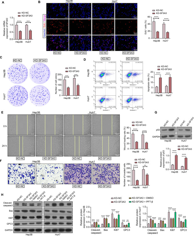
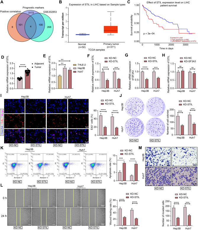
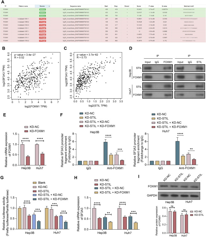
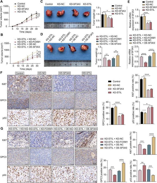
Methods: SF3A3 expression in HCC tissues and cell lines was examined using RT-qPCR. Changes in malignant behavior of HCC cells after downregulation of SF3A3 were assessed by EdU, colony formation, flow cytometry, wound healing, and Transwell invasion assays. Multiple datasets were combined to identify the upstream modifiers of SF3A3. The binding relationship between STIL and FOXM1 was explored by co-IP assay, and the effect of STIL and FOXM1 on the binding of FOXM1 at the SF3A3 promoter was detected by ChIP-qPCR assay. A xenograft tumor model was established to explore the changes of tumors in vivo, and the expression of Ki67, GPC3, and p53 in tumor tissues was detected by immunohistochemistry.

Results: SF3A3 and STIL were overexpressed in HCC tissues and cells, and downregulation of SF3A3 or STIL inhibited the malignant behavior of HCC cells by promoting the expression of p53. An interaction between STIL and FOXM1 regulated the SF3A3 expression in HCC cells. Knockdown of FOXM1 further enhanced the anti-tumor effects of STIL loss on HCC cells in vitro and in vivo, whereas SF3A3 overexpression overturned the impact of STIL loss on HCC cells in vitro and in vivo.

Conclusions: Our findings indicate that STIL/FOXM1 expedites HCC development by activating SF3A3, which highlights the importance of SF3A3 as a promising prognostic marker and therapeutic target for HCC.

Keywords: FOXM1; Hepatocellular carcinoma; P53; SF3A3; STIL.


Conflict of interest statement

Declarations. Ethical approval: The research protocol was approved by the Ethics Committee of the First Hospital of Qiqihar and conducted according to the guidelines of the Declaration of Helsinki. All patients signed a written informed consent. All animal experiments performed were approved by the Ethics Committee of the First Hospital of Qiqihar and conducted in conformance with the Guide for the Care and Use of Laboratory Animals published by the NIH. Consent to participate: Written informed consent for publication was obtained from all participants. Consent for publication: All authors read the guidelines of the journal and agreed with consent for publication. Competing interests: The authors declare no competing interests.

Figures

Fig. 1
Fig. 1
SF3A3 is upregulated and links to unsatisfactory outcomes of HCC patients. (A) Transcription level of SF3A3 (transcript per million) in primary tumor tissues and normal tissues of HCC patients in the UALCAN database. (B) The protein expression of SF3A3 in primary tumor tissues and normal tissues of HCC patients in the UALCAN database. (C) The survival of HCC patients with high SF3A3 expression (n = 90) or medium/low SF3A3 expression (n = 275). (D) SF3A3 mRNA expression in tumor and adjacent tissues from HCC patients using RT-qPCR (n = 30). (E) SF3A3 mRNA expression in HCC cell lines Hep3B and Huh7 and normal hepatocyte cell line THLE-2 using RT-qPCR. All data are expressed as mean ± SD from three experiments. In panel D, significance is determined by paired t-test, and in panel E, significance is determined by one-way ANOVA. **p < 0.01, ****p < 0.0001
Fig. 2
Fig. 2
Downregulation of SF3A3 inhibits malignant behavior in HCC cells. (A) SF3A3 mRNA expression in HCC cell lines Hep3B and Huh7 treated with KD-SF3A3 or KD-NC using RT-qPCR. Evaluation of cell proliferation using EdU assay (B) and colony formation assay (C) in Hep3B and Huh7 cells. (D) Detection of apoptosis using flow cytometry in Hep3B and Huh7 cells. Cell migration and invasion were assessed by wound healing assay (E) and Transwell invasion assay (F) in Hep3B and Huh7 cells. (G) The p53 protein expression in Hep3B and Huh7 cells after KD-SF3A3 was assessed by western blot analysis. (H) The protein expression of Cleaved-caspase3, Bax, Ki67, and GPC3 in Hep3B and Huh7 cells after KD-SF3A3 alone or in combination with the p53 inhibitor was assessed by western blot analysis. All data are expressed as mean ± SD from three experiments. In panels A-H, significance is determined by two-way ANOVA. **p < 0.01, ***p < 0.001, ****p < 0.0001
Fig. 3
Fig. 3
STIL is overexpressed in HCC and knockdown of STIL inhibits the malignant behavior of HCC cells. (A) The intersection of SF3A3-positively correlated genes and prognostic markers in the UALCAN database, and differentially expressed genes in the GSE202853 dataset. (B) Transcription level of STIL (transcript per million) in primary tumor tissues and normal tissues of HCC patients in the UALCAN database. (C) The survival of HCC patients with high STIL expression (n = 90) or medium/low STIL expression (n = 275). (D) STIL mRNA expression in tumor tissues and corresponding adjacent tissues from HCC patients using RT-qPCR (n = 30). (E) STIL mRNA expression in HCC cell lines Hep3B and Huh7 and normal hepatocyte cell line THLE-2 using RT-qPCR. (F) STIL mRNA expression in HCC cell lines Hep3B and Huh7 treated with KD-STIL or KD-NC using RT-qPCR. (G) SF3A3 mRNA expression in HCC cell lines Hep3B and Huh7 treated with KD-STIL or KD-NC using RT-qPCR. (H) STIL mRNA expression in HCC cell lines Hep3B and Huh7 treated with KD-SF3A3 or KD-NC using RT-qPCR. Evaluation of cell proliferation using EdU assay (I) and colony formation assay (J) in Hep3B and Huh7 cells. (K) Detection of apoptosis using flow cytometry in Hep3B and Huh7 cells. Analysis of cell migration and invasion by wound healing assay (L) and Transwell invasion assay (M) in Hep3B and Huh7 cells. All data are expressed as mean ± SD from three experiments. In panel D, significance is determined by paired t-test; in panel E, significance is determined by one-way ANOVA; in panel F-M, significance is determined by two-way ANOVA. **p < 0.01, ***p < 0.001, ****p < 0.0001
Fig. 4
Fig. 4
An interaction between STIL and FOXM1 induces SF3A3 expression. (A) The presence of binding sites for FOXM1 at the promoter of SF3A3 was predicted in the hTFtarget database. The positive correlation between FOXM1 (B) and STIL (C) with SF3A3 expression in the GEPIA database. (D) The binding relationship between STIL and FOXM1 was analyzed using co-IP. (E) FOXM1 mRNA expression in HCC cell lines Hep3B and Huh7 treated with KD-FOXM1 or KD-NC using RT-qPCR. (F) The effect of KD-STIL alone or in combination with KD-FOXM1 on FOXM1 binding at the SF3A3 promoter was analyzed using ChIP. (G) Changes in SF3A3 promoter luciferase activity were analyzed using dual-luciferase reporter assays. (H) SF3A3 mRNA expression after downregulation of STIL alone or combined downregulation of FOXM1 and STIL detected by RT-qPCR. (I) FOXM1 protein expression in Hep3B and Huh7 cells after knockdown of STIL. All data are expressed as mean ± SD from three experiments. In panels E-I, significance is determined by two-way ANOVA. **p < 0.01, ***p < 0.001, ****p < 0.0001, ns, not significant
Fig. 5
Fig. 5
Combined knockdown of STIL and FOXM1 regulates SF3A3 transcription to suppress malignant behavior of HCC cells. (A) SF3A3 mRNA expression in HCC cells treated with KD-STIL + OE-NC or KD-STIL + OE-SF3A3 was assessed using RT-qPCR. Evaluation of cell proliferation using EdU assay (B) and colony formation assay (C) in Hep3B and Huh7 cells. (D) Detection of apoptosis using flow cytometry in Hep3B and Huh7 cells. Analysis of cell migration and invasion by wound healing assay (E) and Transwell invasion assay (F) in Hep3B and Huh7 cells. All data are expressed as mean ± SD from three experiments. In panels A-F, significance is determined by two-way ANOVA. *p < 0.05, **p < 0.01, ***p < 0.001, ****p < 0.0001
Fig. 6
Fig. 6
Combined knockdown of STIL and FOXM1 regulates SF3A3 transcription to suppress HCC cell growth in vivo. (A) Measurement of tumor volume changes in mice injected with KD-NC-, KD-SF3A3-, and KD-STIL-treated Huh7 cells. (B) Measurement of tumor volume changes in mice injected with KD-STIL + KD-NC-, KD-STIL + KD-FOXM1-, KD-STIL + OE-NC-, and KD-STIL + OE-SF3A3-treated Huh7 cells. (C) Changes in tumor weight in the KD-NC, KD-SF3A3, and KD-STIL groups. (D) Changes in tumor weight in the KD-STIL + KD-NC, KD-STIL + KD-FOXM1, KD-STIL + OE-NC, and KD-STIL + OE-SF3A3 groups. (E) The mRNA expression of SF3A3 in the tumor tissues of mice in the seven groups. (F) Expression of Ki67, GPC3, and p53 in the KD-NC, KD-SF3A3, and KD-STIL groups was detected by immunohistochemistry. (G) Expression of Ki67, GPC3, and p53 in the KD-STIL + KD-NC, KD-STIL + KD-FOXM1, KD-STIL + OE-NC, and KD-STIL + OE-SF3A3 groups was detected by immunohistochemistry. All data are expressed as mean ± SD (n = 5). In panels A-B, significance is determined by two-way ANOVA; in panels C-G. *p < 0.05, **p < 0.01, ***p < 0.001, ****p < 0.0001

Similar articles

Cited by

References

    1. Hepatocellular carcinoma. Nat Rev Dis Primers. 2021;7(1):7. 10.1038/s41572-021-00245-6. - PubMed
    1. Pinero F, Dirchwolf M, Pessoa MG. Biomarkers in Hepatocellular Carcinoma: diagnosis, Prognosis and Treatment Response Assessment. Cells. 2020;9(6). 10.3390/cells9061370. - PMC - PubMed
    1. Zheng H, Zhang G, Zhang L, Wang Q, Li H, Han Y, et al. Comprehensive Review of web servers and Bioinformatics Tools for Cancer Prognosis Analysis. Front Oncol. 2020;10:68. 10.3389/fonc.2020.00068. - PMC - PubMed
    1. Nameki N, Takizawa M, Suzuki T, Tani S, Kobayashi N, Sakamoto T, et al. Structural basis for the interaction between the first SURP domain of the SF3A1 subunit in U2 snRNP and the human splicing factor SF1. Protein Sci. 2022;31(10):e4437. 10.1002/pro.4437. - PMC - PubMed
    1. Zhu W, Zhang Q, Liu M, Yan M, Chu X, Li Y. Identification of DNA repair-related genes predicting pathogenesis and prognosis for liver cancer. Cancer Cell Int. 2021;21(1):81. 10.1186/s12935-021-01779-1. - PMC - PubMed

LinkOut - more resources