

BCL-2 overexpression exosomes promote the proliferation and migration of mesenchymal stem cells in hypoxic environment for skin injury in rats
- PMID: 39825412
- PMCID: PMC11740716
- DOI: 10.1186/s13036-024-00471-y
BCL-2 overexpression exosomes promote the proliferation and migration of mesenchymal stem cells in hypoxic environment for skin injury in rats
Abstract
Objective: The direction of this study was to detect and analyze the specific mechanism of anti-apoptosis in mesenchymal stem cells (MSCs) cells caused by high expression of BCL2.
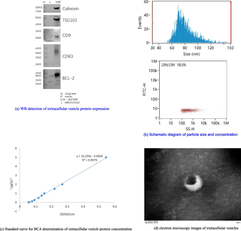
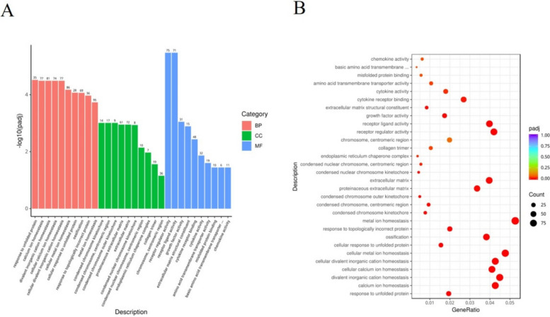
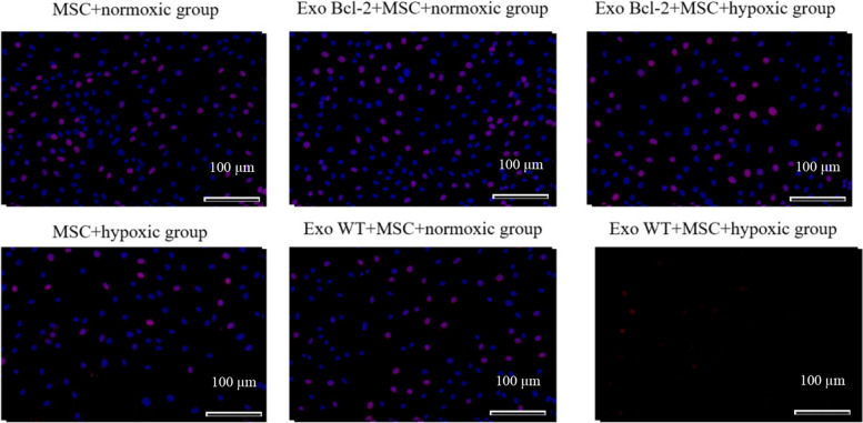
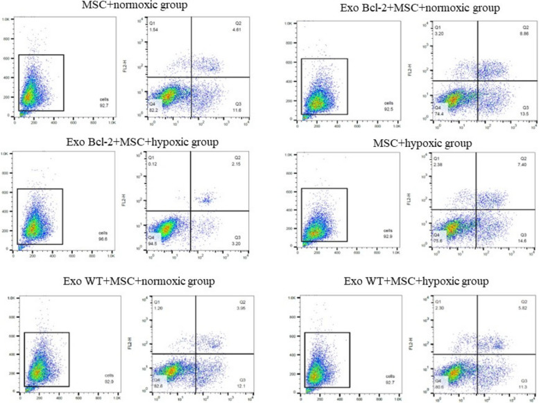
Methods: Bioinformatics was completed in Link omics. GO analysis and KEGG analysis were carried out, and the grope tool of Link omics database was used to evaluate PPI information and other core path analysis information. The cultured cells were divided into MSC + normoxic group (MSCs were cultured in conventional medium, including 10% depleted serum of fetal bovine exosomes, 37 °C, 5% CO2 and 95% air) and Exo-BCL-2 + MSC + normoxic group (a certain concentration of purified BCL-2 exosomes was co-cultured with MSC in conventional medium, 37 °C, 5% CO2 and 95% air), Exo-BCL-2 + MSC + hypoxia group (a certain concentration of purified BCL-2 exosomes and MSC were co-cultured in hypoxia medium at 37 °C, 80% CO2 and 20% air), MSC + hypoxia group (MSCs were cultured in hypoxia medium with 10% depleted serum of fetal bovine exosomes, 37 °C, 80% CO2 and 20% air), exo WT + MSC + normoxic group (co-cultured with MSC in conventional medium at 37 °C, 5% CO2 and 95% air) and exoWT + MSC + hypoxic group (co-cultured with MSC in hypoxic medium at 37 °C, 80% CO2 and 20%). Cell proliferation ability was monitored by cell proliferation test. Cell migration test was used to check the migration capacity of MSCs. The expressions of apoptosis-related proteins BCL-2, caspase3 and caspase9, Runx2, ALP and PPAR-γ were analyzed by western blot. Tissue damage was scored by H&E and Ma Song trichrome staining. Masson staining was used to evaluate the collagen volume fraction of the wound. The expressions of KRT14, α-SMA, CD31 and PCNA in rat trauma tissues were analyzed by immunofluorescence staining. The horizontal of apoptosis-related proteins in skin lesions was checked by Western blot. The horizontal of inflammatory factors TNF-α and IL-6 in traumatic tissue of rats were detected by ELISA.
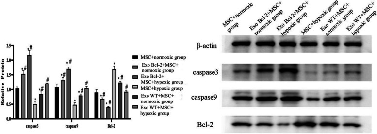
Results: From KEGG's results, we can see that BCL2-2 was closely related to base excision and repair, cell cycle, steroid biosynthesis and other pathways. When cultured for 48h and 72h, the proliferation ability and migration number of MSCs in MSC + hypoxia group were lower than MSC + normoxic group, but the expressions of caspase3 and caspase9 were higher. The proliferation ability and migration number of MSCs in Exo-BCL-2 + MSC + hypoxia group and MSC + hypoxia group were lower than those in Exo-BCL-2 + MSC + normoxic group and MSC + normoxic group, and the horizontal of caspase3 and caspase9 were lower. Exo-BCL-2 + MSC + normoxic group increased the proliferation capacity and migration number of MSCs, but decreased the expression of caspase3 and caspase9. Compared with Exo-BCL-2 + MSC + normoxia and Exo-BCL-2 + MSC + normoxia, the proliferation ability and migration quantity of MSCs in exo WT + MSC + normoxia and exo WT + MSC + hypoxia groups were lower, and the horizontal of caspase3 and caspase9 proteins was higher.
Conclusion: Bioinformatics analysis shows that BCL2-2 plays a worthwhile role in the process of cell apoptosis and proliferation. Exosomes with high expression of BCL-2 can encourage the proliferation of MSC in hypoxic environment. The wound treated with MSCs-BCL-2 promotes the compose of new blood vessels and granulation tissue in the wound, the redifferentiation of epithelial cells and the remodeling of collagen, which has a high therapeutic prospect for chronic wounds and skin regeneration.
Keywords: BCL-2 overexpression; Exosomes; Hypoxia-induced apoptosis; Mesenchymal stem cells.
© 2025. The Author(s).
Conflict of interest statement
Declarations. Ethics approval and consent to participate: All experimental protocols were approved by the Animal Ethics Committee of the Shanghai East Hospital of Tongji University. All methods were carried out in accordance with relevant guidelines and regulations. All methods are reported in accordance with ARRIVE guidelines Consent to publication: Not applicable. Competing interests: The authors declare no competing interests.
Figures


Similar articles
-
[Effects of human umbilical cord mesenchymal stem cells (MSCs)-derived exosomes on pulmonary vascular remodeling in hypoxic pulmonary hypertension].Sheng Li Xue Bao. 2024 Feb 25;76(1):33-44. Sheng Li Xue Bao. 2024. PMID: 38444129 Chinese.
-
[Treatment of intrauterine adhesions in rats with hypoxia-cultured BMSC-derived exosomes].Zhonghua Fu Chan Ke Za Zhi. 2023 Dec 25;58(12):911-921. doi: 10.3760/cma.j.cn112141-20230922-00114. Zhonghua Fu Chan Ke Za Zhi. 2023. PMID: 38123197 Chinese.
-
[Effects of hypoxia-pretreated rat adipose-derived mesenchymal stem cells conditioned medium on wound healing of rats with full-thickness defects].Zhonghua Shao Shang Za Zhi. 2020 Sep 20;36(9):803-812. doi: 10.3760/cma.j.cn501120-20200508-00258. Zhonghua Shao Shang Za Zhi. 2020. PMID: 32972065 Chinese.
-
Exosomal microRNAs from Mesenchymal Stem Cells: Novel Therapeutic Effect in Wound Healing.Tissue Eng Regen Med. 2023 Aug;20(5):647-660. doi: 10.1007/s13770-023-00542-z. Epub 2023 May 2. Tissue Eng Regen Med. 2023. PMID: 37131016 Free PMC article. Review.
-
Mesenchymal Stem Cell-Derived Exosomes Hold Promise in the Treatment of Diabetic Foot Ulcers.Int J Nanomedicine. 2025 May 6;20:5837-5857. doi: 10.2147/IJN.S516533. eCollection 2025. Int J Nanomedicine. 2025. PMID: 40351704 Free PMC article. Review.
Cited by
-
Antimicrobial dual-crosslinked hydrogel synergizes bioengineered extracellular vesicles for enhanced diabetic wound healing.Mater Today Bio. 2025 May 13;32:101870. doi: 10.1016/j.mtbio.2025.101870. eCollection 2025 Jun. Mater Today Bio. 2025. PMID: 40487175 Free PMC article.
References
-
- Walsh GM, Dewson G, Wardlaw AJ, Levi-Schaffer F, Moqbel R. Expression of BCL-2 and Its Homologues in Human Eosinophils. 2019;97:701–9.
-
- Khan WS, Hardingham TE. Mesenchymal stem cells, sources of cells and differentiation potential. J Stem Cells. 2012;7:75–85. - PubMed
LinkOut - more resources
Full Text Sources
Research Materials
Miscellaneous
