. 2025 May 27;9(10):2379-2392.
doi: 10.1182/bloodadvances.2024013651.

PRMT5 inhibition reduces hyperinflammation in a murine model of secondary hemophagocytic lymphohistiocytosis

Affiliations

PRMT5 inhibition reduces hyperinflammation in a murine model of secondary hemophagocytic lymphohistiocytosis

Fiona Brown-Burke et al. Blood Adv. .

Abstract

Hemophagocytic lymphohistiocytosis (HLH) is a rare but aggressive and potentially lethal hyperinflammatory syndrome characterized by pathologic immune activation and excessive production of proinflammatory cytokines leading to tissue damage and multisystem organ failure. There is an urgent need for the discovery of novel targets and development of therapeutic strategies to treat this rare but deadly syndrome. Protein arginine methyltransferase 5 (PRMT5) mediates T-cell-based inflammatory responses, making it a potential actionable target for the treatment of HLH. Using CPG-1826 and anti-interleukin-10R (IL-10R) antibody, we induced murine secondary HLH in vivo with a marked expansion of splenic myeloid cell subsets and concurrent reduction of T- and natural killer (NK)-cell populations. PRMT5 expression was significantly upregulated in splenic T and NK lymphocytes, monocytes, and dendritic cells in mice with HLH (P < .05). Treatment with PRT382, a potent and selective PRMT5 inhibitor, significantly reduced physical signs of secondary HLH, including splenomegaly, hepatomegaly, and anemia (P < .0001 in each case), when compared with untreated mice. Inflammatory cytokines known to drive hyperinflammation in HLH, including interferon-γ and IL-6 were reduced to healthy levels with PRT382 treatment (P > .999 for both). PRT382 treatment also reduced the expansion of myeloid cell populations (P < .0001) in mice with HLH, compared with untreated mice, while restoring T- and NK-cell numbers (P < .001 for both). These results identify PRMT5 as a promising target for the management of secondary HLH and justify further exploration in this and other models of hyperinflammation.


Conflict of interest statement

Conflict-of-interest disclosure: P. Scherle, N.B., and K.V. report employment with Prelude Therapeutics. R.A.B. received research support for PRT drug product from Prelude Therapeutics. The remaining authors declare no competing financial interests.

Figures

None
Graphical abstract
Figure 1.
Figure 1.
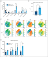
PRMT5 expression is present and increases in myeloid and lymphoid mononuclear cell subsets after HLH induction. (A) Percentage of major immune cell types of all singlets from splenocytes of healthy C57bl/6 mice or those with induced HLH. (B) Mean fluorescent intensity (MFI) of CD44 staining in monocytes (Ly6C+Ly6G) of healthy C57bl/6 mice or those with induced HLH. (C) t-SNE grouping of CD4, CD8, and NK1.1+ splenocytes from a healthy or HLH-induced C57bl/6 mouse. Heat maps reveal expression of key marks that allow for subset identification and PRMT5 expression. (D) MFI of PRMT5 in CD4 T cells, CD8 T cells, and NK cells of healthy C57bl/6 mice or those with induced HLH. Error bars show standard deviation. Multiple unpaired t tests with the Holm-Šídák method were used in panels A and D, whereas a Student t test was used in panel B to determine statistical significance. ∗P < .05; ∗∗P < .01; ∗∗∗P < .001; ∗∗∗∗P < .0001. DCs, dendritic cells; ns, no significance; TX, treatment.
Figure 3.
Figure 3.
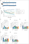
PRMT5 inhibition reduces pathology in a murine model of secondary HLH. (A) Schema of mouse studies. HLH was induced with CPG-1826 and anti–IL-10R on days 0, 2, 4, and 7. PRT382 was dosed daily starting on day 0 (9 days of treatment [TX]), 2 (7 days of TX), or 4 (5 days of TX). Ruxolitinib (Rux) was given twice daily from day 4 to 8. Mice were sacrificed on day 9 and samples were processed for additional analysis. (B) Average weight of each cohort as a percentage change from day 0 in healthy mice, induced HLH with no TX, induced HLH with Rux TX, or HLH with PRT382 TX started on day 0, 2, or 4. Average spleen (C) and liver (D) mass as a percentage of mouse body weight in each cohort determined on necropsy. (E) RBC count, (F) hemoglobin concentration, and (G) lymphocyte count from complete blood counts on blood on day 9. Error bars show standard deviation. A Kruskal-Wallis test with Dunn test for multiple comparisons was used in panel B and 1-way analysis of variances (ANOVAs) with Tukey multiple comparisons were used in panels C-G to determine significance. ∗P < .05; ∗∗P < .01; ∗∗∗P < .001; ∗∗∗∗P < .0001. RBC, red blood cell.
Figure 2.
Figure 2.
PRMT5 expression in monocytes and dendritic cells increases with the induction of HLH. (A) tSNE plot of dendritic cells (D), monocytes (M), and neutrophils (N), with heat maps showing identifying marks. D, CD11c+I-A/I-E+; M, Ly6C+Ly6G; N, Ly6CloLy6G+. (B) Heat map of PRMT5 expression with the same clustering as shown in panel A. (C) Median fluorescent intensity (MFI) of total myeloid cells (D + M + N), monocytes, neutrophils, and dendritic cells positive for PRMT5 expression. Error bars show standard deviation. Multiple unpaired t tests with the Holm-Šídák method were used to determine statistical significance in panel C. ∗P < .05; ∗∗P < .01; ∗∗∗P < .001; ∗∗∗∗P < .0001.
Figure 4.
Figure 4.
Plasma levels of inflammatory cytokines are reduced with PRMT5 inhibition. (A) Concentration of IFN-γ in plasma as determined by ELISA in healthy mice, induced HLH with no TX, induced HLH with Rux TX, or HLH with PRT382 TX started on day 0 (9 days of treatment), 2 (7 days of treatment), or 4 (5 days of TX). Plasma levels of (B) IL-6, (C) IL-1β, and (D) IL-18 as determined by ELISA in healthy mice, induced HLH with no TX, induced HLH with Rux TX, or with PRT382 TX started on day 0. Error bars show standard deviation. One-way ANOVAs with Tukey multiple comparisons were used to determine significance. ∗P < .05; ∗∗P < .01; ∗∗∗P < .001; ∗∗∗∗P < .0001.
Figure 5.
Figure 5.
Proportion of cells returned to normal levels with PRT382 TX. The percentage of all singlets that (A) CD4 T cells, CD8 T cells, and NK cells or (B) myeloid cells (D + M + N), dendritic cells, monocytes, and neutrophils make up in healthy mice, those with induced HLH, with Rux TX, or with PRT382 TX started on day 0. Error bars show standard deviation. A 2-way ANOVA with Sidak's multiple comparisons was used to determine significance. Note: no comparison between HLH no TX and HLH Rux was found to be significant. One outlier was removed from the healthy cohort for myeloid cells. ∗P < .05; ∗∗P < .01; ∗∗∗P < .001; ∗∗∗∗P < .0001. DCs, dendritic cells.
Figure 6.
Figure 6.
PRMT5 inhibition reduces intracellular IFN-γ production in splenocytes derived from mice with HLH. Contours of intracellular IFN-γ in T (A) and NK (B) cells in healthy control mice, mice with HLH and no TX, mice with HLH treated with Rux, and mice with HLH treated with PRT382 started on day 0. Percentage of IFN-γ plus T cells (C) and NK (D) cells in each cohort. Error bars reveal standard deviation. A 1-way ANOVA with Tukey multiple comparisons was used to determine significance. ∗P < .05; ∗∗P < .01; ∗∗∗P < .001; ∗∗∗∗P < .0001.

Comment in

References

    1. George MR. Hemophagocytic lymphohistiocytosis: review of etiologies and management. Hematol Res Rev. 2014;5:69–86. - PMC - PubMed
    1. Bhatt NS, Oshrine B, An Talano J. Hemophagocytic lymphohistiocytosis in adults. Leuk Lymphoma. 2019;60(1):19–28. - PubMed
    1. Ramos-Casals M, Brito-Zerón P, López-Guillermo A, Khamashta MA, Bosch X. Adult haemophagocytic syndrome. Lancet. 2014;383(9927):1503–1516. - PubMed
    1. Gupta S, Weitzman S. Primary and secondary hemophagocytic lymphohistiocytosis: clinical features, pathogenesis and therapy. Expet Rev Clin Immunol. 2010;6(1):137–154. - PubMed
    1. Hines MR, Keenan C, Maron Alfaro G, et al. Hemophagocytic lymphohistiocytosis-like toxicity (carHLH) after CD19-specific CAR T-cell therapy. Br J Haematol. 2021;194(4):701–707. - PMC - PubMed

MeSH terms