. 2025 Jan 28;44(1):115223.
doi: 10.1016/j.celrep.2024.115223. Epub 2025 Jan 17.

Structural development of the HIV-1 apex-directed PGT145-PGDM1400 antibody lineage

Affiliations

Structural development of the HIV-1 apex-directed PGT145-PGDM1400 antibody lineage

Rosemarie D Mason et al. Cell Rep. .

Abstract

Broadly neutralizing antibodies (bNAbs) targeting the apex of the HIV-1-envelope (Env) trimer comprise the most potent category of HIV-1 bNAbs and have emerged as promising therapeutics. Here, we investigate the development of the HIV-1 apex-directed PGT145-PGDM1400 antibody lineage and report cryo-EM structures at 3.4 Å resolution of PGDM1400 and of an improved PGT145 variant (PGT145-R100aS), each bound to the BG505 Env trimer. Cross-species-based engineering improves PGT145 IC80 breadth to near that of PGDM1400. Despite similar breadth and potency, the two antibodies differ in their residue-level interactions with important apex features, including N160 glycans and apex cavity, with residue 100i of PGT145 (sulfated tyrosine) penetrating ∼7 Å farther than residue 100i of PGDM1400 (aspartic acid). While apex-directed bNAbs from other donors use maturation pathways that often converge on analogous residue-level recognition, our results demonstrate that divergent residue-level recognition can occur within the same lineage, thereby enabling improved coverage of escape variants.

Keywords: CAP256-VRC26.25; CP: Immunology; HIV-1 envelope trimer; PCT64; PG9; RHA1.V2.01; V2-apex antibody; antibody evolution; bNAb; broadly neutralizing antibody; tyrosine sulfation.


Conflict of interest statement

Declaration of interests The authors declare no competing interests.

Figures
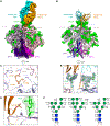
Figure 1.
Figure 1.. Cryo-EM structure of PGDM1400 in complex with BG505 DS-SOSIP.664 reveals an extensive electrostatic interaction network along the trimer axis
(A) Cryo-EM density reconstruction of PGDM1400 in complex with BG505 DS-SOSIP.664. (B) Atomic model of PGDM1400 in complex with BG505 DS-SOSIP.664, with glycans shown as green spheres. Env protomers are identified by subscript A, B, and C. (C) Three Asps at the tip of the CDRH3 of PGDM1400 show electrostatic interactions with Arg166 on each of the three protomers at the apex cavity of the Env trimer. Orientation is rotated 90° from (A). (D) The sulfated tyrosine (Tys)100f orients toward the Fab body with the sulfate moiety contacting K169. (E) Density is shown in detail for neighboring N160 glycans. Orientation is 120° from (B). (F) Glycan interactions are observed with all three N160s, with buried surface area shown on top and the number of hydrogen bonds (HBs) below. See also Figure S1.
Figure 2.
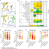
Figure 2.. Cross-species rational design with human bNAb and SIV Env leads to PGT145-R100aS with neutralization breadth similar to PGDM1400
(A) The SIV Env structure bound by human bNAb PGT145 highlights unfavorable interactions at position 169 (top). PGT145 in complex with HIV-1 BG505 shows that 169 adopts an unfavorable rotamer (bottom). (B) Two of four PGT145 variants showed improved neutralization of SIV isolates. The experimental combination of PGT145 with PGT145-R100aS is also shown, with each antibody present at half the total concentration. Gray shading with asterisk denotes isolates where substantial neutralization was detected but did not reach 50% at 50 mg/mL, prohibiting a half-maximal inhibitory concentration calculation. Median is calculated using only strains showing neutralization. (C) IC80 neutralization of a panel of 208 HIV-1 isolates shows improved breadth and potency of PGT145R100aS (left), which is even greater when only considering clade C (right). Median and geometric mean of those strains neutralized are shown as solid and dashed black bars, respectively. (D) Resistant strains show an enrichment for residues other than K/R at position 169. See also Figure S2 and Table S3.
Figure 3.
Figure 3.. Cryo-EM structure of PGT145-R100aS in complex with BG505 DS-SOSIP.664
(A) Cryo-EM density reconstruction of PGT145-R100aS in complex with BG505 DS-SOSIP.664. (B) Atomic model of PGT145-R100aS with BG505 DS-SOSIP.664, with glycans shown as green spheres. (C) Two cryo-EM classes show the flexibility of the PGT145-R100aS Fab body relative to the stable CDRH3 and Env. (D) Glu 100h and one Tys100f show electrostatic interactions with Arg166, with two Env protomers at the apex cavity of the Env trimer, while a second Tys extends to interact with Lys121. Orientation is rotated 90° from (A). (E) Tys100f is oriented away from Lys169 and toward the Env central cavity. The R100aS serine side chain is shown. (F) Density is shown in detail for neighboring N160 glycans. Orientation is the same as (B). (G) Glycan interactions are observed with all three N160s, with buried surface area and HBs shown. See also Figures S3, S4.
Figure 4.
Figure 4.. PGDM1400 and PGT145-R100aS show distinct differences in the details of electrostatic interactions with the Env apex cavity and level of glycan engagement
(A) Overall superposition of the PGDM1400 and PGT145-R100aS structures aligned by the Env trimer shows a high level of overlap in the antibody approach. (B) The CDRH3 sequences differ substantially with 15 of 32 residues diverging. Matched sequences are shown in gray and mismatched residues in the color of the heavy chain. The borders of the CDRH3 are shown with black vertical lines. The Kabat subscript for residue 100 is shown above and below for each antibody. Below is an overlay of the CDRH3 structures from the Env alignment, and the left is oriented as in (A). (C) An overlay of the cavity interactions shows PGT145 penetrating deeper into the cavity through the Tys to reach Lys121 (left). The 90° rotation is shown in the center. (Right) The difference in Tys100f orientations. (D) PGDM1400 shows broader resolvable interactions with N160 than PGT145. (E) Scatterplot of the neutralization overlap shared between the two antibodies.
Figure 5.
Figure 5.. Lineage evolution of the PGT145-PGDM1400 reveals diverse CDRH3 development
(A) A phylogenetic tree of heavy chain was constructed based on maximum likelihood algorithm. D-gene evolution is shown to the right of the corresponding branches in the tree colored by motif. Three sequences total of 187 contain the DDD motif. Filled triangle indicates collapse of branches. Scale bar shows percent of nucleotide substitutions per site. (B) Sequence alignment of CDRH3 (Kabat convention used for antibody numbering, with lowercase letters indicating additional residues). (Top) Sequences arranged by relative percentage of breadth in comparable assays. (Bottom) Sequences arranged by relative median potency.
Figure 6.
Figure 6.. PGT145-like antibodies use diverse evolutionary paths to converge on the same general mechanism of recognition
(A) A sequence alignment, based on structure, of the CDRH3s is shown with Tyss in red. (B) Complex structures are shown for all the antibodies, aligned by Env to illustrate the different angles of approach. The protomer-protomer matching of Env was assessed by the best CDRH3 overlay. Dashed lines from the top of the Env protein surface to just below the deepest penetration depth highlight the difference in CDRH3 depth. (C) Overlay of the heavy chains for each antibody is shown. Alignment was by VH region, excluding the CDRH3s. (D) Overlay of the shared CDRH3 interactions is shown. Alignment of the complexes was done by Env as in (B). Env is represented as a transparent surface shown from the orientation of (B). The CDRH3s do not bend from (C); rather, the angle of approach of the entire Fv region changes to allow alignment of CDRH3 by Env superposition.

References

    1. Georgiev IS, Doria-Rose NA, Zhou T, Kwon YD, Staupe RP, Moquin S, Chuang GY, Louder MK, Schmidt SD, Altae-Tran HR, et al. (2013). Delineating antibody recognition in polyclonal sera from patterns of HIV-1 isolate neutralization. Science 340, 751–756. 10.1126/science.1233989. - DOI - PubMed
    1. Walker LM, Simek MD, Priddy F, Gach JS, Wagner D, Zwick MB, Phogat SK, Poignard P, and Burton DR (2010). A limited number of antibody specificities mediate broad and potent serum neutralization in selected HIV-1 infected individuals. PLoS Pathog. 6, e1001028. 10.1371/journal.ppat.1001028. - DOI - PMC - PubMed
    1. Landais E, Huang X, Havenar-Daughton C, Murrell B, Price MA, Wickramasinghe L, Ramos A, Bian CB, Simek M, Allen S, et al. (2016). Broadly Neutralizing Antibody Responses in a Large Longitudinal Sub-Saharan HIV Primary Infection Cohort. PLoS Pathog. 12, e1005369. 10.1371/journal.ppat.1005369. - DOI - PMC - PubMed
    1. Willis JR, Berndsen ZT, Ma KM, Steichen JM, Schiffner T, Landais E, Liguori A, Kalyuzhniy O, Allen JD, Baboo S, et al. (2022). Human immunoglobulin repertoire analysis guides design of vaccine priming immunogens targeting HIV V2-apex broadly neutralizing antibody precursors. Immunity 55, 2149–2167. 10.1016/j.immuni.2022.09.001. - DOI - PMC - PubMed
    1. Voss JE, Andrabi R, McCoy LE, de Val N, Fuller RP, Messmer T, Su CY, Sok D, Khan SN, Garces F, et al. (2017). Elicitation of Neutralizing Antibodies Targeting the V2 Apex of the HIV Envelope Trimer in a Wild-Type Animal Model. Cell Rep. 21, 222–235. 10.1016/j.celrep.2017.09.024. - DOI - PMC - PubMed

Publication types

MeSH terms

Substances

LinkOut - more resources