. 2025 Jan 16:14:101023.
doi: 10.1016/j.jshs.2025.101023. Online ahead of print.

Reduced PI3K(p110α) induces atrial myopathy, and PI3K-related lipids are dysregulated in athletes with atrial fibrillation

Affiliations

Reduced PI3K(p110α) induces atrial myopathy, and PI3K-related lipids are dysregulated in athletes with atrial fibrillation

Sebastian Bass-Stringer et al. J Sport Health Sci. .

Abstract

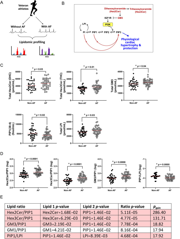
Background: Elucidating mechanisms underlying atrial myopathy, which predisposes individuals to atrial fibrillation (AF), will be critical for preventing/treating AF. In a serendipitous discovery, we identified atrial enlargement, fibrosis, and thrombi in mice with reduced phosphoinositide 3-kinase (PI3K) in cardiomyocytes. PI3K(p110α) is elevated in the heart with exercise and is critical for exercise-induced ventricular enlargement and protection, but the role in the atria was unknown. Physical inactivity and extreme endurance exercise can increase AF risk. Therefore, our objective was to investigate whether too little and/or too much PI3K alone induces cardiac pathology.

Methods: New cardiomyocyte-specific transgenic mice with increased or decreased PI3K(p110α) activity were generated. Multi-omics was conducted in mouse atrial tissue, and lipidomics in human plasma.

Results: Elevated PI3K led to an increase in heart size with preserved/enhanced function. Reduced PI3K led to atrial dysfunction, fibrosis, arrhythmia, increased susceptibility to atrial enlargement and thrombi, and dysregulation of monosialodihexosylganglioside (GM3), a lipid that regulates insulin-like growth factor-1 (IGF1)-PI3K signaling. Proteomic profiling identified distinct signatures and signaling networks across atria with varying degrees of dysfunction, enlargement, and thrombi, including commonalities with the human AF proteome. PI3K-related lipids were dysregulated in plasma from athletes with AF.

Conclusion: PI3K(p110α) is a critical regulator of atrial biology and function in mice. This work provides a proteomic resource of candidates for further validation as potential new drug targets and biomarkers for atrial myopathy. Further investigation of PI3K-related lipids as markers for identifying individuals at risk of AF is warranted. Dysregulation of PI3K may contribute to the association between increased cardiac risk with physical inactivity and extreme endurance exercise.

Keywords: Atrial fibrillation; Atrial myopathy; Exercise; Lipidomics; Proteomics.


Conflict of interest statement

Competing interests The authors declare that they have no competing interests.

Figures

Image, graphical abstract
Graphical abstract
Fig 1
Fig. 1
Dose-dependent increase in heart size of caPI3K Tg mice and normal/enhanced heart function. (A) Hearts of male and female Ntg, heterozygote caPI3K Tg(+/–), and homozygote caPI3K Tg(+/+) mice at dissection. All data in the figure come from mice at ∼20 week of age. (B) Representative ventricular sections. (C–E) HW, AW, and LW normalized to TL. Ntg (male/female): n = 12/11, caPI3K Tg(+/–): n = 22/21, caPI3K Tg(+/+): n = 14/23. (F) M-mode echocardiographic images. LVPW and percentage of fractional shortening (%). Ntg (male/female): n = 8/5, caPI3K Tg(+/–): n = 15/13, caPI3K Tg(+/+): n = 7/10. (G) Left, electrocardiogram traces. Dots and arrows highlight R- and P-waves, respectively. Right, episodes of arrhythmia expressed as a percentage over an interval of ∼5–6 min (further details in Supplementary Methods). Ntg (male/female): n = 8/9, caPI3K Tg(+/–): n = 9/10, caPI3K Tg(+/+): n = 8/7. Data points represent individual mice, presented as mean ± standard error of the mean (SEM). * indicates the one way analysis of variance followed by correction for multiple comparisons using Tukey's method. #p-value by Mann-Whitney U-test vs. caPI3K Tg(+/–) of same sex. AW = atrial weight; caPI3K Tg(+/–) = heterozygote constitutively activated phosphoinositide 3-kinase mutant; caPI3K Tg(+/+) = homozygote constitutively activated phosphoinositide 3-kinase mutant; HW = heart weight; LV = left ventricle; LVPW = left ventricular posterior wall thickness; LW = lung weight; Ntg = non-transgenic; RV = right ventricle; TL = tibia length.
Fig 2
Fig. 2
Distinct morphological phenotypes of heterozygote and homozygote dnPI3K Tg mice. (A) Hearts of male and female Ntg, heterozygote dnPI3K Tg(+/–), and homozygote dnPI3K Tg(+/+) mice at dissection. Images from the homozygote Tg highlight the range of phenotypes (i.e., dilated chambers and enlarged atria). All data presented in the figure come from mice at ∼20 week of age, unless otherwise stated. (B) Ventricular cross-sections. Unlike the Ntg and dnPI3K Tg(+/–), the dnPI3K Tg(+/+) presented with varying phenotypes (A and B—i.e., a subset of dnPI3K Tg(+/+)—had more dilated hearts at dissection (right). (C) HW normalized to TL. (D) Images of isolated ventricular myocytes and quantitation of myocyte area from male Ntg (n = 4), dnPI3K Tg(+/–) (n = 5), dnPI3K Tg(+/+) (n = 3), respectively. AW normalized to (E) TL and (F) HW. (C, E, and F): Ntg (male/female): n = 14/12, dnPI3K Tg(+/–): n = 32/26, dnPI3K Tg(+/+): n = 17/17. (G) LA samples stained with Masson's Trichrome showing atrial fibrosis (blue) in the dnPI3K Tg(+/+) mice and quantitation. Ntg (male/female): n = 5/6, dnPI3K Tg(+/–): n = 9/9, dnPI3K Tg(+/+): n = 6/9. (H) Upper, photos at dissection of atria from female mice (yellow arrows highlight thrombi). Lower, number of mice presenting with atrial thrombi at dissection. (I) M-mode echocardiographic measurements. LVPW thickness and percentage of fractional shortening (%). Ntg (male/female): n = 12/7, dnPI3K Tg(+/–): n = 15/20, dnPI3K Tg(+/+): n = 13/11. (J) P–V loop analysis curves and quantitation of SW in mice at ∼32 week. Ntg (male/female): n = 14/6, dnPI3K Tg(+/–): n = 11/9, dnPI3K Tg(+/+): n = 11/7. (K) Electrocardiogram traces. Dots and arrows highlight R and clear P-waves, respectively. (L) Percentage of mice displaying arrhythmia during an interval of ∼5–6 min. The threshold for selecting mice displaying arrhythmia was set at >4.5% based on data obtained from Ntg mice in Fig. 1H (further details in Supplementary Methods). Numbers within the bar graphs refer to the number of mice displaying arrhythmia. Ntg (male/female): n = 8/5, dnPI3K Tg(+/–): n = 14/13, dnPI3K Tg(+/+): n = 13/12. Data points represent individual mice, presented as mean ± SEM. * indicates the one way analysis of variance followed by Tukey's. † indicates Kruskal-Wallis followed by Dunn's (data not normally distributed). #p value by unpaired t test. AW = atrial weight; dnPI3K Tg(+/–) = heterozygote dominant-negative phosphoinositide 3-kinase mutant; dnPI3K Tg(+/+) = homozygote dominant-negative phosphoinositide 3-kinase mutant; HW = heart weight; LA = left atrium; LVPW = left ventricular posterior wall thickness; Ntg = non-transgenic; SW = stroke work; TL = tibial length.
Fig 3
Fig. 3
Atrial dysfunction in heterozygote and homozygote dnPI3K Tg models. (A) Left panel, echocardiographic image highlighting the location of the mouse LA, LAA, RA, LV, and RV. Right panel, representative images from female mice. Trace from Ntg (white) superimposed onto dnPI3K Tg(+/–)/Tg(+/+) shown in yellow. (B–E) Measurements from (B) LA reservoir function, (C) LA Booster pump fuction, (D) LAA, and (E) RA of male and female mice at ∼30 week. Ntg (male/female): n = 9/6, dnPI3K Tg(+/–): n = 6/8, dnPI3K Tg(+/+): n = 11/9. Data points represent individual mice, presented as mean ± SEM. * indicates the one way analysis of variance followed by Tukey's. dnPI3K Tg(+/–) = heterozygote dominant-negative phosphoinositide 3-kinase mutant; dnPI3K Tg(+/+) = homozygote dominant-negative phosphoinositide 3-kinase mutant; EF = ejection fraction; LA = left atrium; LAA = LA appendage; LV = left ventricular; Ntg = non-transgenic; RA = right atrium; RV = right ventricle.
Fig 4
Fig. 4
Distinct regulation of the atrial proteome in female heterozygote and homozygote dnPI3K Tg mice and comparative analysis with the human AF proteome. (A) Schematic overview of atrial size in each mouse model and proteomic workflow. Blue lines within the atria highlight the presence of fibrosis in the homozygote dnPI3K Tg(+/+) mice. (B) Heatmap of significant proteins (p < 0.01 analysis of variance) in RA from female Ntg, dnPI3K Tg(+/–), and dnPI3K Tg(+/+) divided into 3 subsets based on atrial size (S = small, M = medium, L = large) (728 proteins); mice: ∼20 week. n = 4/group. Proteins were identified in at least 70% of samples in at least 1 group. (C) Venn diagram of the number of proteins identified in each group n = 4 (Ntg/control: 5145, dnPI3K Tg(+/–): 5221, S_dnPI3K Tg(+/+): 5249, M_dnPI3K Tg(+/+): 5265, L_dnPI3K Tg(+/+): 4954). (D) PCA plot of valid values 70% cut off in at least 1 group across all groups. Based on the log2 intensity (LFQ) transformed value of all samples. Missing values were replaced by imputation from a normal distribution (downshift 1.8, width 0.3, Perseus). (E–G) Gene Ontology enrichment of Clusters 723, 724, and 725, respectively, GO:BP, KEGG, REAC using Gprofiler. –log10 p-value. (H) Comparative analysis venn diagram of shared proteins in each cluster across 3 human AF proteome studies (further details in Supplementary Data File 2). AF = atrial fibrillation; B4GALT1-CDG (CDG-2d) = beta-1,4-galactosyltransferase deficiency with carbohydrate deficient glycoprotein syndrome type IId; CHST6 = carbohydrate sulfotransferase 6; dnPI3K Tg(+/–) = heterozygote dominant-negative phosphoinositide 3-kinase mutant; dnPI3K Tg(+/+) = homozygote dominant-negative phosphoinositide 3-kinase mutant; EIEE15 = developmental and epileptic encephalopathy, 15; GO:BP = Gene Ontology: Biological Process; KEGG = Kyoto Encyclopedia of Genes of Genomes; LFQ = label free quantitation; MCT12 = monocarboxylate transporter 12; MCDC1 = macular corneal dystrophy (a condition associated with CHST6 gene); nLC-MS/MS = nano liquid chromatography-tandem mass spectrometry; Ntg = non-transgenic; PCA = principal component analysis; RA = right atrium; REAC = Reactome; ST3GAL3 = ST3 beta-galactoside alpha-2,3-sialyltransferase 3.
Fig 5
Fig. 5
Inflammatory signaling, GM3, and defective physiological signaling in a model of extreme endurance swim exercise and dnPI3K Tg(+/+) mice. (A) mRNAs related to TLR4 (Tlr4), NFκβ (Nfkbia), TNFα (Tnfaip3) signaling, and GM3 (GM3S gene expression, St3gal5), and (B) mRNAs related to the PI3K–Akt1–CEBPβ–Cited4 pathway by microarray in atria from sedentary mice (n = 3), and mice subjected to 6 week of extreme swim exercise (n = 6). Microarray deposited at https://www.ebi.ac.uk/arrayexpress/experiments/E-MTAB-3106/. (C) Schematic showing IGF1R–PI3K signaling in mice in response to aerobic swim training (green) and the differences with extreme swim exercise (red). (D) Gene expression by qPCR of Tlr4 relative to Hprt1 from ventricle tissue of 20-week-old dnPI3K Tg/caPI3K Tg mice and Ntg littermates: Ntg (male/female): n = 11/11, dnPI3K Tg(+/–): n = 11/12, dnPI3K Tg(+/+): n = 13/13; and caPI3K Tg mice: Ntg (male/female): n = 8/9, caPI3K Tg(+/–): n = 10/13, caPI3K Tg(+/+): n = 9/20. (E) Lipidomics was performed on ventricle and RA from 20-week-old dnPI3K Tg and Ntg littermates (male/female): Ntg: n = 5/10, dnPI3K Tg(+/–): n = 12/10, dnPI3K Tg(+/+): n = 7/11 (RA: female dnPI3K Tg(+/+): n = 10). For caPI3K Tg and Ntg littermates (male/female): Ntg: n = 5/7, caPI3K Tg(+/–): n = 9/9, caPI3K Tg(+/+): n = 8/12. Data points represent individual mice, presented as mean ± SEM. A and B: Unpaired t tests. D and E: * indicates one ay analysis of variance followed by Tukey's. †Kruskal-Wallis followed by Dunn's (data not normally distributed). Akt1 = AKT serine/threonine kinase 1; caPI3K Tg(+/–) = heterozygote constitutively activated phosphoinositide 3-kinase mutant; caPI3K Tg(+/+) = homozygote constitutively activated phosphoinositide 3-kinase mutant; CEBPβ = CCAAT/enhancer binding protein beta; Cited4 = Cbp/P300-interacting transactivator with Glu/Asp-rich carboxy-terminal Domain 4; dnPI3K Tg(+/–) = heterozygote dominant-negative phosphoinositide 3-kinase mutant; dnPI3K Tg(+/+) = homozygote dominant-negative phosphoinositide 3-kinase mutant; Hprt1= hypoxanthine phosphoribosyltransferase 1; IGF1R = insulin-like growth factor 1 receptor; NFκβ = nuclear factor kappa-light-chain-enhancer of activated B cells; Ntg = non-transgenic; PC = phosphatidylcholine; qPCR = quantitative polymerase chain reaction; PI3K = phosphoinositide 3-kinase; RA = right atrium; TLR4 = Toll-like receptor 4; TNFα = tumor necrosis factor alpha; GM3 = monosialodihexosylganglioside; GM3S = monosialodihexosylganglioside synthase .
Fig 6
Fig. 6
Dysregulation of GM3 and PI3K-related lipids in veteran athletes with AF. (A) Lipidomics on plasma from veteran athletes with and without AF. (B) Relationship of PI species with PI3K and GM3 and ceramide lipid species. Changes in regulators of the pathway can impact PI3K and the formation of PIP3. (C) Total Hex2Cer (DHC), Hex3Cer (THC), GM3, PIP1, and LPI. (D) Most significant lipid ratios related to the PI3K pathway. Athletes without AF, n = 37; athletes with AF, n = 41. Data presented as scatter plots and mean. (E) Lipid ratios corrected for age with a pgain > 10 (top 5 presented from the total number of ratios (>800); only 6 ratios had a pgain > 10; Supplementary Data File 2). * indicates unpaired-test. † indicates Mann-Whitney U test (data not normally distributed). AF = atrial fibrillation; GM3 = monosialodihexosylganglioside; Hex2Cer (DHC) = dihexosylceramide; Hex3Cer (THC) = trihexosylceramide; IGF1R = insulin-like growth factor 1 receptor; LPI = lysophosphatidylinositol; m/z = mass-to-charge ratio; PI = phosphatidylinositol; PI3K = phosphoinositide 3-kinase; PIP = phosphatidylinositol monophosphate.
Fig 7
Fig. 7
Schematics highlighting cellular processes and signaling pathways during progression of atrial pathology, and regular aerobic exercise vs. Extreme/intense exercise. (A) Summary of differences in atrial size, function, fibrosis, and thrombi formation in Ntg, heterozygote dnPI3K Tg(+/–), and homozygote dnPI3K Tg(+/+). Cellular processes and signaling pathways dysregulated in the atria based on proteomics data. (B) Overview of IGF1–PI3K signaling in the heart with regular and extreme exercise (upper panel). IGF1 is elevated with regular exercise (left side, blue). In a setting of increased PI3K (exercise or caPI3K Tg), PIP2 is converted to PIP3, limiting the availability of PIP2 for TLR4 signaling and subsequently reducing pro-inflammatory cytokines. Extreme/intense exercise is associated with elevated LPS, lower IGF1, and increased TLR4 signaling (right side, red). There is also evidence of increased GM3 in response to increased TNFα, which would inhibit IGF1R–PI3K signaling. Increased GM3 reduces membrane fluidity and disrupts the normal interaction of IGF1R with caveolins to induce downstream protective PI3K signaling. Collectively these signaling alterations will contribute to cell death, fibrosis, and arrhythmia. Lower panel: Potential contribution of PI3K signaling to the reverse J-shaped relationship between physical activity and cardiac risk. An inverse relationship between PI3K activity and cardiac risk is hypothesized. Akt1 = AKT serine/threonine kinase 1; caPI3K Tg(+/–) = heterozygote constitutively activated phosphoinositide 3-kinase mutant; caPI3K Tg(+/+) = homozygote constitutively activated phosphoinositide 3-kinase mutant; C/EBPβ = CCAAT/enhancer binding protein beta; Cited4 = Cbp/P300-interacting transactivator with Glu/Asp-rich carboxy-terminal domain 4; cGMP = cyclic guanosine monophosphate; dnPI3K Tg(+/–) = heterozygote dominant-negative phosphoinositide 3-kinase mutant; dnPI3K Tg(+/+) = homozygote dominant-negative phosphoinositide 3-kinase mutant; GM3 = monosialodihexosylganglioside; IGF1R = insulin-like growth factor 1 receptor; LA = left atrium; LPS = lipopolysaccharide; NFκβ = nuclear factor kappa-light-chain-enhancer of activated B cell; Ntg = non-transgenic; ROS = reactive oxygen species; pAkt = phosphorylated Akt; qPCR = quantitative polymerase chain reaction; PI3K = phosphoinositide 3-kinase; PIP2 = phosphatidylinositol 4,5-bisphosphate; PKG = protein kinase G; PTEN = phosphatase and tensin homolog deleted on chromosome 10; RA = right atrium; ROBO = roundabout; TLR4 = Toll-like receptor 4; TNFα = tumor necrosis factor-alpha.

References

    1. Elliott A.D., Middeldorp M.E., Van Gelder I.C., Albert C.M., Sanders P. Epidemiology and modifiable risk factors for atrial fibrillation. Nat Rev Cardiol. 2023;20:429. doi: 10.1038/s41569-023-00834-w. - DOI - PubMed
    1. Kornej J., Börschel C.S., Benjamin E.J., Schnabel R.B. Epidemiology of atrial fibrillation in the 21st century: Novel methods and new insights. Circ Res. 2020;127:4–20. - PMC - PubMed
    1. Chen Y.C., Voskoboinik A., Gerche A.L., Marwick T.H., McMullen J.R. Prevention of pathological atrial remodeling and atrial fibrillation: JACC State-of-the-Art review. J Am Coll Cardiol. 2021;77:2846–2864. - PubMed
    1. McMullen J.R., Shioi T., Zhang L., et al. Phosphoinositide 3-kinase(p110alpha) plays a critical role for the induction of physiological, but not pathological, cardiac hypertrophy. Proc Natl Acad Sci U S A. 2003;100:12355–12360. - PMC - PubMed
    1. Weeks K.L., Gao X., Du X.J., et al. Phosphoinositide 3-kinase p110alpha is a master regulator of exercise-induced cardioprotection and PI3K gene therapy rescues cardiac dysfunction. Circ Heart Fail. 2012;5:523–534. - PubMed

LinkOut - more resources