

Enhancing elemental release and antibacterial properties of resin-based dental sealants with calcium phosphate, bioactive glass, and polylysine
- PMID: 39827118
- PMCID: PMC11742498
- DOI: 10.1186/s12903-025-05489-2
Enhancing elemental release and antibacterial properties of resin-based dental sealants with calcium phosphate, bioactive glass, and polylysine
Abstract
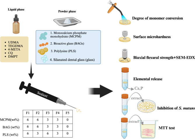
Background: This study aimed to develop ion-releasing and antibacterial resin-based dental sealants comprising 3 to 6 wt% monocalcium phosphate monohydrate (MCPM, M), 3 to 6 wt% bioactive glass (BAG, B), and 3 to 6 wt% polylysine (PLS, P). The physical properties, mechanical performance, cytotoxicity, and inhibition of S. mutans biofilm by these materials were subsequently evaluated.
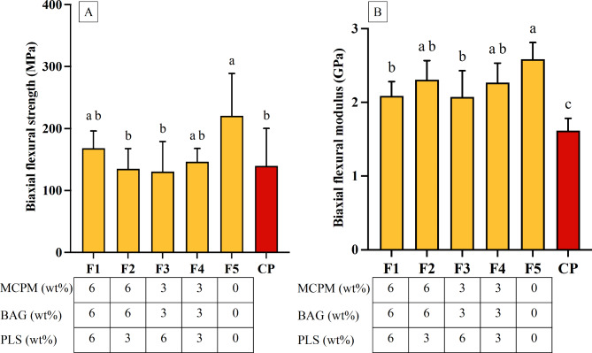
Methods: Five experimental dental sealants were formulated as follows: F1 (M6B6P6), F2 (M6B6P3), F3 (M3B3P6), F4 (M3B3P3), and F5 (M0B0P0, serving as the control). ClinproXT (CP, 3 M, Saint Paul, MN, USA) was used for commercial comparison. The degree of monomer conversion (DC) was determined using attenuated total reflectance-Fourier transform infrared spectroscopy (n = 5). The biaxial flexural strength (n = 6) and Vickers surface microhardness (n = 5) of the materials were evaluated after a 24-hour immersion in water. The element release over 4 weeks was measured using inductively coupled plasma-optical emission spectrometry (ICP-OES) (n = 3). The cell viability of mouse fibrosarcoma cells exposed to the extract was assessed via an MTT assay (n = 3). Additionally, the inhibition of S. mutans biofilm was tested (n = 3). Statistical analysis was conducted using one-way ANOVA and the Tukey HSD test.
Results: The lowest DC among experimental sealants was obtained from F1 (66 ± 4%), which was significantly higher than CP (54 ± 2%, p < 0.001). The lowest biaxial flexural strength was obtained from F3 (131 ± 47 MPa). This was comparable to that of CP (140 ± 58 MPa, p = 0.992). The lowest surface microhardness among experimental materials was detected with F2 (19 ± 2 Vickers hardness number), which was higher than that of CP (12 ± 1 Vickers hardness number, p = 0.003). Furthermore, high cell viability of > 90% after exposure to extracts from the experimental materials was detected, which was similar to that observed with CP. Additionally, the experimental materials exhibited higher Ca and P release compared to CP and showed a potential trend for reducing S. mutans biofilm formation. Increasing additive concentrations exhibited minimal effects on material properties, except for enhanced elemental release and a slight reduction in BFM with higher PLS content.
Conclusion: The experimental sealants provided sufficient physical and mechanical strength and maintained cell viability and bacterial inhibition with higher elemental release than the commercial product.
Keywords: Biaxial flexural strength; Bioactive glass; Calcium phosphate; Degree of monomer conversion; Polylysine; Resin sealant.
© 2025. The Author(s).
Conflict of interest statement
Declarations. Ethics approval and consent to participate: Not applicable. Consent for publication: Not applicable. Competing interests: Anne Young has a patent on the use of MCPM in dental composites licensed to a dental company (Davis Schottlander and Davis Ltd., Letchworth Garden City, UK). The other authors declare no conflicts of interest.
Figures


Similar articles
-
The in vitro assessment of resin coating materials containing calcium phosphate, bioactive glass, and polylysine for glass ionomer cement restorations.Biomater Investig Dent. 2025 Jan 14;12:42783. doi: 10.2340/biid.v12.42783. eCollection 2025. Biomater Investig Dent. 2025. PMID: 40124687 Free PMC article.
-
Assessment of Physical/Mechanical Performance of Dental Resin Sealants Containing Sr-Bioactive Glass Nanoparticles and Calcium Phosphate.Polymers (Basel). 2022 Dec 12;14(24):5436. doi: 10.3390/polym14245436. Polymers (Basel). 2022. PMID: 36559804 Free PMC article.
-
Monomer conversion, dimensional stability, strength, modulus, surface apatite precipitation and wear of novel, reactive calcium phosphate and polylysine-containing dental composites.PLoS One. 2017 Nov 14;12(11):e0187757. doi: 10.1371/journal.pone.0187757. eCollection 2017. PLoS One. 2017. PMID: 29136013 Free PMC article.
-
The effect of an antibacterial monomer on the antibacterial activity and mechanical properties of a pit-and-fissure sealant.J Am Dent Assoc. 2011 Feb;142(2):184-93. doi: 10.14219/jada.archive.2011.0062. J Am Dent Assoc. 2011. PMID: 21282685 Clinical Trial.
-
Synthesis, monomer conversion, and mechanical properties of polylysine based dental composites.J Mech Behav Biomed Mater. 2024 Mar;151:106398. doi: 10.1016/j.jmbbm.2024.106398. Epub 2024 Jan 14. J Mech Behav Biomed Mater. 2024. PMID: 38237205
References
-
- Shoaee S, Ghasemi E, Sofi-Mahmudi A, Shamsoddin E, Tovani-Palone MR, Roshani S, Heydari MH, Yoosefi M, Masinaei M, Azadnaejafabadi S, et al. Global, regional, and national burden and quality of care index (QCI) of oral disorders: a systematic analysis of the global burden of disease study 1990–2017. BMC Oral Health. 2024;24(1):116. - PMC - PubMed
-
- Carvalho JC. Caries process on occlusal surfaces: evolving evidence and understanding. Caries Res. 2014;48(4):339–46. - PubMed
-
- Akinlotan M, Chen B, Fontanilla TM, Chen A, Fan VY. Economic evaluation of dental sealants: a systematic literature review. Community Dent Oral Epidemiol. 2018;46(1):38–46. - PubMed
-
- Quinonez RB, Downs SM, Shugars D, Christensen J, Vann WF Jr. Assessing cost-effectiveness of sealant placement in children. J Public Health Dent. 2005;65(2):82–9. - PubMed
MeSH terms
Substances
Grants and funding
LinkOut - more resources
Full Text Sources
Medical
Molecular Biology Databases
Miscellaneous
