. 2025 Jan 18;24(1):20.
doi: 10.1186/s12933-025-02573-3.

ICAM1 blockade improves ischemic muscle reperfusion in diabetic mice

Affiliations

ICAM1 blockade improves ischemic muscle reperfusion in diabetic mice

Ninon Foussard et al. Cardiovasc Diabetol. .

Abstract

Background: Chronic Limb-Threatening Ischemia (CLTI) represents the most advanced stage of Peripheral Artery Disease (PAD) and is associated with dire prognosis, characterized by a substantial risk of limb amputation and diminished life expectancy. Despite significant advancements in therapeutic interventions, the underlying mechanisms precipitating the progression of PAD to CLTI remain elusive.

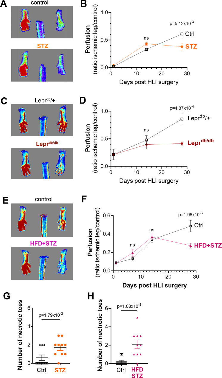
Methods: Considering diabetes is one of the main risk factors contributing to PAD exacerbation into CLTI, we compared hind limb ischemia recovery in HFD STZ vs. non-HFD STZ mice to identify new mechanisms responsible for the exacerbation of PAD.

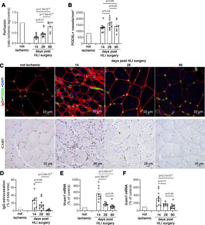
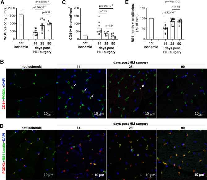
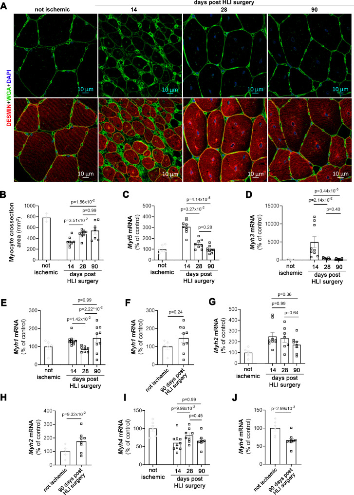
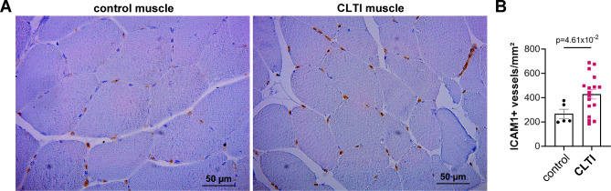
Results: We used three different mouse models of diabetes and found that blood flow recovery in HFD STZ mice is altered only from day 14 post-surgery. Consistent with this kinetics, we found that angiogenesis and myogenesis which typically occur between day five and day 14 post-surgery are not impaired in mice in which diabetes was induced by a high fat diet and streptozotocin injections (HFD STZ mice). On the contrary, we found that capillary functionality e.i. acquisition of functional intercellular junctions and immune quiescence is impaired in HFD + STZ mice. Notably, 28 days after hind limb ischemia surgery, HFD + STZ mice display significantly increased capillary permeability to IgG and significantly increased levels of ICAM1. This was associated with an increased macrophage infiltration and an impaired myocyte differentiation. Importantly, we used ICAM1-blocking antibodies to demonstrate that increased ICAM1 expression in HFD + STZ mice decreases white blood cell circulation velocity within the microcirculation, which impairs its perfusion. Notably anti-ICAM1 therapy did diminish macrophage infiltration and oxidative stress but not myopathy suggesting that myopathy characterized by small myocytes expressing higher level of MYH2 could be responsible for microangiopathy.

Conclusion: ICAM1 expression by the microvasculature impairs ischemic muscle reperfusion in HFD + STZ mice. Importantly, the increase in blood flow between day 14 and day 90 post-HLI surgery is not associated with an increased capillary density but with an improved functionality of capillaries.

Keywords: Endothelial activation; Limb ischemia; Microcirculation; Perfusion.


Conflict of interest statement

Declarations. Competing interests: The authors declare no competing interests.

Figures

Fig. 1
Fig. 1
Blood flow recovery in the ischemic foot of diabetic mice is impaired from day 14 following HLI surgery. AB Diabetes was induced, or not, in 8 week-old C57BL/6 mice via STZ injections, 12 weeks later mice underwent HLI surgery. CD Leprdb/db male mice and their control littermates (Leprdb/+) underwent HLI surgery at 12 weeks of age. EF 8 week-old C57BL/6 mice were fed with HFD or a normal chow diet; one month later HFD-fed mice received STZ administration to induce hyperglycemia. At 20 weeks of age, mice underwent HLI surgery. Ischemic foot perfusion was assessed via laser Doppler perfusion imaging 7, 14, and 28 days after surgery. A, C, E Representative images taken 28 days after surgery are shown. B, D,F Ischemic foot perfusion was quantified as the perfusion ratio of ischemic foot over control foot (n = 10 mice in each group). GH The number of necrotic toes was counted (n = 10 mice in each group). Mann Whitney tests
Fig. 2
Fig. 2
Increased ischemic foot perfusion occurring from day 14 is associated with improved microvessel functionality. 20 week-old C57BL/6 mice underwent HLI surgery. Mice were sacrificed 14, 28, and 90 days post-surgery to harvest their ischemic tibialis anterior muscle. A Ischemic foot perfusion was assessed via laser Doppler perfusion imaging and quantified as the perfusion ratio of ischemic foot over control foot (n = 4, 10, 10, and 7 mice per group mice in each group). B Muscle cross-sections were immunostained with anti-PODXL antibodies to identify endothelial cells. Capillary density was quantified as the number of PODXL + vessels per mm² (n = 10, 10, 10, and 7 mice per group). C Muscle cross-sections were immunostained with anti-IgG antibodies (in red) and anti-PODXL antibodies (in green). Muscle cross-sections were immunostained with anti-ICAM1 antibodies (in brown). D Vessel leakage was quantified as the IgG + area fraction (n = 10, 10, 10, and 7 mice per group). EVcam1 mRNA expression was quantified via RT-qPCR (n = 6, 9, 8 and 10 mice per group). FIcam1 mRNA expression was quantified via RT-qPCR (n = 6, 10, 8, and 10 mice per group). Kruskal-Wallis test followed by Dunn’s multiple comparison test
Fig. 3
Fig. 3
Increased ischemic foot perfusion occurring from day 14 is associated with improved microvessel perfusion. 20 week-old C57BL/6 mice underwent HLI surgery. Mice were sacrificed 14, 28, and 90 days post-surgery to harvest their ischemic tibialis anterior muscle. A Before sacrifice, mice were administered Rhodamine 6G intravenously to label white blood cells (WBCs). WBC movement was visualized using a stereomicroscope and WBC velocity was calculated using ImageJ software (n = 8, 10, 10, and 7 mice per group). BC Muscle cross-sections were immunostained with anti-CD41 antibodies to identify platelets B Representative pictures are shown. C Thrombi density was quantified as the number of CD41 + elements per mm2 (n = 3, 8, 8, and 7 mice per group). DE mice were administered fluorescein isothiocyanate (FITC)-labeled BS1 lectin 2 min before sacrifice. D Ischemic muscle was co-immunostained with anti-PODXL antibodies to identify total capillaries. E The percentage of capillaries perfused with BS1 lectin was calculated (n = 1, 8, 10, and 10). Kruskal-Wallis test followed by Dunn’s multiple comparison test
Fig. 4
Fig. 4
Increased ischemic foot perfusion occurring from day 14 is associated with improved myocyte differentiation. 20 week-old C57BL/6 mice underwent HLI surgery. Mice were sacrificed 14, 28, and 90 days post-surgery to harvest their ischemic tibialis anterior muscle. A Muscle cross-sections were stained with FITC-labelled WGA to stain membranes and anti-Desmin antibodies to identify myocytes. B The mean skeletal muscle cross-section surface area was measured (n = 2, 8, 10 and 7). CMyf5 mRNA expression was quantified via RT-qPCR (n = 6, 9, 8, and 9). DMyh3 mRNA expression was quantified via RT-qPCR (n = 6, 9, 8, and 9). EFMyh1 mRNA expression was quantified via RT-qPCR (E: n = 5, 9, 8 and 9) (F: n = 5 and 9). GHMyh2 mRNA expression was quantified via RT-qPCR (G: n = 5, 9, 7 and 8) (H: n = 5 and 8). IJMyh4 mRNA expression was quantified via RT-qPCR (I: n = 6, 10, 8 and 8) (J: n = 6 and 8). B, C, D, E, G, and I: Kruskal-Wallis test followed by Dunn’s multiple comparison test; F, H and J: Mann Whitney tests
Fig. 5
Fig. 5
HFD STZ mice display impaired capillary functionality. Diabetes was induced or not in 8 week-old C57BL/6 mice by a HFD diet followed by STZ injections. At 20 weeks of age, mice underwent HLI surgery. Mice were sacrificed 28 days post-HLI surgery to harvest their ischemic tibialis anterior muscle. A Muscle cross-sections were immunostained with anti-PODXL antibodies to identify endothelial cells. B Capillary density was quantified as the number of PODXL + vessels/mm2 (n = 10 and 10 mice per group). C Muscle cross-sections were immunostained with anti-IgG antibodies (in red) and anti-PODXL antibodies (in green). D Vessel leakage was quantified as the IgG + area fraction (n = 10, and 9 mice per group). E Muscle cross-sections were immunostained with anti-CD41 antibodies to identify platelets and anti-PODXL antibodies to identify endothelial cells. F Thrombi density was quantified as the number of CD41 + elements per mm2 (n = 3, 8, 8, and 7 mice per group). GVcam1 mRNA expression was quantified via RT-qPCR (n = 10 and 10 mice per group). HIcam1 mRNA expression was quantified via RT-qPCR (n = 10 and 10 mice per group). ISele mRNA expression was quantified via RT-qPCR (n = 9 and 8 mice per group). J Muscle cross-sections were immunostained with anti-ICAM1 antibodies (in red) and anti-CD31 antibodies (in green). K ICAM1 level was quantified as the percentage of ICAM1 positive area (n = 10 and 9 mice per group). (L) Before sacrifice, mice were administered Rhodamine 6G intravenously to label white blood cells (WBCs). WBC movement was visualized using a stereomicroscope and WBC velocity was calculated using ImageJ software (n = 9 and 10 mice per group). M Mice were administered fluorescein isothiocyanate (FITC)-labeled BS1 lectin 2 minutes before sacrifice. Ischemic muscle was co-immunostained with anti-PODXL antibodies to identify total capillaries. The percentage of capillaries perfused with BS1 lectin was calculated (n = 10 and 10 mice per group). Mann Whitney tests
Fig. 6
Fig. 6
ICAM1 blockade improved capillary perfusion and ischemic foot function. Diabetes was induced in 8 week-old C57BL/6 mice by a HFD diet followed by STZ injections. At 20 weeks of age, mice underwent HLI surgery. From Day 14 to Day 28 post surgery mice were treated with anti-ICAM1 antibodies or isotype controls. Mice were sacrificed 28 days post-HLI surgery to harvest their ischemic tibialis anterior muscle. A Muscle cross-sections were immunostained with anti-PODXL antibodies to identify endothelial cells. Capillary density was quantified as the number of PODXL + vessels/mm2 (n = 10 mice per group). B Muscle cross-sections were immunostained with anti-IgG antibodies. Vessel leakage was quantified as the IgG + area fraction (n = 10 mice per group). C Before sacrifice, mice were administered Rhodamine 6G intravenously to label white blood cells (WBCs). WBC movement was visualized using a stereomicroscope and WBC velocity was calculated using ImageJ software (n = 10 mice per group). D Mice were administered fluorescein isothiocyanate (FITC)-labeled BS1 lectin 2 minutes before sacrifice. Ischemic muscle was co-immunostained with anti-PODXL antibodies (in red) to identify total capillaries. E The percentage of capillaries perfused with BS1 lectin was calculated (n = 10 mice per group). F, G Ischemic foot perfusion was assessed via laser Doppler perfusion imaging 28 days after surgery. F Representative images taken 28 days after surgery are shown. G Ischemic foot perfusion was quantified as the perfusion ratio of ischemic foot over control foot (n = 6 and 7 mice in each group). H Ischemic foot function was assessed using the grip strength test (n = 6 and 9 mice per group). Mann Whitney tests
Fig. 7
Fig. 7
Capillaries within the ischemic muscles of patients with CLTI express higher levels of ICAM1. A Muscle sections from human biopsies were immunostained with anti-ICAM1 antibodies. B The number of ICAM1 positive vessels per mm2 was counted (n = 5 and 17). Mann Whitney test
Fig. 8
Fig. 8
HFD STZ mice display myopathy. Diabetes was induced or not in 8 week-old C57BL/6 mice by a HFD diet followed by STZ injections. At 20 weeks of age, mice underwent HLI surgery. Mice were sacrificed 28 days post-HLI surgery to harvest their ischemic tibialis anterior muscle. A Muscle cross-sections were stained with FITC-labelled WGA to stain membranes and anti-Desmin antibodies to identify myocytes. B Myogenesis was quantified as the Desmin positive surface area (n = 10 mice per group). C The mean skeletal muscle cross-section surface area was measured ((n = 10 and 9 mice per group). DMyh3 mRNA expression was quantified via RT-qPCR (n = 10 mice per group). EMyh2 mRNA expression was quantified via RT-qPCR (n = 10 mice per group). FMyh1 mRNA expression was quantified via RT-qPCR (n = 10 mice per group). GMyh4 mRNA expression was quantified via RT-qPCR (n = 10 mice per group). HMyh7 mRNA expression was quantified via RT-qPCR (n = 10 mice per group). I Muscle cross-sections were immunostained with anti-MYH2 antibodies to identify Type IIa fibers. J The number of MYH2 positive fibers per mm2 was counted (n = 10 mice per group). Mann Whitney tests

References

    1. Criqui MH, Matsushita K, Aboyans V, Hess CN, Hicks CW, Kwan TW, McDermott MM, Misra S, Ujueta F, on behalf of the American Heart Association Council on Epidemiology and Prevention; Council on Arteriosclerosis, Thrombosis and Vascular Biology; Council on Cardiovascular Radiology and Intervention; Council on Lifestyle and Cardiometabolic Health; Council on Peripheral Vascular Disease; and Stroke Council. Lower Extremity Peripheral Artery Disease: Contemporary Epidemiology, Management Gaps, and Future Directions: A Scientific Statement From the American Heart Association. Circulation [Internet]; 144, (2021). Accessed 24 Jul 2022. 10.1161/CIR.0000000000001005 - PMC - PubMed
    1. Lin J, Chen Y, Jiang N, Li Z, Xu S. Burden of peripheral artery disease and its attributable risk factors in 204 countries and territories from 1990 to 2019. Front Cardiovasc Med. 2022;9:868370. - PMC - PubMed
    1. Song P, Rudan D, Zhu Y, Fowkes FJI, Rahimi K, Fowkes FGR, Rudan I. Global, regional, and national prevalence and risk factors for peripheral artery disease in 2015: an updated systematic review and analysis. Lancet Global Health. 2019;7:e1020–30. - PubMed
    1. Roth GA, Mensah GA, Johnson CO, Addolorato G, Ammirati E, Baddour LM, Barengo NC, Beaton AZ, Benjamin EJ, Benziger CP, Bonny A, Brauer M, Brodmann M, Cahill TJ, Carapetis J, Catapano AL, Chugh SS, Cooper LT, Coresh J, Criqui M, DeCleene N, Eagle KA, Emmons-Bell S, Feigin VL, Fernández-Solà J, Fowkes G, Gakidou E, Grundy SM, He FJ, Howard G, Hu F, Inker L, Karthikeyan G, Kassebaum N, Koroshetz W, Lavie C, Lloyd-Jones D, Lu HS, Mirijello A, Temesgen AM, Mokdad A, Moran AE, Muntner P, Narula J, Neal B, Ntsekhe M, Moraes de Oliveira G, Otto C, Owolabi M, Pratt M, Rajagopalan S, Reitsma M, Ribeiro ALP, Rigotti N, Rodgers A, Sable C, Shakil S, Sliwa-Hahnle K, Stark B, Sundström J, Timpel P, Tleyjeh IM, Valgimigli M, Vos T, Whelton PK, Yacoub M, Zuhlke L, Murray C, Fuster V, Roth GA, Mensah GA, Johnson CO, Addolorato G, Ammirati E, Baddour LM, Barengo NC, Beaton A, Benjamin EJ, Benziger CP, Bonny A, Brauer M, Brodmann M, Cahill TJ, Carapetis JR, Catapano AL, Chugh S, Cooper LT, Coresh J, Criqui MH, DeCleene NK, Eagle KA, Emmons-Bell S, Feigin VL, Fernández-Sola J, Fowkes FGR, Gakidou E, Grundy SM, He FJ et al. Global Burden of cardiovascular diseases and risk factors. J Am Coll Cardiol. 2020;76:2982–3021. - PMC - PubMed
    1. Prompers L, Huijberts M, Apelqvist J, Jude E, Piaggesi A, Bakker K, Edmonds M, Holstein P, Jirkovska A, Mauricio D, Ragnarson Tennvall G, Reike H, Spraul M, Uccioli L, Urbancic V, Van Acker K, van Baal J, van Merode F, Schaper N. High prevalence of ischaemia, infection and serious comorbidity in patients with diabetic foot disease in Europe. Baseline results from the Eurodiale study. Diabetologia. 2007;50:18–25. - PubMed

MeSH terms

Substances

LinkOut - more resources