. 2025 Jan 18;15(1):2413.
doi: 10.1038/s41598-024-83424-1.

TCTEX1D2 is essential for sperm flagellum formation in mice

Affiliations

TCTEX1D2 is essential for sperm flagellum formation in mice

Ryua Harima et al. Sci Rep. .

Abstract

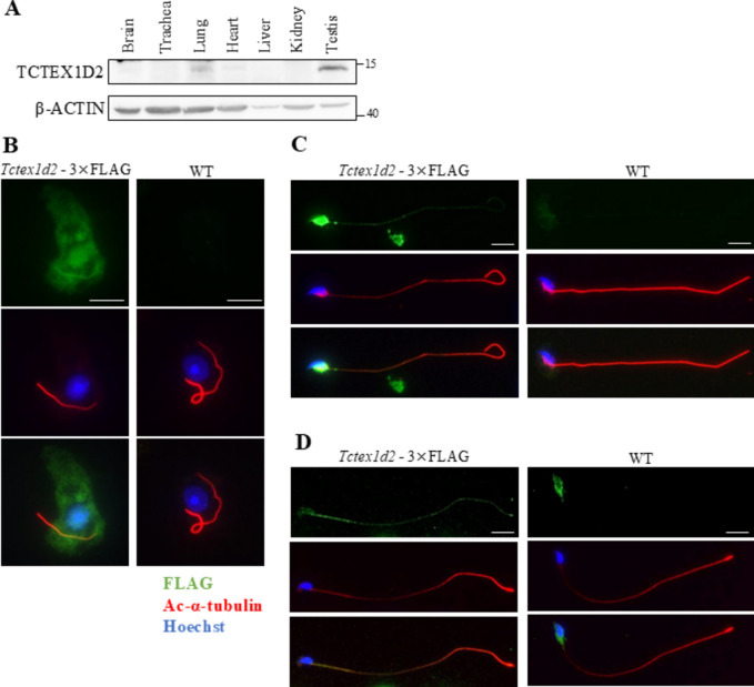
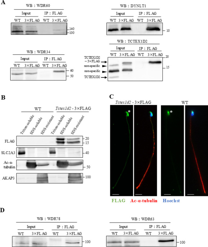
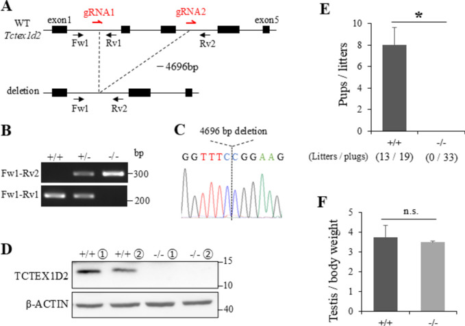
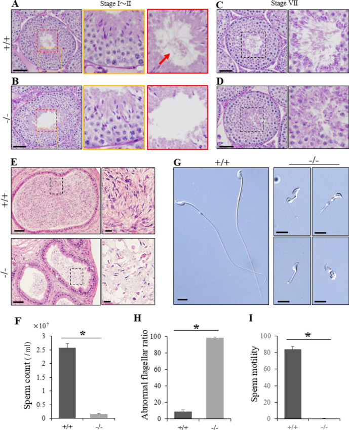
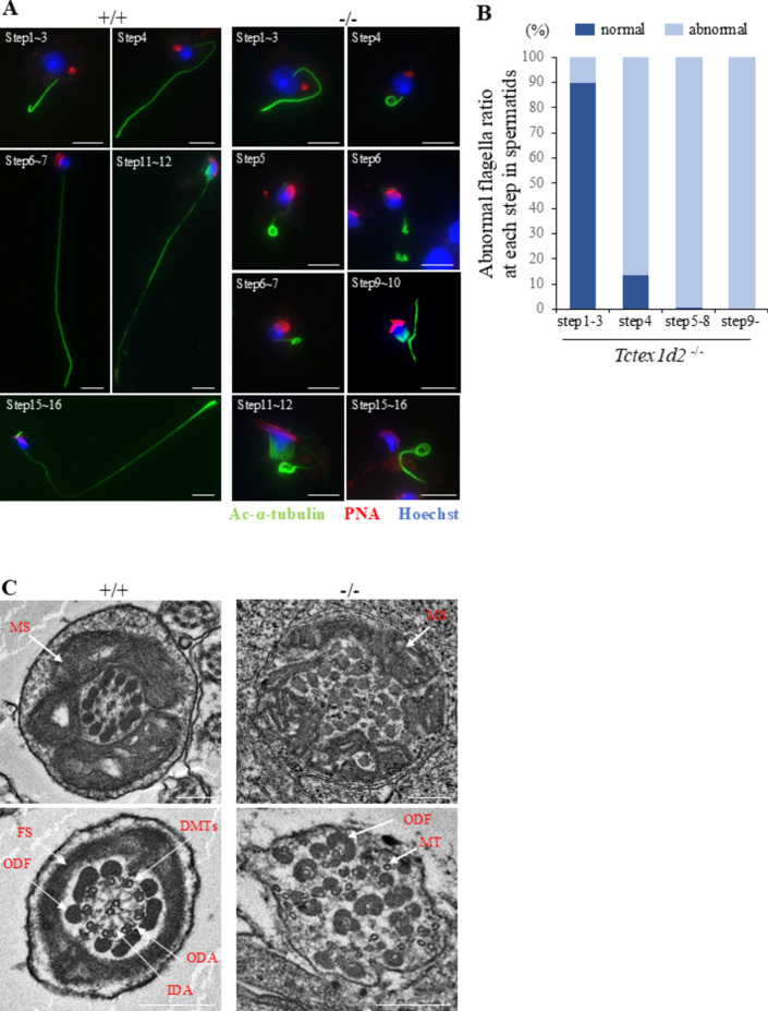
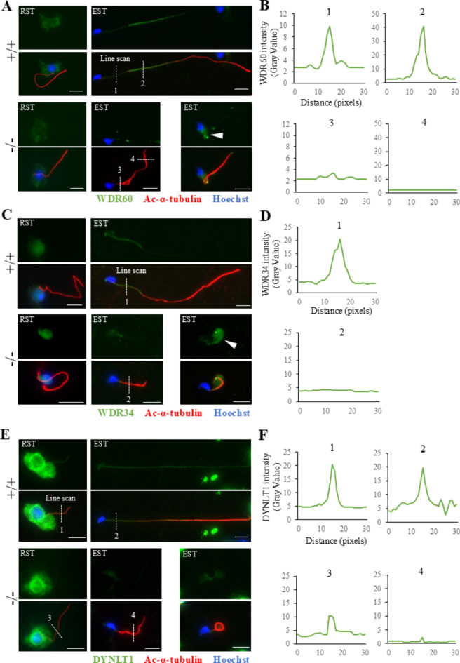
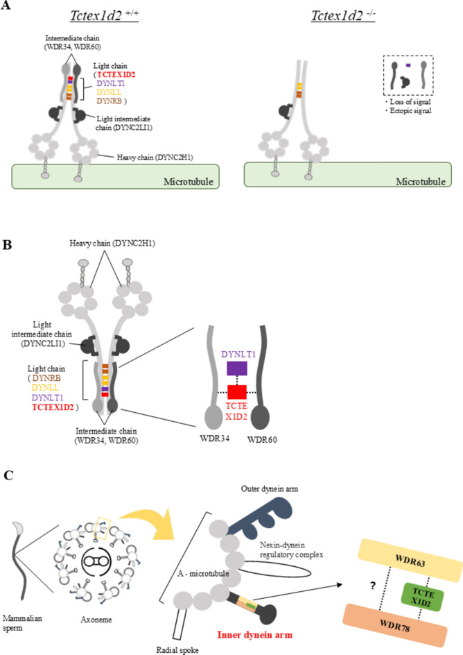
Flagella and cilia are widely conserved motile structures, in mammalian, sperm possess flagella. Large protein complexes called dynein, including cytoplasmic dynein 2 and axonemal dynein, play a role in the formation of cilia and flagella. The function of each subunit component of dynein complexes in sperm flagellum formation remains unclear. One such subunit is TCTEX1D2. Co-immunoprecipitation studies showed that TCTEX1D2 interacted with cytoplasmic dynein 2 subunits WDR34, WDR60, and DYNLT1 in the testes. Furthermore, TCTEX1D2 also interacted with WDR63 and WDR78, subunits of inner dynein arm, which is axonemal dynein. Tctex1d2-/- mice generated in this study exhibited male infertility due to flagellar dysplasia, and the axonemal structures were disrupted inside the flagella. Further, the localization of cytoplasmic dynein 2 subunits was abnormal in in Tctex1d2-/- mice. In contrast, the motile cilia of Tctex1d2-/- mice were normal. Overall, we revealed that TCTEX1D2 is important for the assembly of cytoplasmic dynein 2 and inner dynein arm and functions in two distinct dynein complexes during mouse sperm flagellum formation. This is only in sperm flagellum formation, not in cilia formation.

Keywords: Cytoplasmic dynein 2; Inner dynein arm; Male infertility; Sperm flagella; Spermatogenesis; TCTEX1D2.


Conflict of interest statement

Competing interests: The authors declare no competing interests.

Figures

Fig. 1
Fig. 1
TCTEX1D2 was high expression in the testis and localized along the flagella from round spermatids to elongated spermatids and the manchette. (A) Protein expression analysis of mouse TCTEX1D2 in various organs using western blotting (WB); TCTEX1D2 was highly expressed in the testes. (BD) ICC in WT and Tctex1d2 - 3 × FLAG germ cells to analyze the localization of TCTEX1D2 (green: anti-FLAG, red: anti-acetylated-α-tubulin, blue: Hoechst33342). TCTEX1D2 localized to flagella from early round spermatids to elongated spermatids. In addition, TCTEX1D2 localized to the manchette. (B) Round spermatids in steps 1–3. (C) Elongated spermatids of steps 10–12. (D) Elongated spermatids of steps 15–16. Scale bars are 10 µm.
Fig. 2
Fig. 2
TCTEX1D2 interacted with the subunits of cytoplasmic dynein 2, and localized within axoneme and interacted with axonemal dynein. (A) Co-immunoprecipitation in the testes of Tctex1d2 - 3 × FLAG mice. In the testes, TCTEX1D2 interacted with WDR60, WDR34, and DYNLT1. (B) WB of cauda epididymis sperm fractionated into Triton X-100 soluble, SDS-soluble, and SDS-insoluble fractions from WT and Tctex1d2 - 3 × FLAG mice. TCTEX1D2 detected in SDS-soluble and SDS-insoluble fractions. (C) ICC of cauda epididymis sperm from WT and Tctex1d2 - 3 × FLAG mice (green: FLAG, red: acetylated-α-tubulin, blue: Hoechst33342). TCTEX1D2 localized along the sperm flagella. Asterisks indicate non-specific signals. Scale bars are 10 µm. (D) Co-immunoprecipitation with WDR63 and WDR78 in the testes of Tctex1d2 - 3 × FLAG mice. TCTEX1D2 interacted with WDR63 and WDR78. The used membrane was same as that in (A).
Fig. 3
Fig. 3
TCTEX1D2 was required for male fertility. (A) Schematic representation of the generation of Tctex1d2−/− mice. The gRNA was designed between exons 1 and 2 and between exons 3 and 4. Genotyping primer pairs were Fw1 and Rv1 for Tctex1d2+/+ mice, Fw1 and Rv2 for Tctex1d2−/− mice. (B) Genotyping and (C) Sanger sequencing showed that Tctex1d2−/− mice had a 4696 bp deletion within the target region. (D) WB of tissue from mouse testes confirmed that TCTEX1D2 protein expression was completely lost in Tctex1d2−/− mice. (E) Male Tctex1d2+/+ and Tctex1d2−/− mice were mated with two female WT mice. We observed 33 plugs in Tctex1d2−/− mice; however, Tctex1d2−/− mice were unable to sire any offspring. Error bars indicate means ± S.D. Two-tailed Student’s t-test, Tctex1d2+/+ mice (n = 3), Tctex1d2−/− mice (n = 4), *p < 0.05. (F) The ratio of testes weight / body weight. There were no differences between Tctex1d2+/+ and Tctex1d2−/− mice. Error bars indicate means ± S.D. Two-tailed Student’s t-test, n = 3, n.s. : no significant.
Fig. 4
Fig. 4
Tctex1d2 is important for sperm flagellum formation. (AD) Periodic acid Schiff hematoxylin (PAS-H) stain of seminiferous tubules of Tctex1d2+/+ and Tctex1d2−/− mice. In stage I–II seminiferous tubules, Tctex1d2−/− mice showed layered structures of germ cells (B; yellow box), similar to that in Tctex1d2+/+ mice (A; yellow box). However, the flagella were barely observed in the lumen in Tctex1d2−/− mice (B; red box) compared to Tctex1d2+/+ mice (A; red box arrow). In the lumen of stage VII seminiferous tubules, only a few elongated spermatids were observed in Tctex1d2−/− mice (D; black box) in contrast to Tctex1d2+/+ mice (C; black box). Scale bars are 50 µm. (E) Hematoxylin and eosin (H&E) stain of the cauda epididymis. Many sperm were observed in Tctex1d2+/+ mice, whereas only very few sperm were observed in Tctex1d2−/− mice. Scale bars are 50 µm. (F) Sperm counts in the cauda epididymis. Tctex1d2−/− mice had significantly reduced sperm counts compared to Tctex1d2+/+ mice. Error bars are means ± S.D. Two-tailed Student’s t-test, n = 3, *p < 0.05. (G) Cauda epididymis sperm observed using phase contrast microscopy. Sperm flagella of Tctex1d2−/− mice showed morphological abnormalities. Scale bars are 10 µm. (H) The ratio of sperm with abnormal flagella in the cauda epididymis. Almost all sperm showed abnormal flagella in Tctex1d2−/− mice. Error bars are means ± S.D. Two-tailed Student’s t-test, n = 3, *p < 0.05. (I) Sperm motility analysis. There was no motility in Tctex1d2−/− mice sperm. Error bars are means ± S.D. Two-tailed Student’s t-test, n = 3, *p < 0.05.
Fig. 5
Fig. 5
Abnormal assembly of sperm flagella in Tctex1d2−/− mice occurred from step 4 round spermatids. (A) The analysis of flagellum formation in spermiogenesis using ICCs (green: anti-acetylated-α-tubulin, red: PNA lectin, blue: Hoechst33342). Scale bars are 10 µm. (B) Abnormal flagella ratio at each step spermatids in Tctex1d2−/− mice. N = 125 cells for step1-3, n = 22 cells for step4, n = 416 cells for step5-8, n = 230 cells for after step9 were counted in three different mice. In Tctex1d2+/+ mice, flagella elongated as the spermatids differentiated. However, Tctex1d2−/− mice showed markedly abnormal flagella after step 4 spermatids. (C) Observation of the internal structure of the flagella of spermatids using transmission electron microscopy. In Tctex1d2+/+ mice, microtubule 9 + 2 structures and other structures were neatly aligned in the axonemes. In contrast, in Tctex1d2−/− mice, the internal structure of the axonemes were disorganized. The 9 + 2 structures, inner dynein arm (IDA), outer dynein arm (ODA), outer dense fiber (ODF), and fibrous sheath (FS) were not aligned properly. Scale bars are 250 nm. MS, mitochondrial sheath; DMTs, doublet microtubules; ODF, outer dense fibers; FS, fibrous sheath; ODA, outer dynein arm; IDA, inner dynein arm; MTs, microtubule (not doublet).
Fig. 6
Fig. 6
TCTEX1D2 played a role in the assembly of cytoplasmic dynein 2 subunits. (A) Expression analysis of WDR60 in Tctex1d2−/− mice using ICC (green: WDR60, red: acetylated-α-tubulin, blue: Hoechst33342). There was no expression of WDR60 in flagella of round spermatids in Tctex1d2+/+ mice. In elongated spermatids, abnormal expression was observed in Tctex1d2−/− mice (left: loss of signal, right arrow heads: ectopic expression). (B) The line scan analysis of WDR60 intensity in elongated spermatids of Tctex1d2+/+ mice (line 1, 2) and Tctex1d2−/− mice (line 3, 4). (C) Expression analysis of WDR34 in Tctex1d2−/− mice using ICC (green: WDR34, red: acetylated-α-tubulin, blue: Hoechst33342). There was no expression of WDR34 was in flagella of round spermatids, as seen with WDR60. In elongated spermatids, abnormal expression was observed in Tctex1d2−/− mice (left: loss of signal, right arrow heads: ectopic expression). (D) The line scan analysis of WDR34 intensity in elongated spermatids of Tctex1d2+/+ mice (line 1) and Tctex1d2−/− mice (line 2). (E) Expression analysis of DYNLT1 in Tctex1d2−/− mice using ICC (green: DYNLT1, red: acetylated-α-tubulin, blue: Hoechst33342). The signal was decreased in round spermatids and absent in elongated spermatids in Tctex1d2−/− mice. (F) The line scan analysis of DYNLT1 intensity in elongated spermatids of Tctex1d2+/+ mice (line 1, 2) and Tctex1d2−/− mice (line 3, 4). RST, round spermatid; EST, elongated spermatid.
Fig. 7
Fig. 7
The disorder of cytoplasmic dynein 2 in Tctex1d2−/− mice and predicted schematic diagram of the surrounding structure of TCTEX1D2. (A) Schematic diagram of the defect of cytoplasmic dynein 2 subunits in Tctex1d2−/− mice. In Tctex1d2−/− mice elongated spermatids, DYNC2LI1, WDR60, WDR34 and DYNLT1 showed abnormal localization and were not assembled in the cytoplasmic dynein 2 complex. (B) Schematic diagram of the cytoplasmic dynein 2 complex in sperm flagellum formation. The dashed lines indicate protein—protein interaction. (C) Model of TCTEX1D2 localization within inner dynein arm in mouse sperm flagella. The dashed lines indicate predicted protein—protein interaction.

Similar articles

References

    1. Gibbons, I. R. Cilia and flagella of eukaryotes. J Cell Biol91, 107s–124s. 10.1083/jcb.91.3.107s (1981). - PMC - PubMed
    1. Carvalho-Santos, Z., Azimzadeh, J., Pereira-Leal, J. B. & Bettencourt-Dias, M. Evolution: Tracing the origins of centrioles, cilia, and flagella. J Cell Biol194, 165–175. 10.1083/jcb.201011152 (2011). - PMC - PubMed
    1. Lindemann, C. B. & Lesich, K. A. Functional anatomy of the mammalian sperm flagellum. Cytoskeleton (Hoboken)73, 652–669. 10.1002/cm.21338 (2016). - PubMed
    1. Satir, P. & Christensen, S. T. Overview of structure and function of mammalian cilia. Annu Rev Physiol69, 377–400. 10.1146/annurev.physiol.69.040705.141236 (2007). - PubMed
    1. Allen, R. D. A reinvestigation of cross-sections of cilia. J Cell Biol37, 825–831. 10.1083/jcb.37.3.825 (1968). - PMC - PubMed

Publication types

LinkOut - more resources