. 2025 May;97(5):894-906.
doi: 10.1002/ana.27181. Epub 2025 Jan 19.

Adult Neurogenesis in the Subventricular Zone of Patients with Huntington's and Parkinson's Diseases and following Long-Term Treatment with Deep Brain Stimulation

Affiliations

Adult Neurogenesis in the Subventricular Zone of Patients with Huntington's and Parkinson's Diseases and following Long-Term Treatment with Deep Brain Stimulation

Marta Snapyan et al. Ann Neurol. 2025 May.

Abstract

Objective: Parkinson's and Huntington's diseases are characterized by progressive neuronal loss. Previous studies using human postmortem tissues have shown the impact of neurodegenerative disorders on adult neurogenesis. The extent to which adult neural stem cells are activated in the subventricular zone and whether therapeutic treatments such as deep brain stimulation promote adult neurogenesis remains unclear. The goal of the present study is to assess adult neural stem cells activation and neurogenesis in the subventricular zone of patients with Huntington's and Parkinson's diseases who were treated or not by deep brain stimulation.

Methods: Postmortem brain samples from Huntington's and Parkinson's disease patients who had received or not long-term deep brain stimulation of the subthalamic nucleus were used.

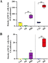
Results: Our results indicate a significant increase in the thickness of the subventricular zone and in the density of proliferating cells and activated stem cells in the brain of Huntington's disease subjects and Parkinson's disease patients treated with deep brain stimulation. We also observed an increase in the density of immature neurons in the brain of these patients.

Interpretation: Overall, our data indicate that long-term deep brain stimulation of the subthalamic nucleus promotes cell proliferation and neurogenesis in the subventricular zone that are reduced in Parkinson's disease. Taken together, our results also provide a detailed characterization of the cellular composition of the adult human subventricular zone and caudate nucleus in normal condition and in Parkinson's and Huntington's diseases and demonstrate the plasticity of these regions in response to neurodegeneration. ANN NEUROL 2025;97:894-906.


Conflict of interest statement

Nothing to report.

Figures

FIGURE 1
FIGURE 1
Measurements of the thickness of the hypocellular gap (HG) and subventricular zone (SVZ) from control brains and from patients who suffered from Huntington's (HD) and Parkinson's diseases (PD), with and without deep brain stimulation (DBS) treatment. (A) Schematic diagram of a coronal section taken through the adult human brain region that contains the lateral ventricle (LV), ependymal cell layer (EL), HG, SVZ, and caudate nucleus (Cd). (B) Transverse sections from a control brain (upper panel) and brains obtained from a HD patient (lower panel). Astrocytes and neural stem cells (NSCs) are stained with glial fibrillary acidic protein (GFAP) (red), proliferative cells are stained with proliferating cell nuclear antigen (PCNA) (green), and cell nuclei are stained with DAPI (blue). (C) Transverse sections from a control brain (upper panel) and brains obtained from PD patients with (middle panel) and without (lower panel) long‐term DBS treatment. (D, E) Box plot diagrams representing the thickness of the HG (D) and SVZ (E) in the different conditions. (F) Box plot diagrams representing the density of PCNA+ cells in the SVZ of postmortem human tissues under different conditions. **p < 0.01 using the Mann–Whitney test (D–F). Scale bars: 20 μm (B, C). [Color figure can be viewed at www.annalsofneurology.org]
FIGURE 2
FIGURE 2
Increased density of putative activated neural stem cells (NSCs) in the subventricular zone (SVZ) of Huntington's disease (HD) and deep brain stimulation (DBS) brains. (A) High magnification image of the SVZ showing proliferating cell nuclear antigen (PCNA) (green) and glial fibrillary acidic protein (GFAP) (red) labeled cells in a DBS brain. (B) Box plot diagrams representing the density of putative activated NSCs (GFAP+/PCNA+) in the SVZ of HD and Parkinson's disease (PD) brains, with or without DBS, and in the brains of matched controls. **p < 0.01 using the Mann–Whitney test. Scale bar: 5 μm (A). [Color figure can be viewed at www.annalsofneurology.org]
FIGURE 3
FIGURE 3
Increased density of immature doublecortin (DCX+) neurons in the subventricular zone (SVZ) and the caudate nucleus (Cd) of Huntington's disease (HD) and deep brain stimulation (DBS) brains. (A) Box plot diagrams representing the density of immature DCX+ neurons in the SVZ under different conditions. A greater density of immature neurons was observed in the SVZ of DBS brains. This density was even higher in the SVZ of HD brains. No DCX+ cells were observed in the brains of Parkinson's disease (PD) subjects who had not received DBS treatment. (B) Box plot diagrams representing the density of immature neurons (DCX+) in the Cd. No DCX+ cells were observed in the Cd of PD and control brains. Some DCX+ cells were observed in the Cd of PD subjects who had received DBS. A significant number of DCX+ cells were observed in the Cd of HD brains. **p < 0.01 using the Mann–Whitney test. [Color figure can be viewed at www.annalsofneurology.org]
FIGURE 4
FIGURE 4
Morphological features of doublecortin (DCX+) cells observed in the subventricular zone (SVZ) and the caudate nucleus (Cd) of Huntington's disease (HD) brains. (A‐C) High magnification images showing DCX+ cells (red) in the SVZ of a control brain (A), in the SVZ of a HD brain (B) and in the Cd of the same HD brain (C). Note the extended processes of the DCX+ cell shown in the Cd, compared to the one observed in the SVZ. DCX+ cells do not stain for glial fibrillary acidic protein (GFAP) (shown in white). Cell nuclei are stained with DAPI (4′,6‐diamidino‐2‐phenylindole) (blue). Scale bar: 10 μm (A‐C). (D) Box plot diagrams representing cell body diameters of DCX+ immature neurons observed in the SVZ of control and HD brains, and in the Cd of HD brain. **p < 0.01 using the Mann–Whitney test. (E) Sholl analysis indicating the number of intersections with DCX+ processes found at increasing distances from their cell body in the SVZ of control (purple) and HD (red) brains, and in the Cd of HD brain (blue). $$$p < 0.001, p < 0,0001 for HD SVZ vs. Ctrl SVZ and ###p < 0.001 for HD Cd vs. HD SVZ, with 2‐way repeated measures analysis of variance (ANOVA) followed by a Bonferroni's multiple comparisons test. (F) Box plot diagrams representing the total length of the processes for DCX+ cells observed in the SVZ of control and HD brains, and in the Cd of HD brains. **p < 0.01 using the Mann–Whitney test. [Color figure can be viewed at www.annalsofneurology.org]

Comment in

References

    1. Marymonchyk A, Malvaut S, Saghatelyan A. In vivo live imaging of postnatal neural stem cells. Development 2021;148:1–13. - PubMed
    1. Ming GL, Song H. Adult neurogenesis in the mammalian brain: significant answers and significant questions. Neuron 2011;70:687–702. - PMC - PubMed
    1. Obernier K, Alvarez‐Buylla A. Neural stem cells: origin, heterogeneity and regulation in the adult mammalian brain. Development 2019;146:1–15. - PMC - PubMed
    1. Sorrells SF, Paredes MF, Cebrian‐Silla A, et al. Human hippocampal neurogenesis drops sharply in children to undetectable levels in adults. Nature 2018;555:377–381. - PMC - PubMed
    1. Boldrini M, Fulmore CA, Tartt AN, et al. Human hippocampal neurogenesis persists throughout aging. Cell Stem Cell 2018;22:589–599. e5. - PMC - PubMed