. 2025 May 30;66(5):722-737.
doi: 10.1093/pcp/pcaf012.

Evidence for peroxisomal redundancy among the glucose-6-phosphate dehydrogenase isoforms of Arabidopsis thaliana

Affiliations

Evidence for peroxisomal redundancy among the glucose-6-phosphate dehydrogenase isoforms of Arabidopsis thaliana

Loreen Linnenbrügger et al. Plant Cell Physiol. .

Abstract

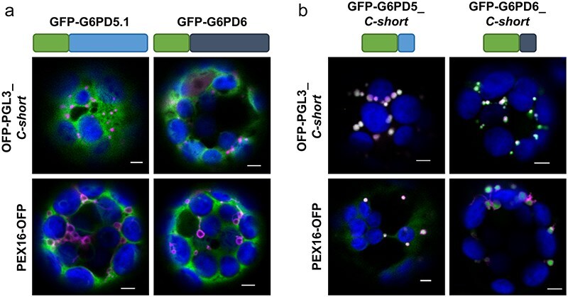
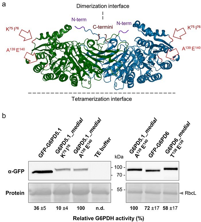
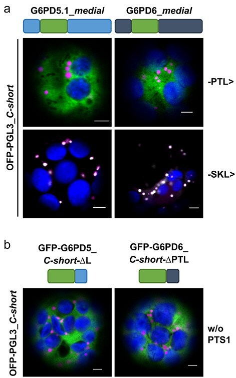
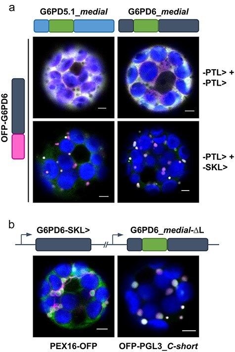
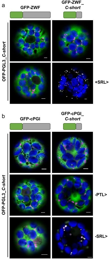
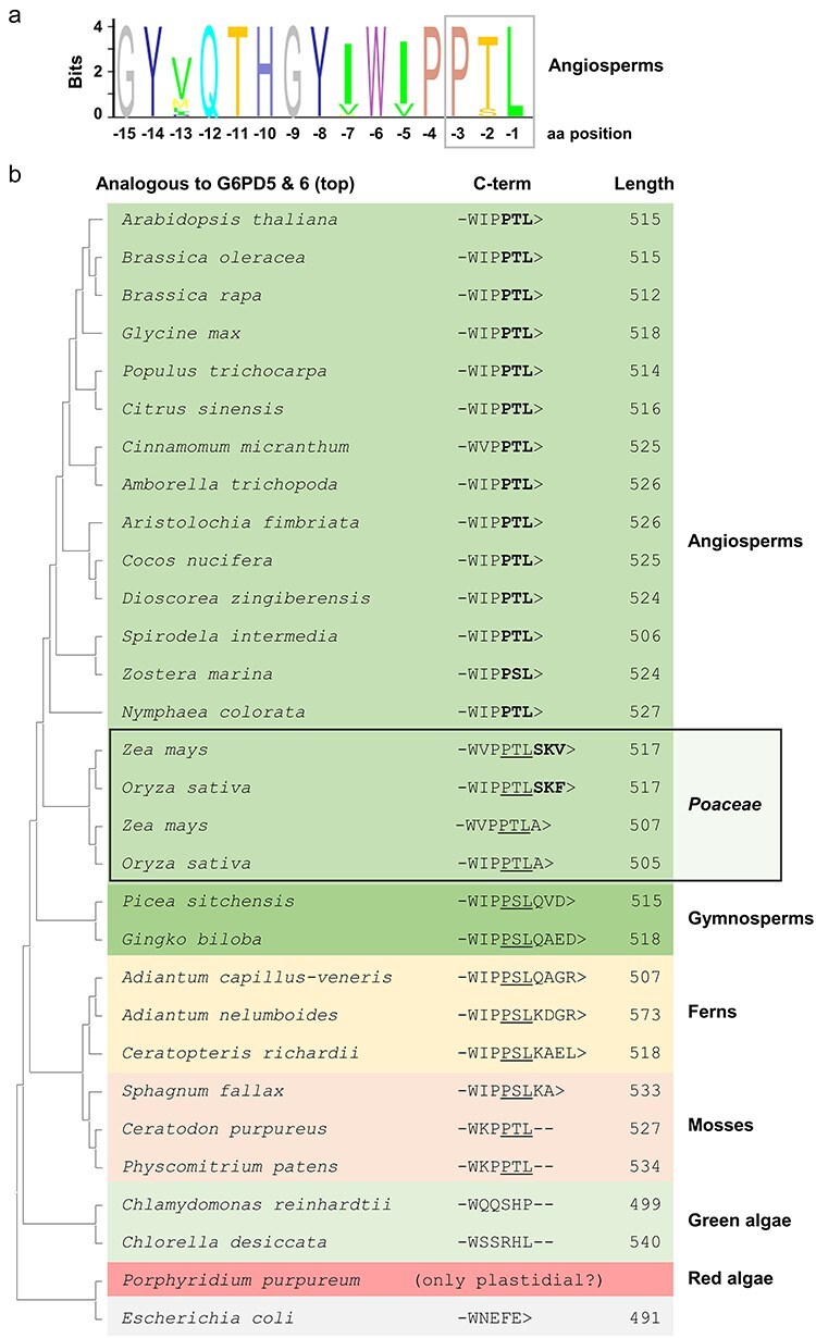
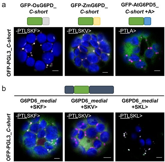
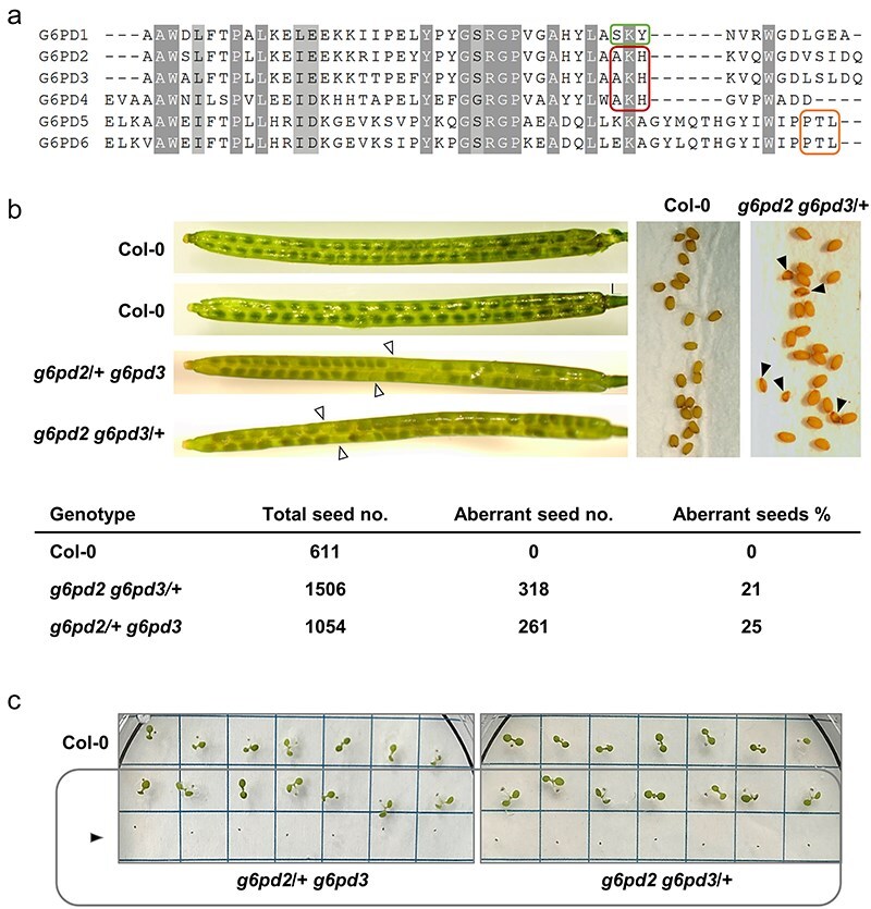
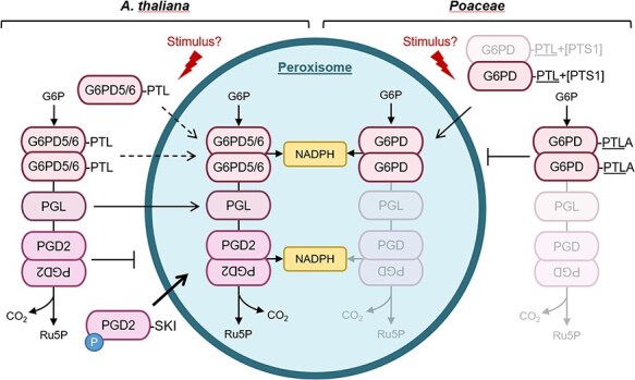
The oxidative pentose phosphate pathway (OPPP) plays an important role in the generation of reducing power in all eukaryotes. In plant cells, the OPPP operates in several cellular compartments, but as full cycle only in the plastid stroma where it is essential. As suggested by our recent results, OPPP reactions are also mandatory inside peroxisomes, at least during fertilization. For the first enzyme of the OPPP, glucose-6-phosphate dehydrogenase (G6PD), we previously showed that one Arabidopsis isoform (G6PD1) can be directed to peroxisomes under specific circumstances. Since g6pd1 knock-out plants are viable, we aimed at elucidating potential redundancy regarding peroxisomal targeting among the other G6PD isoforms. Localization studies of so far cytosolic annotated G6PD5 and G6PD6 (both ending -PTL>) using different reporter fusions of full-length versus the last 50 amino acids revealed that GFP-C-short versions are efficiently imported into peroxisomes. Modification of the final tripeptide to a canonical peroxisomal targeting signal type 1 (PTS1) also resulted in peroxisomal localization of the full-length versions and revealed that G6PD5/6 import may occur as homo- or heterodimer. Interestingly, the new noncanonical PTS1 motif is highly conserved among the cytosolic G6PD isoforms of the Angiosperms, whereas members of the Poaceae (rice and maize) possess two variants, one ending with an additional amino acid (-PTLA>) and the other one extended by a stronger PTS1 motif. From both evolutionary and physiological perspectives, we postulate that G6PD import as homo- and heterodimer restricted the acquisition of more efficient peroxisomal targeting motifs to leave some G6PDH activity in the cytosol.

Keywords: G6PD; OPPP; dimeric protein import; dual targeting; peroxisomes; targeting motifs.


Conflict of interest statement

None declared.

Figures

Figure 1.
Figure 1.
The last 50 amino acids of the cytosolic Arabidopsis G6PD isoforms confer peroxisomal targeting. Coexpression in Arabidopsis wild-type (Col-0) mesophyll protoplasts. (a) G6PD5.1 and G6PD6 full-length or (b) only the part coding for the last 50 amino acids (ending -PTL>) N-terminally fused to GFP (schematically depicted above) with two different peroxisomal markers: top panels, soluble marker OFP-PGL3_C-short (the last 50 amino acids of PGL3 ending -SKL>); bottom panels, membrane marker PEX16-OFP. Note that the signals of the GFP-G6PD full-length fusions are evenly distributed in the cytosol, showing no overlap with the peroxisomal markers. Only the corresponding G6PD_C-short versions accumulate in dot-like structures that colocalize with both markers (bright dot-like signals). The images show single optical sections as the merger of all fluorescent channels (for single-channel images, see Supplementary Fig. S1). GFP is depicted in green, OFP in magenta, and chlorophyll autofluorescence in blue; white signals indicate colocalization or very close signals (<200 nm) of GFP and OFP. Scale bars, 3 μm.
Figure 2.
Figure 2.
One of two G6PD5.1_medial GFP fusions proved to be highly active. (a) G6PD5 dimer (PDB file obtained from Alphafold, modified with Protean 3D), with dimerization and tetramerization interfaces (dashed lines) based on human G6PD enzymes (Supplementary Fig. S2). The positions selected for insertion of the GFP reporter into G6PD5 lie in outer loop regions (after 75 and 139 amino acids, respectively), as indicated by white arrows with red outlines. Note that 3D structure prediction for the N-terminus (N-term) is weak. (b) Immunoblot analysis of protein extracts prepared from g6pd5-1 g6pd6 double-mutant protoplasts upon transfection with the indicated GFP constructs or TE buffer (control), using anti-GFP antibodies (α-GFP). Protein refers to signals on the Ponceau S-stained blot with RubisCO large subunit (RbcL) as loading reference. G6PDH activity was determined ∼24 h post-transfection with at least three measurements per extract from three independent experiments. Relative activities were calculated (±SD) based on corresponding band intensities on the α-GFP immunoblots, with G6PD5.1_medial (A139 E140) set to 100%. Abbreviation: n.d., not detectable.
Figure 3.
Figure 3.
Peroxisomal import of G6PD5 and G6PD6 depends on a C-terminal PTS1 motif. Coexpression in Arabidopsis wild-type (Col-0) mesophyll protoplasts. (a) The new medial GFP fusions of G6PD5.1 and G6PD6 (schematically depicted above) ending either -PTL> (top) or with canonical PTS1 motif -SKL> (bottom) like the soluble peroxisomal marker (OFP-PGL3_C-short ending -SKL>). While the original versions show no overlap with the OFP signals, the engineered -SKL> versions show colocalization (bright signals) in dot-like structures. (b) Coexpression with the corresponding C-short constructs (schematically depicted above) lacking a PTS1 motif, either upon deletion of only the last amino acid (ΔL) or the entire PTS1 motif (ΔPTL). Note that both result in a cytosolic localization pattern. The images show single optical sections as the merger of all fluorescent channels (for single-channel images, see Supplementary Fig. S3). GFP is depicted in green, OFP in magenta, and chlorophyll autofluorescence in blue; white signals indicate colocalization or very close signals (<200 nm) of GFP and OFP. Scale bars, 3 μm.
Figure 4.
Figure 4.
Peroxisomal import of G6PD5 and G6PD6 may occur as hetero- or homo-dimer. Coexpression in Arabidopsis wild-type (Col-0) mesophyll protoplasts: (a) The medial GFP fusions of G6PD5.1 (left) and G6PD6 (right, both ending -PTL>) were coexpressed with OFP-G6PD6 ending either -PTL> (top) or with engineered canonical PTS1 motif -SKL> (bottom). In both cases of the latter, a clear change in colocalization (white signals) occurred. (b) Localization study using a double cassette construct, containing G6PD6 ending -SKL> (without fluorescent reporter) and the G6PD6_medial GFP fusion without the last amino acid (ΔL, to destroy the PTS1 motif), upon coexpression with the peroxisomal membrane marker PEX16-OFP (left) or soluble peroxisomal marker OFP-PGL3_C-short (right). In both cases, dot-like accumulations of the GFP signal were observed, either surrounded by OFP (left) or colocalizing with OFP (white signals, right). The images show single optical sections as the merger of all fluorescent channels (for single-channel images, see Supplementary Fig. S6). GFP is depicted in green, OFP in magenta, and chlorophyll autofluorescence in blue; white signals indicate colocalization or very close signals (<200 nm) of GFP and OFP. Scale bars, 3 μm.
Figure 5.
Figure 5.
Peroxisomal import of G6PD variants is governed by more than the last three amino acids. Coexpression of the soluble peroxisomal marker (OFP-PGL3_C-short, ending -SKL>) in Arabidopsis wild-type (Col-0) mesophyll protoplasts, (a) with the G6PD homolog of E. coli (ZWF, ‘Zwischenferment’, ending -EFE>) fused to GFP (left), or only the last 50 amino acids (right). Wild-type versions (top panels), plus canonical PTS1 motif -SRL> (bottom panels). (b) Coexpression of OFP-PGL3_C-short with cPGI (ending -PQM>) fused to GFP (left), or only the last 54 amino acids (right). Wild-type versions (top panels), or similar to G6PD5 and G6PD6 with engineered -PTL> (centre panels), and with canonical PTS1 motif -SRL> (lower panels). Note that only the short versions with additional or engineered canonical PTS1 motif colocalize with the peroxisomal marker. The images show single optical sections as the merger of all fluorescent channels (for single-channel images, see Supplementary Fig. S8). GFP is depicted in green, OFP in magenta, and chlorophyll autofluorescence in blue; white signals indicate colocalization or very close signal (<200 nm) of GFP and OFP. Scale bars, 3 μm.
Figure 6.
Figure 6.
Phylogenetic analyses hint at a conserved function of weak PTS1 signal -PTL> in the Angiosperms. (a) ‘Sequence logo’ analysis (created with MATLAB) of the last 15 amino acids of AtG6PD6 homologs derived from 41 Angiosperm species, showing high conservation of the novel PTS1 motif -PTL>. (b) The maximum likelihood tree of AtG6PD6 homologs selected from different species was created with Mega (see also Supplementary Fig. S9). Subtrees were flipped to present A. thaliana on top. The C-terminal 6–9 amino acids highlight conservation of the PTL motif in the Angiosperm sequences with exceptions in the grasses (Poaceae) and PSL or PTL occurring internally in the sequences of ferns and mosses (motifs underlined).
Figure 7.
Figure 7.
Isoforms of the Poaceae rice and maize could also be located inside peroxisomes. (a) Coexpression of C-short constructs of G6PD isoforms from rice (Os, O. sativa, left panels), maize (Zm, Z. mays, centre panels), and AtG6PD5 with additional alanine (+A, right panels) in Arabidopsis Col-0 mesophyll protoplasts with the soluble peroxisomal marker. Note that the short rice and maize fusions show peroxisomal localization, while the AtG6PD5 version ending -PTLA> labels the cytosol. (b) Coexpression of G6PD6_medial ending either like rice (-PTLSKF>, left panels), maize (-PTLSKV>, centre panels), or with attached canonical PTS1 motif (-PTLSKL>, right panels). Marker, soluble OFP-PGL3_C-short (ending -SKL>). The images show single optical sections as the merger of all channels (for single-channel images, see Supplementary Fig. S10). GFP is depicted in green, OFP in magenta, and chlorophyll autofluorescence in blue; white signals indicate colocalization or very close signals (<200 nm) of GFP and OFP. Scale bars, 3 μm.
Figure 8.
Figure 8.
Defective seed development of g6pd2 g6pd3/+ and g6pd2/+ g6pd3 double-mutant plants. (a) Alignment of the C-terminal amino acid sequences of the six Arabidopsis G6PD isoforms. The internal PTS1 motif of P1 isoform G6PD1 is outlined in green, the corresponding tripeptides of the P2 isoforms G6PD2 and G6PD3, including catalytically inactive P0 isoform G6PD4, are outlined in red, and the exposed weak PTS1 of cytosolic isoforms G6PD5 and G6PD6 is outlined in orange. (b) Analysis of siliques and seeds from g6pd2 g6pd3/+ and g6pd2/+ g6pd3 double-mutant plants compared to A. thaliana wild-type (Col-0). Immature siliques of g6pd2 g6pd3/+ and g6pd2/+ g6pd3 plants contained 21% and 25% whitish seeds, respectively (left, white triangles), with shrunken appearance after desiccation (right, black triangles). (c) Note that the shrunken seeds did not germinate (black triangle) when placed below ‘normal’ seeds of each genotype on the same agar plate.
Figure 9.
Figure 9.
Summary scheme of cytosolic G6PD isoform contribution to OPPP activity in peroxisomes. Based on the results obtained in this and our previous studies on OPPP reactions in plant cells, we propose that members of the Angiosperms developped two different ways to ensure dual cytosolic and peroxisomal G6PDH activity. Left, the situation in A. thaliana (as an example for Angiosperms without the Poaceae), where both G6PD5 and G6PD6 possess a ‘weak’ PTS1 motif (-PTL>) and get imported in small quantities (dashed arrows), likely also as homo- or heterodimers so that a basic level of activity is guaranteed inside peroxisomes. Right, the situation in the Poaceae, where two different isoforms are present. One solely cytosolic (-PTLA>), and one with a stronger PTS1 motif attached to the conserved ‘PTL’ (-PTL+[PTS1]), allowing for a basal level of G6PD import into peroxisomes (possibly also as dimers, shaded). Upon an unknown stimulus (red flash sign), stronger import of these isoforms may be achieved. Alternatively, G6PD1 import upon cellular redox changes (Meyer et al. 2011) may be sufficient. For the second OPPP step, there is extensive redundancy for the peroxisomal part, since all five isoforms were able to enter peroxisomes - some via piggybacking (Lansing et al. 2020), but PGL3 seems to be the most important isoform in case of high metabolic flux requirements. PGD2 is present in peroxisomes at low levels, but likely not imported as dimer. Upon N-terminal phosphorylation mimicry (T6E or T6D), monomeric import was drastically enhanced (bold arrow; Doering et al. 2024), likely to promote NADPH provision by the OPPP in peroxisomes. For the last two enzymes of the Poaceae, the situation may be very similar but this was not tested experimentally in our study.

Similar articles

References

    1. Andriotis V.M.E. and Smith A.M. (2019) The plastidial pentose phosphate pathway is essential for postglobular embryo development in Arabidopsis. Proc. Natl. Acad. Sci. 116: 15297–15306. - PMC - PubMed
    1. Apanasets O., Grou C.P., Van Veldhoven P.P., Brees C., Wang B., Nordgren M., et al. (2014) PEX5, the shuttling import receptor for peroxisomal matrix proteins, is a redox-sensitive protein: PTS1 protein import and oxidative stress. Traffic 15: 94–103. - PubMed
    1. Baune M.-C., Lansing H., Fischer K., Meyer T., Charton L., Linka N., et al. (2020) The Arabidopsis plastidial glucose-6-phosphate transporter GPT1 is dually targeted to peroxisomes via the endoplasmic reticulum. Plant Cell 32: 1703–1726. - PMC - PubMed
    1. Buchanan B.B. (1991) Regulation of CO2 assimilation in oxygenic photosynthesis: the ferredoxin/thioredoxin system. Arch. Biochem. Biophys. 288: 1–9. - PubMed
    1. Bunkelmann J.R. and Trelease R.N. (1996) Ascorbate peroxidase (a prominent membrane protein in oilseed glyoxysomes). Plant Physiol. 110: 589–598. - PMC - PubMed

MeSH terms