

A review of feline infectious peritonitis virus infection
- PMID: 39829669
- PMCID: PMC11736369
- DOI: 10.14202/vetworld.2024.2417-2432
A review of feline infectious peritonitis virus infection
Abstract
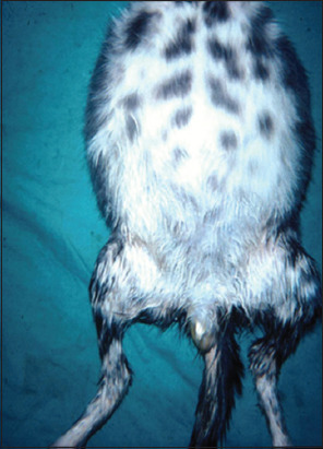
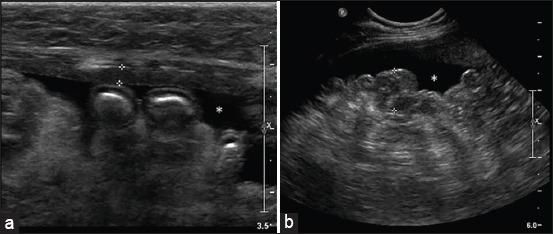
Feline infectious peritonitis (FIP) is an infectious disease characterized by non-specific laboratory changes and clinical signs. Clinical symptoms include anorexia, jaundice, fever, and weight loss. Moreover, some lesions are found in the digestive and respiratory systems. FIP, whose virulence varies, cannot be distinguished using several diagnostic methods. Moreover, feline coronaviruses (FCoVs) can be classified into two serotypes based on differences in their amino acid sequences, spike (S) protein sequences, and antibody (Ab) neutralization. There are two pathotypes, namely those caused by FCoV, which are often referred to as feline enteric coronavirus and FIP virus (FIPV). Furthermore, FIPV infection can be caused by sub-neutralizing levels of anti-FIPV S Abs. Therefore, a supporting diagnosis is needed to confirm FIP because there are no specific symptoms. This review aimed to provide updated information on FIP, including epizootiology, clinical and pathological characteristics, pathogenesis, hematology, clinicopathological and imaging features, pathological features, experimental infection, treatment and prevention, infection and immunity, animal and public health considerations.
Keywords: clinical; feline coronavirus; feline infectious peritonitis virus; infectious disease.
Copyright: © Solikhah, et al.
Conflict of interest statement
The authors declare that they have no competing interests.
Figures
Similar articles
-
Establishment of Full-Length cDNA Clones and an Efficient Oral Infection Model for Feline Coronavirus in Cats.J Virol. 2021 Oct 13;95(21):e0074521. doi: 10.1128/JVI.00745-21. Epub 2021 Aug 18. J Virol. 2021. PMID: 34406859 Free PMC article.
-
Sensitivity and specificity of a real-time reverse transcriptase polymerase chain reaction detecting feline coronavirus mutations in effusion and serum/plasma of cats to diagnose feline infectious peritonitis.BMC Vet Res. 2017 Aug 2;13(1):228. doi: 10.1186/s12917-017-1147-8. BMC Vet Res. 2017. PMID: 28768514 Free PMC article.
-
Reverse Genetics for Type I Feline Coronavirus Field Isolate To Study the Molecular Pathogenesis of Feline Infectious Peritonitis.mBio. 2018 Jul 31;9(4):e01422-18. doi: 10.1128/mBio.01422-18. mBio. 2018. PMID: 30065095 Free PMC article.
-
Feline Coronaviruses: Pathogenesis of Feline Infectious Peritonitis.Adv Virus Res. 2016;96:193-218. doi: 10.1016/bs.aivir.2016.08.002. Epub 2016 Aug 31. Adv Virus Res. 2016. PMID: 27712624 Free PMC article. Review.
-
Feline infectious peritonitis: still an enigma?Vet Pathol. 2014 Mar;51(2):505-26. doi: 10.1177/0300985814522077. Vet Pathol. 2014. PMID: 24569616 Review.
Cited by
-
The use of high-resolution melting analysis to distinguish feline enteric and feline infectious peritonitis coronaviruses.Braz J Microbiol. 2025 Sep;56(3):2081-2086. doi: 10.1007/s42770-025-01742-6. Epub 2025 Jul 21. Braz J Microbiol. 2025. PMID: 40691754
-
Detection of Feline Coronavirus Membrane Gene Based on Conventional Revere Transcription-Polymerase Chain Reaction, Nested Reverse Transcription-Polymerase Chain Reaction, and Reverse Transcription-Quantitative Polymerase Chain Reaction: A Comparative Study.Int J Mol Sci. 2025 Jul 17;26(14):6861. doi: 10.3390/ijms26146861. Int J Mol Sci. 2025. PMID: 40725108 Free PMC article.
References
Publication types
LinkOut - more resources
Full Text Sources