[Preprint]. 2025 Jan 6:2025.01.06.631470.
doi: 10.1101/2025.01.06.631470.

Transcriptional Integration of Meiotic Prophase I Progression and Early Oocyte Differentiation

Affiliations

Transcriptional Integration of Meiotic Prophase I Progression and Early Oocyte Differentiation

Kimberly M Abt et al. bioRxiv. .

Abstract

Female reproductive senescence results from the regulated depletion of a finite pool of oocytes called the ovarian reserve. This pool of oocytes is initially established during fetal development, but the oocytes that comprise it must remain quiescent for decades until they are activated during maturation in adulthood. In order for developmentally competent oocytes to populate the ovarian reserve they must successfully initiate both meiosis and oogenesis. As the factors that regulate the timing and fidelity of these early events remain elusive, we assessed the precise function and timing of the transcriptional regulator TAF4b during meiotic prophase I progression in mouse fetal oocytes. Compared to matched controls, E14.5 Taf4b-deficient oocytes enter meiosis I in a timely manner however, their subsequent progression through the pachytene-to-diplotene transition of meiotic prophase I is compromised. Moreover, this disruption of meiotic progression is associated with the reduced ability of Taf4b-deficient oocytes to repair double-strand DNA breaks. Transcriptional profiling of Taf4b-deficient oocytes reveals that between E16.5 and E18.5 these oocytes fail to coordinate the reduction of meiotic gene expression and the induction of oocyte differentiation genes. These studies reveal that TAF4b promotes the formation of the ovarian reserve in part by orchestrating the timely transition to meiosis I arrest and oocyte differentiation, which are often perceived as separate events.


Figures
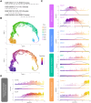
Figure 1 –
Figure 1 –. Integration of scRNA-seq data from oocytes during ovarian reserve establishment.
(A) Schematic of datasets used for integration. (B) Uniform Manifold Approximation and Projection (UMAP) of female germ cells colored by time (upper panel) and pseudotime (lower panel). (C) Expression of candidate genes plotted in terms of pseudotime. Color of gene labels correspond to approximate prophase I stage, Figure 1 (D) Expression of germ-cell enriched TFIID components plotted in terms of pseudotime.
Figure 2 –
Figure 2 –. TAF4b is enriched in pachytene oocytes at E16.5 and E18.5.
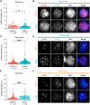
Prophase I chromatin spreads were prepared from wildtype ovaries at E16.5 and E18.5 using the drying down technique. Images of oocytes stained with TAF4b (red), SYCP3 (green), and DAPI (blue) at each prophase I stage present at E16.5 (A) or E18.5 (C), scalebar represents 10 μm. Quantification of TAF4b signal intensity in oocytes grouped by stage at E16.5 (B) or E18.5 (D), au= arbitrary units. Spreads were collected from 3 animals at E16.5 and 4 animals at E18.5, and n refers to the number of spreads used in analysis. Dots represent individual spreads, bar height represents the sample mean, and error bars represent standard error of the mean. Statistical significance for stage specific comparison at E16.5 was determined using an ordinary one-way ANOVA with multiple comparisons. For E18.5 samples a two-tailed T-test was used, ns=not significant, * p<0.05, ** p <0.01, and *** p <0.001.
Figure 3 –
Figure 3 –. Taf4b/− ovaries initiate meiosis normally at E14.5.
Images of E14.5 Taf4b+/+ (A) and Taf4b/− (B) ovary sections stained with TRA98 (green) as a marker of total germ cells, STRA8 (red) as a marker of meiotic germ cells, and DAPI (blue) as a marker of cell nuclei. Scalebar represents 100 μm. Quantification of TRA98 (C) or STRA8 (D) cell density of tissue sections per genotype. Cell density was calculated by dividing the number of positively marked cells by the area of the tissue section. Sections were collected from three E14.5 ovary pairs per genotype. Dots represent individual sections, bar height represents the sample mean, and error bars represent standard error of the mean. Statistical significance for genotype comparisons was determined using a two-tailed T-test, ns=not significant.
Figure 4 –
Figure 4 –. Taf4b/− ovaries experience delays in prophase I progression at E16.5, E18.5 and PND0.
(A) Proportion of Taf4b+/+ and Taf4b/− chromatin spreads found at each substage of prophase I at E16.5, E18.5, and PND0. Substage was determined by spatial configuration of SYCP3 and representative images of spreads at each stage are shown in (B). Spreads were stained with SYCP3 (green) and DAPI (blue) and scalebar represents 10 μm. Color of labels in stacked bar chart corresponds to prophase I stage, leptonema (purple), zygonema (blue), pachynema (teal), diplonema (orange). Spreads were collected from three animals per genotype at each timepoint and n refers to the number of spreads analyzed per stage. Statistical significance was calculated using a Chi-square test, **** p<0.0001.
Figure 5 –
Figure 5 –. Taf4b/− oocytes have elevated levels of γH2AX during pachynema and diplonema.
Quantification of γH2AX signal intensity in chromatin spreads during zygonema (A) pachynema (C) and diplonema (E). Spreads were pooled from three E16.5, E18.5, and PND0 mice per genotype, n = the number of spreads analyzed, au = arbitrary units. Images of spreads stained with γH2AX (red) SYCP3 (green) and DAPI (blue) from each genotype during zygonema (B) pachynema (D) and diplonema (F). Scalebar represents 10 μm. Dots in graphs represent individual spreads, bar height represents the sample mean, and error bars represent standard error of the mean. Statistical significance for stage specific comparisons was determined using a two-tailed T-test, ns=not significant, and **** p <0.0001.
Figure 6 –
Figure 6 –. Bulk RNA-seq of E18.5 Taf4b+/+, +/−, and −/− oocytes.
(A) PCA plot of E18.5 samples labelled based on genotype. Dashed circles represent samples compared in differential gene expression analysis. (B) Volcano plot of differentially expressed genes (protein-coding, padj <0.05, avg TPM > 1, log2FoldChange > l0.6l). The top 10 most significant DEGs are labelled. Dashed lines represent padj and log2FoldChange cutoffs. (C) Dotplot of GO biological process analysis of all 1551 DEGs.
Figure 7 –
Figure 7 –. Dysregulation of genes involved in meiosis and oogenesis in E18.5 Taf4b/− oocytes.
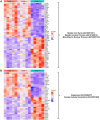
(A) Heatmap of genes in meiotic cell cycle, meiotic nuclear division, or male meiotic nuclear division GO categories that were differentially expressed in E18.5 Taf4b/− oocytes. (B) Heatmap of genes in oogenesis or female gamete generation GO categories that were differentially expressed in E18.5 Taf4b/− oocytes. Heatmaps were generated using based normalized counts output from DESeq2.
Figure 8 –
Figure 8 –. Model of defective ovarian reserve establishment in Taf4b/− mice.
Schematic timeline of major events that occur during ovarian reserve establishment in mice (top). Estimations of oocyte numbers in Taf4b+/+ (red line) and Taf4b/− (blue line) mice throughout time course are depicted below schematic. In the absence of TAF4b, there is a reduced number of oocytes at PND1. Taf4b/− oocytes at E18.5 and PND0 are unable efficiently reach diplotene and are largely stuck in pachytene. Taf4b/− oocytes do not reach pachytene in a timely manner at E16.5 but initiation of meiosis occurs normally at E14.5. At E16.5 and E18.5 Taf4b/− oocytes downregulate genes essential for oocyte development, display reduced expression of the X chromosome, and dysregulate chromatin remodeling genes. In addition, expression of genes required for successful completion of prophase is highly perturbed in Taf4b/− oocytes at E18.5.

Similar articles

References

    1. Abby E., Tourpin S., Ribeiro J., Daniel K., Messiaen S., Moison D., Guerquin J., Gaillard J. C., Armengaud J., Langa F., Toth A., Martini E., & Livera G. (2016). Implementation of meiosis prophase I programme requires a conserved retinoid-independent stabilizer of meiotic transcripts. Nat Commun, 7, 10324. 10.1038/ncomms10324 - DOI - PMC - PubMed
    1. Alexander A. K., Rice E. J., Lujic J., Simon L. E., Tanis S., Barshad G., Zhu L., Lama J., Cohen P. E., & Danko C. G. (2023). A-MYB and BRDT-dependent RNA Polymerase II pause release orchestrates transcriptional regulation in mammalian meiosis. Nat Commun, 14(1), 1753. 10.1038/s41467-023-37408-w - DOI - PMC - PubMed
    1. Anderson E. L., Baltus A. E., Roepers-Gajadien H. L., Hassold T. J., de Rooij D. G., van Pelt A. M., Page D. C., Baudat F., Imai Y., & de Massy B. (2008). Stra8 and its inducer, retinoic acid, regulate meiotic initiation in both spermatogenesis and oogenesis in mice Meiotic recombination in mammals: localization and regulation. Proc Natl Acad Sci U S A, 105(39), 14976–14980. 10.1073/pnas.080729710510.1038/nrg3573 - DOI - PMC - PubMed
    1. Ayhan O., Balkan M., Guven A., Hazan R., Atar M., Tok A., & Tolun A. (2014). Truncating mutations in TAF4B and ZMYND15 causing recessive azoospermia. J Med Genet, 51(4), 239–244. 10.1136/jmedgenet-2013-102102 - DOI - PubMed
    1. Bolcun-Filas E., & Handel M. A. (2018). Meiosis: the chromosomal foundation of reproduction. Biol Reprod, 99(1), 112–126. 10.1093/biolre/ioy021 - DOI - PubMed

Publication types