[Preprint]. 2025 Jan 8:2025.01.08.632016.
doi: 10.1101/2025.01.08.632016.

Multi-omics and biochemical reconstitution reveal CDK7-dependent mechanisms controlling RNA polymerase II function at gene 5'- and 3'-ends

Affiliations

Multi-omics and biochemical reconstitution reveal CDK7-dependent mechanisms controlling RNA polymerase II function at gene 5'- and 3'-ends

Olivia Luyties et al. bioRxiv. .

Update in

Abstract

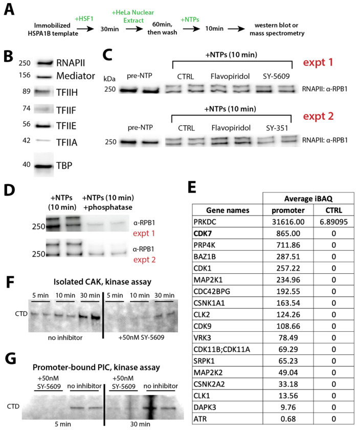
CDK7 regulates RNA polymerase II (RNAPII) initiation, elongation, and termination through incompletely understood mechanisms. Because contaminating kinases precluded CDK7 analysis with nuclear extracts, we completed biochemical assays with purified factors. Reconstitution of RNAPII transcription initiation showed CDK7 inhibition slowed and/or paused RNAPII promoter-proximal transcription, which reduced re-initiation. These CDK7-regulatory functions were Mediator- and TFIID-dependent. Similarly in human cells, CDK7 inhibition reduced transcription by suppressing RNAPII activity at promoters, consistent with reduced initiation and/or re-initiation. Moreover, widespread 3'-end readthrough transcription was observed in CDK7-inhibited cells; mechanistically, this occurred through rapid nuclear depletion of RNAPII elongation and termination factors, including high-confidence CDK7 targets. Collectively, these results define how CDK7 governs RNAPII function at gene 5'-ends and 3'-ends, and reveal that nuclear abundance of elongation and termination factors is kinase-dependent. Because 3'-readthrough transcription is commonly induced during stress, our results further suggest regulated suppression of CDK7 activity may enable this RNAPII transcriptional response.


Conflict of interest statement

DJT received some funding support for this project from Syros Pharmaceuticals, Inc. R.D.D. is a founder of Arpeggio Biosciences. The remaining authors declare no competing interests.

Figures

Figure 1.
Figure 1.. Biochemical reconstitution can specifically address CDK7-dependent functions, whereas nuclear extracts cannot
(A) Overview of immobilized template experiments. (B) Western blots showing all PIC factors bound to the HSPA1B promoter template, as expected. (C) RNAPII western blots show shifted band following NTP addition, consistent with RPB1 CTD phosphorylation. Technical replicates shown side-by-side for each condition, and data from 2 different biological replicates are shown (expt 1 & expt 2). Expt 2 used a covalent CDK7 inhibitor, SY-351. (D) The shifted RPB1 band was phosphorylated, as expected, based upon incubation with λ-phosphatase. (E) Summary of kinases bound to the native HSPA1B promoter after washing, based upon mass spectrometry analysis. Although CDK7 was a top hit, many other contaminating kinases were present. The CTRL experiment used beads only (no immobilized template). (F) Purified CAK module (CDK7, CCNC, MNAT1) phosphorylates full-length mammalian GST-CTD in vitro; phosphorylation is blocked by 50nM SY-5609. Some phosphorylation occurred at 30min, a timeframe longer than in vitro transcription reactions. Note is 20,000-fold higher than [SY-5609] in these experiments (1mM vs. 50nM). (G) Purified reconstituted PICs assembled on the HSPA1B promoter contain only one kinase, CDK7. These PICs phosphorylate the RNAPII CTD, as expected, but phosphorylation is blocked by SY-5609, although some phosphorylation can be detected after 30min.
Figure 2.
Figure 2.. CDK7 inhibition blocks promoter escape and pause release, and reduces overall transcription output
(A) Schematic of the human pre-initiation complex (PIC). (B) Overview of the in vitro transcription pausing assays. (C) Representative gel images of pausing assays, with pre-escape, paused, and runoff/elongated regions indicated. Each region was independently contrast-enhanced to aid visualization and each region was adjusted equally across comparisons (e.g. CTRL vs. SY-5609). Quantitation was completed with uniform exposure across the entire lane; gel lanes with uniform exposure are shown in Figure S1B. (D) CDK7 inhibition decreases and slows promoter escape. Each point (n=6) represents the ratio of pre-escape transcripts (5-15nt length) to runoff products (150-216nt) from reactions treated with 50nM SY-5609, normalized to no inhibitor controls. (E) CDK7 inhibition increases pausing. Plot shows the pause index of reactions (n=6) treated with SY-5609, normalized to no inhibitor controls. (F) Overview of in vitro transcription runoff assays. (G) CDK7 inhibition reduces overall RNAPII transcriptional output (n=7). Inset: representative gel images of runoff transcription ±SY-5609. See also Figure S1C. (H) CDK7 inhibition under single-round conditions (sarkosyl added 1-min post-NTP; n=5). Plot shows quantification of runoff transcription +SY-5609, normalized to no inhibitor controls. Representative gels shown in Figure S1D.
Figure 3.
Figure 3.. Mediator and TFIID cooperate with CDK7 to enhance promoter escape and pause release, respectively
(A) Quantitation of runoff transcription +SY-5609, normalized to no inhibitor controls (dashed line), comparing PICs with and without Mediator (n=6). Representative gel lanes in Figure S1F. (B) TFIID affects runoff transcription as a function of CDK7 kinase activity. Data from PICs ±TFIID shown, +SY-5609, normalized to no inhibitor controls (dashed line). No IID = TBP instead of TFIID (n=7). PIC data (left column, dark purple dots) are the same as in panel A. Representative gel lanes in Figure S1G. (C) Representative gel images from TBP PIC pausing assays, with pre-escape, paused, and runoff/elongated regions indicated. Each region was independently contrast-enhanced to aid visualization and each region was adjusted equally across comparisons (e.g. CTRL vs. SY-5609). Quantitation completed with uniform exposure across the entire lane; gel lanes with uniform exposure are shown in Figure S1H. (D) In contrast to TFIID PICs, pausing does not occur with TBP PICs and CDK7-dependent effect on pausing is lost (n=7). Purple points are the same as those in 2E, shown again here for comparison.
Figure 4.
Figure 4.. CDK7 inhibition reduces RNAPII transcription genome-wide, including at promoter-proximal regions, but increases promoter-proximal RNAPII occupancy
(A) Violin plots of the PRO-seq normalized count data. The interquartile range (IQR) counts are plotted (i.e. the middle 50% of data), showing a reduction in SY-5609-treated cells. (B) CDK7 inhibition increases RNAPII pause index (n=2400 genes; p = 1.26E−11). (C) Metagene plot of normalized PRO-seq reads at highly expressed genes (n=453), focused on promoter-proximal region ±SY-5609. (D) Metagene analysis of ChIP-seq data (normalized to read depth), focused on promoter-proximal region ±SY-5609 at high-occupancy RNAPII genes (n=185). Elevated RNAPII occupancy +SY-5609 coupled with reduced PRO-seq reads +SY-5609 (panel C) suggests increased levels of inactive RNAPII around the TSS. As expected, levels of RNAPII CTD ser5-P and ser7-P decrease at the TSS in SY-5609-treated cells.
Figure 5.
Figure 5.. Readthrough defects and depletion of RNAPII elongation and termination factors in CDK7-inhibited cells
(A) Representative PRO-seq gene traces demonstrating increased 3′-end readthrough transcription +SY-5609. (B) Metagene analysis at long genes ( >120kb, n=684) shows extensive 3′-end readthrough transcription with SY-5609 treatment. Position 0 indicates the polyA site. (C) Heatmap summarizing quantitative TMT-MS data from OV90 and HCT116 cells treated with 50nM SY-5609 for 120min (vs. DMSO controls). Proteins shown were identified as significantly changing in both OV90 and HCT116 cells (p<0.01). Asterisk: RNAPII elongation/termination factors. (D) GSEA results (GOBP set), based on TMT-MS data ±SY-5609, plotted by normalized enrichment score (NES) for OV90 and HCT116 cells. Significantly upregulated or downregulated pathways (FDR q < 0.05) are highlighted in color, and a representative subset of pathways are listed.
Figure 6.
Figure 6.. CDK7 inhibition (CDK7AS line) depletes RNAPII elongation and termination/3′-end processing factors, many of which are phosphorylated by CDK7
(A) Heatmap showing significantly changing (p < 0.01) protein abundance from quantiative TMT-MS experiments in CDK7AS OV90 cells ±3MB-PP1 (10μM, t=4h) or OV90 cells ±SY-5609 (50nM, t=2h). Proteins associated with transcription elongation, mRNA splicing, and termination are starred. (B) GSEA results (GOBP set), based on TMT-MS data ±SY-5609 (y-axis; OV90) or ±3MB-PP1 (x-axis; CDK7AS OV90), plotted by their normalized enrichment score (NES). Significantly upregulated or downregulated pathways (FDR q < 0.05) are highlighted in color, and a subset of pathways are listed at right. (C) Volcano plot showing proteins with phosphorylation changes ±SY-5609 (50nM, 1h) in HCT116 cells. For simplicity, only proteins that were also nuclear depleted +SY-5609 are included here. Among all proteins depleted with statistical confidence, about 20% showed phospho-site changes (93/483; Venn diagram inset).
Figure 7.
Figure 7.. Model: CDK7 regulates RNAPII transcription at gene 5′-ends and 3′-ends through diverse mechanisms
At 5′-ends, CDK7 inhibition slows or stalls RNAPII in the promoter escape and promoter-proximal pause regions; this prevents re-initiation and may contribute to RNAPII inactivation at promoters in CDK7-inhibited cells. Separately, CDK7 inhibition causes nuclear depletion of RNAPII elongation and termination factors, which is sufficient to trigger 3′-readthrough transcription. CDK7 inhibition also causes phospho-site changes in many elongation and termination factors, which may independently contribute to the 3′-readthrough defects. PIC factors are represented as globular in the promoter escape and pause regions based upon cryo-EM data that indicate structural dynamics post-initiation.,,

References

    1. Schier A.C., and Taatjes D.J. (2020). Structure and mechanism of the RNA polymerase II transcription machinery. Genes Dev 34, 465–488. 10.1101/gad.335679.119. - DOI - PMC - PubMed
    1. Abdella R., Talyzina A., Chen S., Inouye C.J., Tjian R., and He Y. (2021). Structure of the human Mediator-bound transcription preinitiation complex. Science 372, 52–56. 10.1126/science.abg3074. - DOI - PMC - PubMed
    1. Chen X., Yin X., Li J., Wu Z., Qi Y., Wang X., Liu W., and Xu Y. (2021). Structures of the human Mediator and Mediator-bound preinitiation complex. Science 372, eabg0635. 10.1126/science.abg0635. - DOI - PubMed
    1. Rengachari S., Schilbach S., Aibara S., Dienemann C., and Cramer P. (2021). Structure of human Mediator-RNA polymerase II pre-initiation complex. Nature 594, 129–133. 10.1038/s41586-021-03555-7. - DOI - PubMed
    1. Naar A.M., Taatjes D.J., Zhai W., Nogales E., and Tjian R. (2002). Human CRSP interacts with RNA polymerase II CTD and adopts a specific CTD-bound conformation. Genes Dev 16, 1339–1344. - PMC - PubMed

Publication types

LinkOut - more resources