[Preprint]. 2025 Jan 10:2025.01.10.632119.
doi: 10.1101/2025.01.10.632119.

The Q226L mutation can convert a highly pathogenic H5 2.3.4.4e virus to bind human-type receptors

Affiliations

The Q226L mutation can convert a highly pathogenic H5 2.3.4.4e virus to bind human-type receptors

María Ríos Carrasco et al. bioRxiv. .

Update in

Abstract

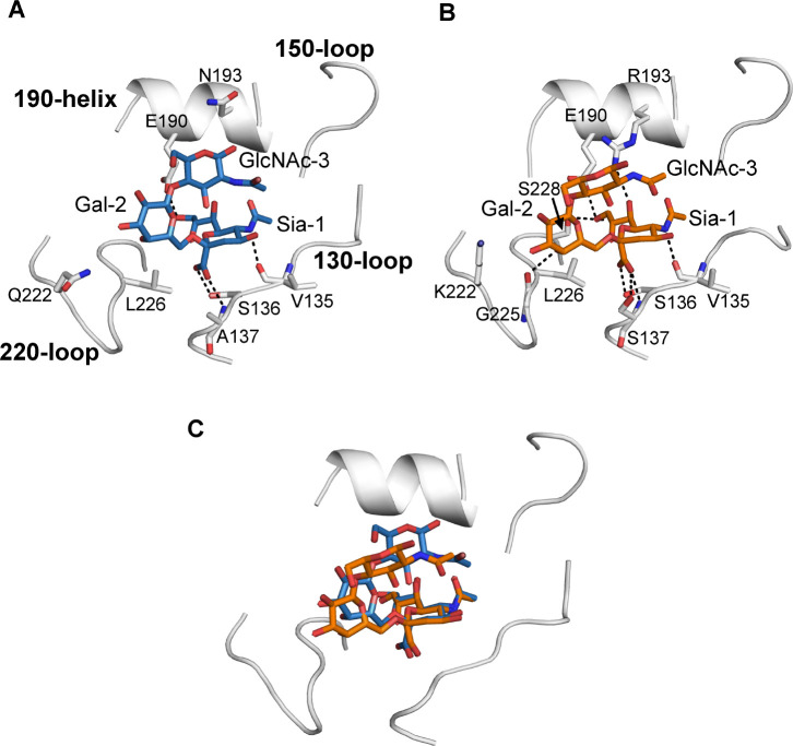
H5Nx viruses continue to wreak havoc in avian and mammalian species worldwide. The virus distinguishes itself by the ability to replicate to high titers and transmit efficiently in a wide variety of hosts in diverse climatic environments. Fortunately, transmission to and between humans is scarce. Yet, if such an event were to occur, it could spark a pandemic as humans are immunologically naïve to H5 viruses. A significant determinant of transmission to and between humans is the ability of the influenza A virus hemagglutinin (HA) protein to shift from an avian-type to a human-type receptor specificity. Here, we demonstrate that a 2016 2.3.4.4e virus HA can convert to human-type receptor binding via a single Q226L mutation, in contrast to a cleavage-modified 2016 2.3.4.4b virus HA. Using glycan arrays, x-ray structural analyses, tissue- and direct glycan binding, we show that L133aΔ and 227Q are vital for this phenotype. Thus, whereas the 2.3.4.4e virus HA only needs a single amino acid mutation, the modified 2.3.4.4b HA was not easily converted to human-type receptor specificity.


Figures

Figure 1.
Figure 1.. Glycan microarray analyses of H5 hemagglutinins including Q226L and G228S mutants.
Synthetic bi-antennary N-glycans printed on the microarray, either without sialic acid (structures 1–3, gray), with α2,6-linked NeuAc (4–6, red), or α2,3-linked NeuAc (7–9, white). Structures 1, 4, and 7, contain one LacNAc repeat, while structures 2, 5, and 8 have two repeats and structures 3, 6, and 9 contain three repeats (A). Indo05 and the Q226L mutant (B). France16 2.3.4.4b as wt, with the single Q226L and G228S added (C). Akita16 as wt, with Q226L and G228S added (D). Akita16 with the 2.3.4.4b specific mutations L133a & R227Q, and Q226L added (E). A/Netherlands/761/09 as the human-type receptor specific control (F). Bars represent the mean ± SD (n= 4)
Figure 2.
Figure 2.. Tissue binding properties of different H5 proteins on chicken and human tracheal slides reveal different phenotypes conferred by Q226L.
The binding to chicken and human tracheal tissue was investigated for different influenza A H5 HAs at two different concentrations. From top to bottom Indo05, Akita16 and France16 including their Q226L mutant. AEC staining was used to visualize tissue binding.
Figure 3.
Figure 3.. Direct receptor binding assay using biotinylated linear avian- and human-type receptors.
3SLN3 (top) and 6SLN3 (bottom) coated streptavidin plates interrogated with A/Indonesia/05/05 (left), A/black swan/Akita/1/16 (middle) and A/duck/France/161108h/16 (right) WT proteins as shown with orange circles and the Q226L mutants using red squares.
Fig. 4.
Fig. 4.. Crystal structure of A/black swan/Akita/1/16 and A/Indonesia/5/2005 H5 HA with avian receptor analog LSTa.
The receptor binding site (RBS) is shown as a backbone cartoon. HA residues involved in the interaction with the receptor analog are presented as sticks. Hydrogen bonds are indicated as black dashes. Avian-type receptor analog LSTa (NeuAcα2-3Galβ1-3GlcNAcβ1-3Galβ1-4Glc) is used for both HA. (A) Akita16-WT HA with LSTa (C). (B) Indo05-WT HA with LSTa (PDB: 4K63). (C) Superposition of LSTa in Akita16-WT HA (yellow) and Indo05-WT HA (cyan).
Fig. 5.
Fig. 5.. Crystal structure of A/black swan/Akita/1/1utant with human receptor analog LSTc and comparison with other H5 HAs with LSTc.
Receptor binding site (RBS) is shown as back bone cartoon. Residues that involved in the interaction between HA and receptor analog are presented as sticks. Hydrogen bonds are indicated as black dashes. LSTc (NeuAcα2-6Galβ1-4GlcNAcβ1-3Galβ1-4Glc ) is used as Human-type receptor analog. (A) Akita16-Q226L mutant with LSTc. (B) Indo05mut HA with LSTc (PDB: 4K67). (C) Superposition of LSTc in Akita16-Q226L mutant with Indo05mut HA (orange).
Fig. 6.
Fig. 6.. Crystal structure of A/duck/France/161108h/2016 Q226L mutant with avian receptor analog LSTa and the comparison with other H7 HA and H5 HA with LSTa.
(A) France16-Q226L mutant with LSTa. (B) Superposition of LSTa in France16-Q226L (cyan) mutant with LSTa in the Akita16-WT (magenta). (C) A/Shanghai/02/2013 (H7N9) HA with LSTa (PDB: 4N5K).

Similar articles

References

    1. Joseph U., Su Y. C., Vijaykrishna D., Smith G. J., The ecology and adaptive evolution of influenza A interspecies transmission. Influenza Other Respir Viruses 11, 74–84 (2017). - PMC - PubMed
    1. Karakus U. et al., H19 influenza A virus exhibits species-specific MHC class II receptor usage. Cell Host Microbe 32, 1089–1102.e (2024). - PMC - PubMed
    1. Caserta L. C. et al., Spillover of highly pathogenic avian influenza H5N1 virus to dairy cattle. Nature 634, 669–676 (2024). - PMC - PubMed
    1. Shi J., Zeng X., Cui P., Yan C., Chen H., Alarming situation of emerging H5 and H7 avian influenza and effective control strategies. Emerg Microbes Infect 12, 2155072 (2023). - PMC - PubMed
    1. Xu X., Subbarao, Cox N. J., Guo Y., Genetic characterization of the pathogenic influenza A/Goose/Guangdong/1/96 (H5N1) virus: similarity of its hemagglutinin gene to those of H5N1 viruses from the 1997 outbreaks in Hong Kong. Virology 261, 15–19 (1999). - PubMed

Publication types

LinkOut - more resources