. 2024 Dec 21;16(3):1289-1303.
doi: 10.1039/d4md00850b. eCollection 2025 Mar 19.

Design and synthesis of cyclic lipidated peptides derived from the C-terminus of Cx43 for hemichannel inhibition and cardiac endothelium targeting

Affiliations

Design and synthesis of cyclic lipidated peptides derived from the C-terminus of Cx43 for hemichannel inhibition and cardiac endothelium targeting

Debora Iaculli et al. RSC Med Chem. .

Abstract

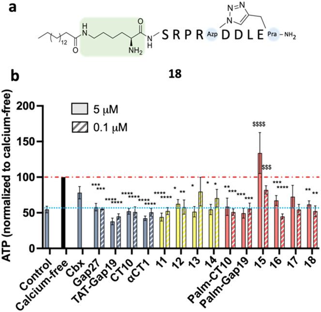
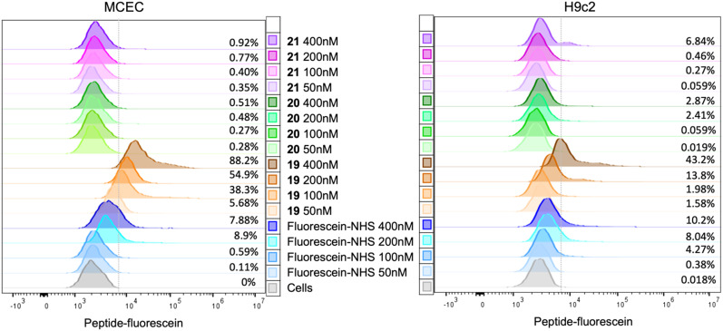
A peptide segment that is 10 residues long at the C-terminal (CT) region of Cx43 is known to be involved in interactions, both with the Cx43 protein itself and with other proteins, that result in hemichannel (HC) activity regulation. Previously reported mimetic peptides based on this region (e.g., αCT1, CT10) have been revealed to be promising therapeutic agents in the context of cardiovascular diseases. In this work, novel approaches, such as C- and N-terminal modification and cyclization, to improve the proteolytic stability and bioavailability of the CT10 peptide are presented. These efforts resulted in a set of unprecedented potent cyclic inhibitors of HC-mediated ATP release with a half-life largely exceeding 24 hours. Additionally, the introduction of a lipophilic moiety with different solubilizing linkers led to the generation of a novel series of water-soluble and lipidated peptides that exhibited high inhibitory capacity in in vitro assays at submicromolar concentrations. A cardiac endothelium targeting strategy was also adopted, exploiting the ability of the CRPPR peptide to selectively deliver the peptides to endothelial cells.


Conflict of interest statement

There are no conflicts to declare.

Figures

Fig. 1
Fig. 1. Structure of a hexameric Cx43 hemichannel. EC: extracellular domain. IC: intracellular domain. A single Cx43 monomer is highlighted in dark blue. The transmembrane Cx43 protein is represented in the box. EL1 and EL2: extracellular loops. CL: cytoplasmic loop. NT and CT: cytoplasmic N- and C-terminal tails. The reference sequences of CT10 and αCT1 are shown, with the mimicked region of the CT highlighted in grey.
Fig. 2
Fig. 2. a) Structure of the CT domain of Cx43, based on earlier NMR studies (PDB1R5S). The α-helical regions H1 and H2 are indicated in red. b) Zoomed image of the C-terminal extremity. The residues are numbered according to their relative positions in Cx43.
Fig. 3
Fig. 3. a) Sequences of linear and cyclic CT10 analogues. The substitutions in the linear sequences are shown in red. b) Cyclic peptides based on the CT10 sequence. Azp, Pra and Azk stand for (4S)-4-azidoproline, propargylglycine and azidolysine, respectively. c) Solid-phase synthesis of cyclic peptides. i. 4-Methylpiperidine 20% in DMF, RT, 1 × 5 min, 1 × 15 min; ii. Fmoc-AA(PG)-OH (1.5 or 3 eq.), HBTU (1.5 or 3 eq.), DIPEA (5 eq.), DMF, RT, 1–2 h; iii. CuBr (3 eq.), Na-ascorbate (3 eq.) in H2O, 2,6-lutidine (10 eq.), DIPEA (10 eq.), DMF, RT, 18 h; iv. Ac2O (10 eq.), DIPEA (5 eq.), DCM, RT, 45 min; v. TFA : TIPS : H2O = 90 : 5 : 5, RT, 3 h.
Fig. 4
Fig. 4. Selected lipidated peptide sequences based on Gap19 and CT10 compounds. The different linkers applied are shown in green.
Fig. 5
Fig. 5. a) Setup of controls for ATP release experiments in B16-BL6 cells. Control represents basal ATP release in HBSS (HC closed condition; blue line), while calcium-free is maximal ATP release through connexin HCs induced by calcium depletion (red line). The nonspecific reference compound carbenoxolone (Cbx) was used at 25 μM and the reference peptides TAT-Gap19, Gap19, Gap27 and αCT1 at concentrations ranging from 0.1 μM to 100 μM. Triton X-100 (1%) was used as a cell lysis control. b) ATP release assay in B16-BL6 cells. HCs were opened by calcium depletion (calcium-free, 30 min). Cbx was used at 25 μM and all reference peptides and modified analogues were used at 5 μM. The orange bars represent the peptides in which alanine substitutions or C- and/or N-terminal modifications were introduced. The yellow bars represent the cyclic peptides. The red bars represent the lipidated peptides. Data are shown as mean ± SEM $P value compared to control conditions, *P value compared to calcium-free conditions. N = 3–9.
Fig. 6
Fig. 6. a) Structure of cyclic lipidated peptide 18. b) ATP release assay in B16-BL6 cells. Hemichannels were opened by calcium depletion (calcium-free, 30 min). Cbx was used at 25 μM and all reference peptides and modified analogues were used at 5 μM (solid bars) or 0.1 μM (hatched bars). The yellow bars represent the cyclic peptides. The red bars represent the lipidated peptides. Data are shown as mean ± SEM. $P value compared to control conditions, *P value compared to calcium-free conditions. N = 5–10.
Fig. 7
Fig. 7. In vitro plasma stability of peptides CT10 and 11. Recovery (%) over time after incubation in human plasma at 37 °C.
Fig. 8
Fig. 8. a) Western blot showing the presence or absence of Cx43 in each cell line used for the selectivity experiments. Mouse microvascular cardiac endothelial cells (MCECs) were used as the positive control. GAPDH served as the loading control. NA: non-applicable. b) Communication-incompetent parental HeLa cells lacking functional connexin HCs neither responded to calcium-free conditions nor to any of the Cx43 HC inhibiting compounds, while the HeLa–Cx43 transfectants showed a higher level of ATP release under control conditions, which was 3–4 times increased after calcium removal. Cbx (25 μM), control peptides and 10 (all at 5 μM) inhibited the ATP release induced by calcium removal to a varying extent. c) Parental SK-HEP-1 cells, lacking Cx43 but containing Cx45, responded to calcium removal with increased ATP release, which was inhibited by Cbx (25 μM), control peptides and 10 (all at 5 μM), showing that the control peptides and 10 are not specific for Cx43 HCs but also affect Cx45 HCs. SK-HEP-1–Cx43 transfectants showed a higher level of ATP release under control conditions, which was 3 times increased after calcium removal. Cbx (25 μM), control peptides and 11 (all at 5 μM) inhibited the ATP release induced by calcium removal. Data are shown as mean ± SEM $P value compared to control conditions, *P value compared to calcium-free conditions. N = 3.
Fig. 9
Fig. 9. Convergent synthesis strategy adopted for peptides 19 and 21. The sequences of the peptides are shown, together with the sequence of 20, synthesized linearly. The cleavage of the fully protected peptide was performed in 20% HFIP in DCM for 2 h. i) HATU 1.2 eq., DIPEA (2 eq.), DMF, 18 h; ii. PhSiH3 (24 eq.), Pd(PPh3)4 (0.2 eq.), DCM, 2 × 30 min; iii. Fluorescein-NHS (1.05 eq.), DIPEA (2 eq.), DMF, 18 h; iv. TFA : TIPS : H2O = 90 : 5 : 5, RT, 30 020 h.
Fig. 10
Fig. 10. Representative examples of flow cytometry experiments in MCEC and H9c2 cells. The grey curve represents a parallel dish of cells without any staining to measure the level of autofluorescence, which denotes the cut-off value. The percentage noted at the right side of the curves indicates the percentage of fluorescent cells for each condition.

References

    1. Sorgen P. L. Duffy H. S. Sahoo P. Coombs W. Delmar M. Spray D. C. J. Biol. Chem. 2004;279:54695–54701. doi: 10.1074/jbc.M409552200. - DOI - PubMed
    1. Leybaert L. Lampe P. D. Dhein S. Kwak B. R. Ferdinandy P. Beyer E. C. Laird D. W. Naus C. C. Green C. R. Schulz R. Pharmacol. Rev. 2017;69:396–478. doi: 10.1124/pr.115.012062. - DOI - PMC - PubMed
    1. Rusiecka O. M. Montgomery J. Morel S. Batista-Almeida D. Campenhout R. V. Vinken M. Girao H. Kwak B. R. Biomolecules. 2020;10:1–22. doi: 10.3390/biom10091225. - DOI - PMC - PubMed
    1. Kwak B. R. and Nielsen M. S., in Zipes and Jalife's Cardiac Electrophysiology: From Cell to Bedside, ed. J. Jalife and W. G. Stevenson, Elsevier, Philadelphia, PA, USA, 8th edn, 2021, ch. 15, pp. 168–177
    1. Delvaeye T. Vandenabeele P. Bultynck G. Leybaert L. Krysko D. V. Trends Mol. Med. 2018;24:1036–1053. doi: 10.1016/j.molmed.2018.10.005. - DOI - PubMed

LinkOut - more resources