

Imaging Carotid Plaque Burden in Living Mice via Hybrid Semiconducting Polymer Nanoparticles-Based Near-Infrared-II Fluorescence and Magnetic Resonance Imaging
- PMID: 39830010
- PMCID: PMC11740978
- DOI: 10.34133/research.0186
Imaging Carotid Plaque Burden in Living Mice via Hybrid Semiconducting Polymer Nanoparticles-Based Near-Infrared-II Fluorescence and Magnetic Resonance Imaging
Abstract
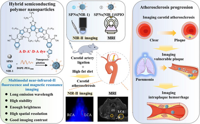
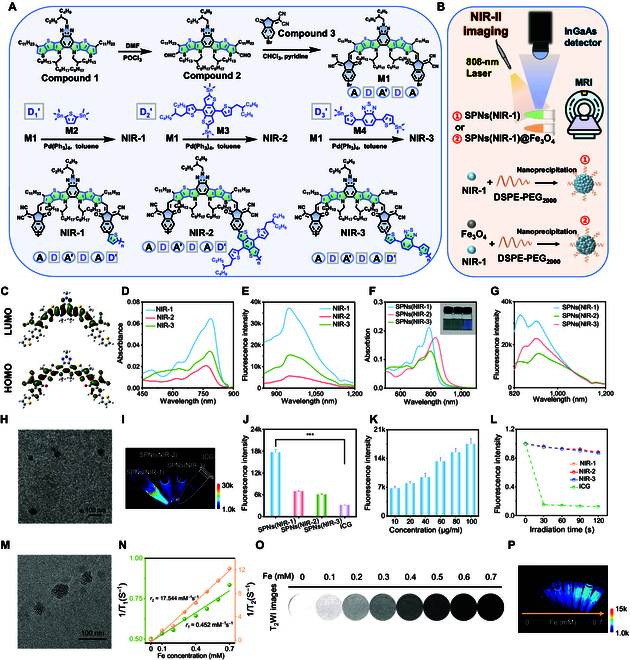
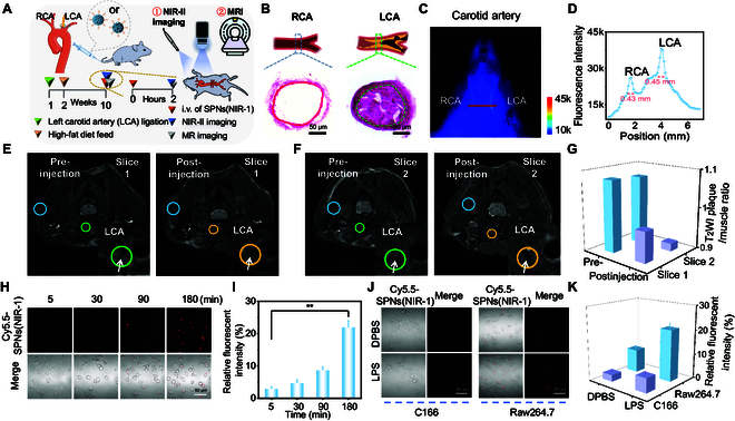
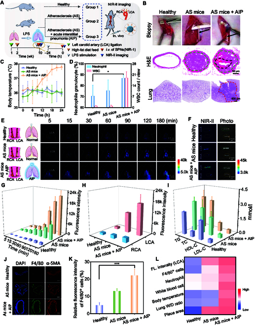
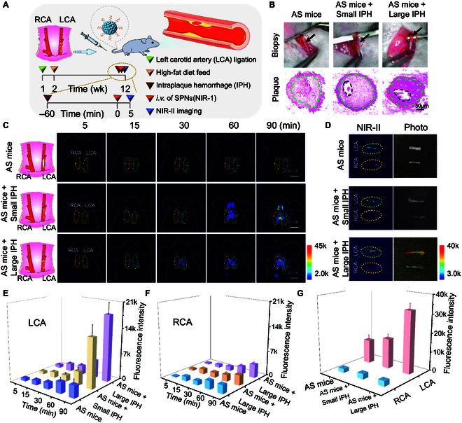
The majority of atherothrombotic events (e.g., cerebral or myocardial infarction) often occur as a result of plaque rupture or erosion in the carotid, and thereby it is urgent to assess plaque vulnerability and predict adverse cerebrovascular events. However, the monitoring evolution from stable plaque into life-threatening high-risk plaque in the slender carotid artery is a great challenge, due to not enough spatial resolution for imaging the carotid artery based on most of reported fluorescent probes. Herein, copolymerizing with the small molecules of acceptor-donor-acceptor-donor-acceptor (A-D-A'-D-A) and the electron-donating units (D'), the screened second near-infrared (NIR-II) nanoprobe presents high quantum yield and good stability, so that it enables to image slender carotid vessel with enough spatial resolution. Encouragingly, NIR-II nanoprobe can effectively target to intraplaque macrophage, meanwhile distinguishing vulnerable plaque in carotid atherosclerosis in living mice. Moreover, the NIR-II nanoprobe can dynamically monitor the fresh bleeding spots in carotid plaque, indicating the increased risk of plaque instability. Besides, magnetic resonance imaging is integrated with NIR-II fluorescence imaging to provide contrast for subtle structure (e.g., narrow lumen and lipid pool), via incorporating ultrasmall superparamagnetic iron oxide into the NIR-II nanoprobe. Thus, such hybrid NIR-II/magnetic resonance imaging multimodal nanoprobe provides an effective tool for assessing carotid plaque burden, selecting high-risk plaque, and imaging intraplaque hemorrhage, which is promising for reducing cerebral/ myocardial infarction-associated morbidity and mortality.
Copyright © 2023 Li Xu et al.
Figures
Similar articles
-
Contemporary carotid imaging: from degree of stenosis to plaque vulnerability.J Neurosurg. 2016 Jan;124(1):27-42. doi: 10.3171/2015.1.JNS142452. Epub 2015 Jul 31. J Neurosurg. 2016. PMID: 26230478 Review.
-
Gadolinium-Chelated Conjugated Polymer-Based Nanotheranostics for Photoacoustic/Magnetic Resonance/NIR-II Fluorescence Imaging-Guided Cancer Photothermal Therapy.Theranostics. 2019 May 31;9(14):4168-4181. doi: 10.7150/thno.34390. eCollection 2019. Theranostics. 2019. PMID: 31281539 Free PMC article.
-
Lipid Core Burden Index Assessed by Near-Infrared Spectroscopy of Symptomatic Carotid Plaques: Association with Magnetic Resonance T1-Weighted Imaging.Cerebrovasc Dis. 2021;50(5):597-604. doi: 10.1159/000516888. Epub 2021 Jun 18. Cerebrovasc Dis. 2021. PMID: 34148038
-
Carotid Plaque Lipid Content and Fibrous Cap Status Predict Systemic CV Outcomes: The MRI Substudy in AIM-HIGH.JACC Cardiovasc Imaging. 2017 Mar;10(3):241-249. doi: 10.1016/j.jcmg.2016.06.017. JACC Cardiovasc Imaging. 2017. PMID: 28279371 Free PMC article. Clinical Trial.
-
Thiadiazoloquinoxaline-Based Semiconducting Polymer Nanoparticles for NIR-II Fluorescence Imaging-Guided Photothermal Therapy.Front Bioeng Biotechnol. 2021 Nov 3;9:780993. doi: 10.3389/fbioe.2021.780993. eCollection 2021. Front Bioeng Biotechnol. 2021. PMID: 34805127 Free PMC article. Review.
Cited by
-
Enhanced Near-Infrared-Excitable Organic Afterglow Nanoparticles for Deep-Tissue Multimodal Imaging via Singlet Oxygen-Mediated Energy Transfer.Research (Wash D C). 2025 Aug 14;8:0834. doi: 10.34133/research.0834. eCollection 2025. Research (Wash D C). 2025. PMID: 40822121 Free PMC article.
-
Ultrabright difuranfluoreno-dithiophen polymers for enhanced afterglow imaging of atherosclerotic plaques.Sci Adv. 2025 Mar 28;11(13):eads4646. doi: 10.1126/sciadv.ads4646. Epub 2025 Mar 26. Sci Adv. 2025. PMID: 40138402 Free PMC article.
References
-
- Libby P. The changing landscape of atherosclerosis. Nature. 2021;592(7855):524. - PubMed
-
- Donnan GA, Fisher M, Macleod M, Davis SM. Stroke. Lancet. 2008;371(9624):1612–1623. - PubMed
-
- Sakakura K, Nakano M, Otsuka F, Ladich E, Kolodgie FD, Virmani R. Pathophysiology of atherosclerosis plaque progression. Heart Lung Circ. 2013;22(6):399–411. - PubMed
-
- Arbab-Zadeh A, Fuster V. From detecting the vulnerable plaque to managing the vulnerable patient: JACC state-of-the-art review. J Am Coll Cardiol. 2019;74(12):1582–1593. - PubMed
LinkOut - more resources
Full Text Sources
Research Materials
Miscellaneous
