. 2025 Jan 3:18:1424137.
doi: 10.3389/fncel.2024.1424137. eCollection 2024.

Cycloastragenol promotes dorsal column axon regeneration in mice

Affiliations

Cycloastragenol promotes dorsal column axon regeneration in mice

Tao Zihan et al. Front Cell Neurosci. .

Abstract

Introduction: Cycloastragenol (CAG) has a wide range of pharmacological effects, including anti-inflammatory, antiaging, antioxidative, and antitumorigenic properties. In addition, our previous study showed that CAG administration can promote axonal regeneration in peripheral neurons. However, whether CAG can activate axon regeneration central nervous system (CNS) remains unknown.

Methods: Here, we established a novel mouse model for visualizing spinal cord dorsal column axon regeneration involving the injection of AAV2/9-Cre into the lumbar 4/5 dorsal root ganglion (DRG) of Rosa-tdTomato reporter mice. We then treated mice by intraperitoneal administration of CAG.

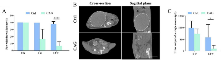
Results: Our results showed that intraperitoneal CAG injections significantly promoted the growth of vitro-cultured DRG axons as well as the growth of dorsal column axons over the injury site in spinal cord injury (SCI) mice. Our results further indicate that CAG administration can promote the recovery of sensory and urinary function in SCI mice.

Conclusion: Together, our findings highlight the therapeutic potential of CAG in spinal cord injury repair.

Keywords: TERT; axon growth; cycloastragenol; dorsal column; functional recovery; nerve injury; p53; spinal cord injury.


Conflict of interest statement

The authors declare that the research was conducted in the absence of any commercial or financial relationships that could be construed as a potential conflict of interest.

Figures

FIGURE 1
FIGURE 1
Cycloastragenol (CAG) promotes axon regeneration in peripheral sensory neurons. (A) Representative images of Tuj1 (green) immunofluorescence staining in dorsal root ganglion (DRG) neurons obtained from mice after 7 days of CAG administration and subsequently cultured for 48 h. Scale bar = 100 μm. (B) Axon length measurements showed that the in vivo administration of CAG promoted axon growth in in vitro-cultured DRG neurons (n = 3, ***p < 0.001). (C) CAG administration did not affect the survival of DRG neurons after 3 days of culture (n = 3).
FIGURE 2
FIGURE 2
A novel model for the visualization of dorsal column axon regeneration. (A) Immunofluorescence staining of dorsal root ganglion (DRG) and sciatic nerve sections showed a high DRG sensory neuron transfection rate 4 weeks after the injection of AAV2/9-Cre into the lumbar (L) 4/5 DRG of ROSA-tdTomato floxed mice. Green: Tuj1 staining, red: transfected sensory neurons, blue: DAPI staining. Scale bar = 100 μm (n = 3). (B) Quantification of the transfection rate in panel (A). (C) Immunofluorescence staining of a spinal cord cross-section showing the presence of numerous tdTomato-labeled axons in the dorsal column 6 weeks after AAV2/9-Cre injection into the L4–5 DRG of ROSA-tdTomato floxed mice. Red: tdTomato-positive axons, blue: DAPI staining. Scale bar = 200 μm. (D) Immunofluorescence staining of a sagittal section of the spinal cord showing the presence of numerous tdTomato-positive axons in the dorsal column 6 weeks after AAV2/9-Cre injection into the L4–5 DRG of ROSA-tdTomato floxed mice; red: tdTomato-positive axons, blue: DAPI staining. Scale bar = 35 μm.
FIGURE 3
FIGURE 3
Cycloastragenol (CAG) promotes axon regeneration in the dorsal column of mice after spinal cord injury (SCI). (A) After 6 weeks of intraperitoneal CAG injection, immunofluorescence staining of sagittal sections of the spinal cord showed that relatively few tdTomato-positive axons had grown over the lesion site in the dorsal column. All the tdTomato-positive axons stopped in the caudal part of the injury site in both control and CAG-treated mice. Green: GFAP staining. Scale bar = 100 μm. (B) Statistics for the axon length across the lesion site in panel (A) (n = 6). (C) After 12 weeks of intraperitoneal CAG injection, immunofluorescence staining of sagittal sections of the spinal cord showed that a greater number of tdTomato-positive axons had grown over the lesion site in the dorsal column and had entered the rostral part of the lesioned spinal cord compared with that seen in the control group. Green: GFAP staining. Scale bar = 100 μm. (D) Statistics for the axon length across the lesion site in panel (C) (n = 6, *p < 0.05, **p < 0.01, ****p < 0.0001).
FIGURE 4
FIGURE 4
Cycloastragenol (CAG) promotes the recovery of both sensory functions in spinal cord injury (SCI) mice. (A) The results of the hot plate experiment showed that CAG administration markedly promoted the recovery of sensory function in the hind limbs of mice 6 and 12 weeks after SCI (n = 6, *p < 0.05, ***p < 0.001, ****p < 0.0001). (B) Representative micro-CT image showing that, compared with control mice, bladder size was significantly reduced in CAG-treated mice after 12 weeks of treatment (n = 5), Scale bar = 10 mm. (C) Quantification results showed that the urine retention volume was significantly decreased in SCI mice after 6 and 12 weeks of CAG administration, (n = 6, *p < 0.05).
FIGURE 5
FIGURE 5
Cycloastragenol (CAG) upregulates TERT and p53 expression. (A) Representative western blot showing that TERT and p53 expression levels were increased in sensory neurons after 7 days of intraperitoneal CAG injection. (B) Quantification of TERT and p53 protein contents in panel (A), (n = 3, *p < 0.05, **p < 0.01). (C) Quantitative PCR analysis showed that TERT and p53 mRNA levels in sensory neurons were increased after 7 days of intraperitoneal CAG injection, (n = 3, **p < 0.01, ****p < 0.0001). (D) Immunofluorescence staining showed that TERT expression was increased in dorsal root ganglion (DRG) sensory neurons after 7 days of intraperitoneal CAG injection. Scale bar = 100 μm (n = 3, **p < 0.01).
FIGURE 6
FIGURE 6
Cycloastragenol (CAG) suppressed the inflammatory response at the site of spinal cord injury (SCI). (A) Immunofluorescence staining showed that reactive oxygen species (ROS) contents at the site of SCI were decreased after 7 days of intraperitoneal CAG injection, (n = 3, **p < 0.01). (B) Immunofluorescence staining showed that TNF-α and iNOS expressions at the site of injury was decreased after 7 days of intraperitoneal CAG administration, (n = 3, *p < 0.05). (C) The mRNA expression of Tnf-α and Il-1β was decreased at the site of injury after 7 days of intraperitoneal CAG injection, (n = 3, **p < 0.01). (D) The mRNA expression of Tgf-β and Il10 at the site of SCI was elevated after 7 days of intraperitoneal CAG injection, (n = 3, *p < 0.05, **p < 0.001, ***p < 0.001). (E) Immunofluorescence staining showed that TGF-β expression at the site of injury was elevated after 7 days of intraperitoneal CAG administration, (n = 3, **p < 0.01).

Similar articles

References

    1. Bagalagel A., Diri R., Noor A., Almasri D., Bakhsh H. T., Kutbi H. I., et al. (2022). The therapeutic effects of cycloastragenol in ulcerative colitis by modulating SphK/MIP-1alpha/miR-143 signalling. Basic Clin. Pharmacol. Toxicol. 131 406–419. 10.1111/bcpt.13788 - DOI - PubMed
    1. Bernardes de Jesus B., Blasco M. A. (2013). Telomerase at the intersection of cancer and aging. Trends Genet. 29 513–520. 10.1016/j.tig.2013.06.007 - DOI - PMC - PubMed
    1. Bodnar C. N., Watson J. B., Higgins E. K., Quan N., Bachstetter A. D. (2021). Inflammatory regulation of CNS barriers after traumatic brain injury: a tale directed by interleukin-1. Front. Immunol. 12:688254. 10.3389/fimmu.2021.688254 - DOI - PMC - PubMed
    1. Cheng L., Zhang H., Cui H., Wang W., Yuan Q. (2020). Efficient production of the anti-aging drug Cycloastragenol: insight from two Glycosidases by enzyme mining. Appl. Microbiol. Biotechnol. 104 9991–10004. 10.1007/s00253-020-10966-5 - DOI - PubMed
    1. Chiang J. Y. L., Ferrell J. M. (2020). Bile acid receptors FXR and TGR5 signaling in fatty liver diseases and therapy. Am. J. Physiol. Gastrointest. Liver Physiol. 318 G554–G573. 10.1152/ajpgi.00223.2019 - DOI - PMC - PubMed

LinkOut - more resources