[Preprint]. 2025 Jan 6:2024.09.16.24313343.
doi: 10.1101/2024.09.16.24313343.

Serum, Cell-Free, HPV-Human DNA Junction Detection and HPV Typing for Predicting and Monitoring Cervical Cancer Recurrence

Affiliations

Serum, Cell-Free, HPV-Human DNA Junction Detection and HPV Typing for Predicting and Monitoring Cervical Cancer Recurrence

Anne Van Arsdale et al. medRxiv. .

Abstract

Almost all cervical cancers are caused by human papillomaviruses (HPVs). In most cases, HPV DNA is integrated into the human genome. We found that tumor-specific, HPV-human DNA junctions are detectable in serum cell-free DNA of a fraction of cervical cancer patients at the time of initial treatment and/or at six months following treatment. Retrospective analysis revealed these junctions were more frequently detectable in women in whom the cancer later recurred. We also found that cervical cancers caused by HPV types outside of phylogenetic clade α9 had a higher recurrence frequency than those caused by α9 types in both our study and The Cancer Genome Atlas cervical cancer database, despite the higher prevalence of α9 types including HPV16 in cervical cancer. Thus, HPV-human DNA junction detection in serum cell-free DNA and HPV type determination in tumor tissue may help predict recurrence risk. Screening serum cell-free DNA for junctions may also offer an unambiguous, non-invasive means to monitor absence of recurrence following treatment.

Keywords: DNA integration; HPV; cancer recurrence; cell-free DNA; cervical cancer; cfDNA; human papilloma virus.


Figures

Figure 1.
Figure 1.
Time course characterizations of the 16 patients in the cohort with individual patients (P), type of cancer (C) either squamous carcinoma (S) or adenocarcinoma (A), diagnostic stage (stage), and HPV type identified in the tumor tissue (HPV). Panel A shows analysis of HPV-human DNA junctions in serum cfDNA at initial examination and at the 6-month post-treatment follow-up. Panel B shows detection of the cognate HPV E7 DNA in serum cfDNA at the same time points. Descriptions of the symbols are in the box included in each panel. Panel C shows the Integrated Genome Viewer (IGV) plots of sequence reads obtained by HPV DNA hybridization capture for each sample as a function of the standard HPV genome numbering system. Panel D shows the integrated HPV genomic structures for the ten tumors that contained only subgenomic segments of the viral genome. Viral DNAs were centered on the URR-E6-E7 stretch of the viral genomes. HPV open reading frame structure is shown at the top. The 5’ splice site (5’ss) shown in the viral genome is just inside the 5’ end of the E1 open reading frame. As ORF sizes and precise positions vary slightly among HPV types, integrated DNA segments may be slightly off-scale. Some of the HPV DNA insertions had more than two junctions with human DNA clustered within stretches up to tens of kilobase pairs of the human genome, a well-established phenomenon most likely due to instability of inserted HPV DNA including extra-chromosomal circularization of HPV-human DNA heterocatemer segments, and nearby reintegration of such structures–,–. In those instances, the junctions shown were evident as read gap borders in Panel C, and they also had the largest number of PCR-confirmed, hybridization capture + sequencing reads. Panel E shows the presumed genomic structures of integrated HPV DNAs in six tumors containing viral segments that were longer than unit length. All were from phylogenetic clade α9, five HPV16 and one HPV35. Arrows show the viral 5’ to 3’ transcriptional orientation of the HPV protein coding strands.
Figure 2.
Figure 2.
PCR detection of HPV-human DNA junctions in human genomic DNAs isolated from biopsy tissue of the cancer recurrences that occurred in the five indicated patients. PCRs for each patient included a sample of DNA from the recurrence and a positive control using the primary tumor tissue DNA from the individual patients. The junctions in the five recurrences were also detected by hybridization capture plus Illumina sequencing on the recurrence biopsies, which likewise confirmed clonal derivation from the cognate primary tumors.
Figure 3.
Figure 3.
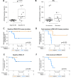
DNA concentration determinations and Kaplan-Meier analyses performed using cfDNA from patients. Panel A shows total cfDNA concentrations in early- vs. late-stage cancers. Panel B shows the same DNA concentration determinations plotted from patients who did not have a recurrence vs. those that did. Panels C and D show Kaplan-Meier analyses of recurrence-free survival in patients where HPV-human DNA junctions were detected in serum vs. those where it was not detected at initial examination (Panel C) or at 6-months post-treatment (Panel D). Panels E and F show Kaplan-Meier analyses of recurrence-free survival in patients where HPV E7 DNA was detected in serum vs. those where it was not detected at initial examination (Panel E) or at 6-months post-treatment (Panel F). In all four plots, squares show left-censoring at the last patient visits.
Figure 4.
Figure 4.
Analyses of HPV DNAs in Patient 16. Panel A shows PCR analysis for HPV16 E7 DNA in serum cfDNA plus DNA from the primary tumor as a positive control. Panel B shows PCR analysis for HPV18 E7 DNA in serum cfDNA plus DNA from the primary tumor as a positive control. Panel C shows the IGV plots of sequence reads following HPV DNA hybridization capture from the primary tumor and cfDNA. Sequence reads for HPV16 and HPV18 in serum cfDNA were plotted separately.
Figure 5.
Figure 5.
Kaplan-Meier recurrence-free survival plot of 297 TCGA cervical cancer patients.

Similar articles

References

    1. Sung H., et al. Global Cancer Statistics 2020: GLOBOCAN Estimates of Incidence and Mortality Worldwide for 36 Cancers in 185 Countries. CA Cancer J Clin 71, 209–249 (2021). - PubMed
    1. Siegel R.L., Giaquinto A.N. & Jemal A. Cancer statistics, 2024. CA Cancer J Clin 74, 12–49 (2024). - PubMed
    1. Girda E., et al. Cervical cancer treatment update: A Society of Gynecologic Oncology clinical practice statement. Gynecol Oncol 179, 115–122 (2023). - PMC - PubMed
    1. Rossi N.M., et al. Extrachromosomal Amplification of Human Papillomavirus Episomes Is a Mechanism of Cervical Carcinogenesis. Cancer Res 83, 1768–1781 (2023). - PMC - PubMed
    1. Van Arsdale A., et al. Structure and transcription of integrated HPV DNA in vulvar carcinomas. NPJ Genom Med 9, 35 (2024). - PMC - PubMed

Publication types

LinkOut - more resources