This is a preprint.
Metastatic spine disease alters spinal load-to-strength ratios in patients compared to healthy individuals
- PMID: 39830276
- PMCID: PMC11741471
- DOI: 10.1101/2025.01.06.25320075
Metastatic spine disease alters spinal load-to-strength ratios in patients compared to healthy individuals
Abstract
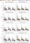
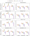
Pathologic vertebral fractures (PVF) are common and serious complications in patients with metastatic lesions affecting the spine. Accurate assessment of cancer patients' PVF risk is an unmet clinical need. Load-to-strength ratios (LSRs) evaluated in vivo by estimating vertebral loading from biomechanical modeling and strength from computed tomography imaging (CT) have been associated with osteoporotic vertebral fractures in older adults. Here, for the first time, we investigate LSRs of thoracic and lumbar vertebrae of 135 spine metastases patients compared to LSRs of 246 healthy adults, comparable by age and sex, from the Framingham Heart Study under four loading tasks. Findings include: (1) Osteolytic vertebrae have higher LSRs than osteosclerotic and mixed vertebrae; (2). In patients' vertebrae without CT observed metastases, LSRs were greater than healthy controls. (3) LSRs depend on the spinal region (Thoracic, Thoracolumbar, Lumbar). These findings suggest that LSRs may contribute to identifying patients at risk of incident PVF in metastatic spine disease patients. The lesion-mediated difference suggests that risk thresholds should be established based on spinal region, simulated task, and metastatic lesion type.
Keywords: Framingham Heart Study cohort; Metastatic Spinal disease; Patient cohort; load-to-strength ratio; musculoskeletal models; vertebral strength.
Conflict of interest statement
Competing Interests The authors declare that the research was conducted in the absence of any commercial or financial relationships that could be construed as a potential conflict of interest.
Figures


Similar articles
-
Evaluation of Load-To-Strength Ratios in Metastatic Vertebrae and Comparison With Age- and Sex-Matched Healthy Individuals.Front Bioeng Biotechnol. 2022 Aug 5;10:866970. doi: 10.3389/fbioe.2022.866970. eCollection 2022. Front Bioeng Biotechnol. 2022. PMID: 35992350 Free PMC article.
-
Patterns of Load-to-Strength Ratios Along the Spine in a Population-Based Cohort to Evaluate the Contribution of Spinal Loading to Vertebral Fractures.J Bone Miner Res. 2021 Apr;36(4):704-711. doi: 10.1002/jbmr.4222. Epub 2020 Dec 13. J Bone Miner Res. 2021. PMID: 33253414 Free PMC article.
-
Improved estimates of strength and stiffness in pathologic vertebrae with bone metastases using CT-derived bone density compared with radiographic bone lesion quality classification.J Neurosurg Spine. 2021 Sep 3;36(1):113-124. doi: 10.3171/2021.2.SPINE202027. Print 2022 Jan 1. J Neurosurg Spine. 2021. PMID: 34479191 Free PMC article.
-
Correlation analysis of the vertebral compression degree and CT HU value in elderly patients with osteoporotic thoracolumbar fractures.J Orthop Surg Res. 2023 Jun 26;18(1):457. doi: 10.1186/s13018-023-03941-z. J Orthop Surg Res. 2023. PMID: 37365576 Free PMC article. Review.
-
Traumatic Fractures of the Thoracic Spine.Z Orthop Unfall. 2021 Aug;159(4):373-382. doi: 10.1055/a-1144-3846. Epub 2020 May 11. Z Orthop Unfall. 2021. PMID: 32392598 Review. English, German.
References
-
- U.S. Cancer Statistics Working Group. U.S. Cancer Statistics Data Visualizations Tool, b. o. s. d. (ed Centers for Disease Control and Prevention and National Cancer Institute; ) (U.S. Department of Health and Human Services, 2022).
-
- Eleraky M., Papanastassiou I. & Vrionis F. D. Management of metastatic spine disease. Curr Opin Support Palliat Care 4, 182–188 (2010). - PubMed
-
- Siegel R. et al. Cancer treatment and survivorship statistics, 2012. CA Cancer J Clin 62, 220–241 (2012). - PubMed
-
- Tamada T. et al. Three-dimensional trabecular bone architecture of the lumbar spine in bone metastasis from prostate cancer: comparison with degenerative sclerosis. Skeletal Radiol. 34, 149–155 (2005). - PubMed
Publication types
Grants and funding
LinkOut - more resources
Full Text Sources
