. 2024 Nov 26;12(2):nwae426.
doi: 10.1093/nsr/nwae426. eCollection 2025 Feb.

A tactfully designed photothermal agent collaborating with ascorbic acid for boosting maxillofacial wound healing

Affiliations

A tactfully designed photothermal agent collaborating with ascorbic acid for boosting maxillofacial wound healing

Yuxin Qian et al. Natl Sci Rev. .

Abstract

Maxillofacial injuries that may cause severe functional and aesthetic damage require effective and immediate management due to continuous exposure to diverse microbial populations. Moreover, drug resistance, biofilm formation, and oxidative stress significantly impede timely bacterial removal and immune function, making the exploration of advanced materials for maxillofacial wound healing an appealing yet highly challenging task. Herein, a near-infrared photothermal sterilization agent was designed, encapsulated with liposomes and coated with ascorbic acid known for its antioxidant and immune-regulatory functions. The resulting nanoparticles, 4TPE-C6T-TD@AA, effectively neutralize reactive oxygen species generated by lipopolysaccharides, facilitate the conversion of pro-inflammatory M1 macrophages to anti-inflammatory M2 macrophages, and eliminate >90% of Staphylococcus aureus and Escherichia coli by disrupting bacterial physiological functions upon exposure to 808 nm laser irradiation. In vivo experiments demonstrate that 4TPE-C6T-TD@AA rapidly eliminates bacteria from infected wounds in the maxillofacial region of rats, and significantly promotes healing in S. aureus-infected wounds by enhancing collagen formation and modulating the inflammatory microenvironment. In conclusion, this study presents a promising therapeutic strategy for effectively combating bacterial infections and excessive inflammation in treating maxillofacial injuries.

Keywords: anti-inflammation; maxillofacial wound healing; photothermal sterilization.


Figures

Scheme 1.
Scheme 1.
(A) Chemical structures of DSPE-PEG2000@AA and 4TPE-C6T-TD. (B) Flow chart illustrating the synthesis of 4TPE-C6T-TD@AA. (C) Schematic diagram depicting the application of 4TPE-C6T-TD@AA in treating maxillofacial infected wounds, promoting wound healing through its antibacterial, antioxidant, and anti-inflammatory effects.
Figure 1.
Figure 1.
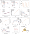
(A) Chemical structures of 4TPA-C6T-TD and 4TPE-C6T-TD. (B) Optimized S0 geometries of 4TPA-C6T-TD and 4TPE-C6T-TD. (C) Illustration of the frontier molecular orbitals (LUMOs and HOMOs) determined at the B3LYP/6–31G(d) level of theory. (D) The absorption spectra of obtained compounds (10 × 10−6 M) dissolved in THF solution. (E) Comparison of the photothermal conversion behavior of the two molecules (100 μM) in DMSO solution upon an 808 nm laser irradiation (1.0 W/cm2). (F) FTIR spectra of AA, DSPE- PEG2000, and DSPE- PEG2000@AA. (G) TEM image of 4TPE-C6T-TD@AA dispersed in water. Scale bar, 30 μm. (H) The absorption spectra of 4TPE-C6T-TD@AA at different concentrations. (I) TEM image and particle size analysis of 4TPE-C6T-TD@AA degraded in water for 7 days. Scale bar, 30 μm.
Figure 2.
Figure 2.
The photothermal and antioxidant properties of 4TPE-C6T-TD@AA. (A) Photothermal activity of 4TPE-C6T-TD@AA with different concentrations under 808 nm laser irradiation (1 W/cm2). (B) Photothermal activity of 4TPE-C6T-TD@AA (100 μg/mL) with different power densities of 808 nm laser irradiation. (C) Heating and cooling curves and (D) absorption spectra of the photothermal conversion cycling test of 4TPE-C6T-TD@AA. (E) Linear fitting of time versus -ln(θ) during cooling of 4TPE-C6T-TD@AA. (F) The absorption spectra and (G) kinetic curve analysis of the DPPH test evaluating the total ROS scavenging capacity of 4TPE-C6T-TD@AA. (H) The absorption spectra and (I) kinetic curve analysis of ABTS test evaluating the total ROS scavenging capacity of 4TPE-C6T-TD@AA. (J) Absorption spectra of Fenton reaction examining the ·OH scavenging capacity of 4TPE-C6T-TD@AA. (K) Electron spin resonance (ESR) measurement of ·O2− scavenging capacity of 4TPE-C6T-TD@AA. (L) Proposed mechanism of 4TPE-C6T-TD@AA to scavenge ROS (n = 3). Statistical analysis was performed using one-way ANOVA with Tukey's post-test. *p < 0.05, **p < 0.01, ***p < 0.001.
Figure 3.
Figure 3.
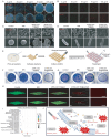
In vitro antibacterial behavior of 4TPE-C6T-TD@AA. (A) Typical photographs of bacterial colonies on agar plates and (B) Live/Dead staining images of S. aureus and E. coli after treatment with different concentrations of 4TPE-C6T-TD@AA under 808 nm laser irradiation. Scale bar, 100 μm. (C) Representative SEM images of bacterial morphologies of S. aureus. Scale bar, 500 nm. (D) Representative SEM images of bacterial morphologies of E. coli. Scale bar, 1 μm. (E) Schematic diagram of biofilm culture. Crystal violet staining images of 7-day (F) S. aureus and (G) E. coli biofilms. (H) 3D confocal Live/Dead staining images of 7-day biofilms. Scale bar, 100 μm. (I) KEGG enrichment analysis of differentially expressed genes (DEGs) in S. aureus after being treated with 4TPE-C6T-TD@AA/NIR compared to an untreated control. (J) Schematic diagram of the mechanism of 4TPE-C6T-TD@AA killing S. aureus with NIR (n = 3). Statistical analysis was performed using one-way ANOVA with Tukey's post-test. *p < 0.05, **p < 0.01, ***p < 0.001.
Figure 4.
Figure 4.
In vitro anti-inflammatory properties of 4TPE-C6T-TD@AA. (A) Light microscopic and DCFH-DA fluorescence images of RAW 264.7 macrophages. Scale bar, 100 μm. (B) Flow cytometry analysis of DCFH-DA expression in RAW 264.7 macrophages. (C) Light microscopic and DCFH-DA fluorescence images of L929 cells. Scale bar, 100 μm. (D) Flow cytometry analysis of DCFH-DA expression in L929 cells. (E) Flow cytometry assessment of CD86 expression, a macrophage marker, in RAW 264.7 macrophages. (F) Quantitative analysis of CD86/CD206 ratio in RAW 264.7 macrophages. (G) Quantitative analysis of immune factor expression in RAW 264.7 macrophages using qRT-PCR (n = 3). Statistical analysis was performed using one-way ANOVA with Tukey's post-test. *p < 0.05, **p < 0.01, and ***p < 0.001.
Figure 5.
Figure 5.
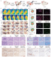
Synergistic PTT with 4TPE-C6T-TD@AA in a rat maxillofacial infection wound model. (A) Establishment of the rat maxillofacial infection wound model and its treatment schematic diagram. (B) Infrared thermal images of NIR and 4TPE-C6T-TD@AA/NIR groups for 5 min NIR laser irradiation, (C) photos of maxillofacial wounds during treatment and (D) visual images of preoperative and postoperative areas of wounds. (E) H&E staining images. Scale bar, 50 μm. (F) Masson staining images. Scale bar, 50 μm. (G) Immunofluorescence staining images, where CD86 shows red fluorescence, and CD206 shows green fluorescence. Scale bar, 50 μm. (H) Immunohistochemical staining images of IL-6 and (I) TNF-α. Scale bar, 50 μm.

Similar articles

Cited by

References

    1. Rivera-Hidalgo F, Stanford TW. Oral mucosal lesions caused by infective microorganisms. I. Viruses and bacteria. Periodontol 2000 1999; 21: 106–24.10.1111/j.1600-0757.1999.tb00171.x - DOI - PubMed
    1. Huang X, Lou Y, Duan Y et al. Biomaterial scaffolds in maxillofacial bone tissue engineering: a review of recent advances. Bioact Mater 2024; 33: 129–56. - PMC - PubMed
    1. Werdin F, Tenenhaus M, Rennekampff HO. Chronic wound care. Lancet 2008; 372: 1860–2.10.1016/S0140-6736(08)61793-6 - DOI - PubMed
    1. Guan W, Gong C, Wu S et al. Instant protection spray for anti-infection and accelerated healing of empyrosis. Adv Mater 2024; 36: 2306589.10.1002/adma.202306589 - DOI - PubMed
    1. Liu L, Zhang H, Xing S et al. Copper-zinc bimetallic single-atom catalysts with localized surface plasmon resonance-enhanced photothermal effect and catalytic activity for melanoma treatment and wound-healing. Adv Sci 2023; 10: e2207342.10.1002/advs.202207342 - DOI - PMC - PubMed

LinkOut - more resources