

A rare case of bone lesion: Mandible's fibrous dysplasia
- PMID: 39830478
- PMCID: PMC11737581
- DOI: 10.4103/njms.njms_35_23
A rare case of bone lesion: Mandible's fibrous dysplasia
Abstract
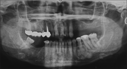
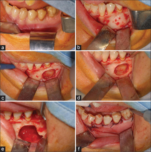
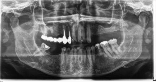
Fibrous dysplasia is a rare genetic syndrome that affects bone tissue. This pathology replaces the mineralized matrix of the bone affected with connective and fibrous tissue. This article describes a mandibular fibrous osseous dysplasia case and its surgical treatment. A 45-year-old woman complained about a slow development of swelling of the left mandibular bone. The orthopantomography (OPT) and the cone beam computed tomography (CBCT) revealed a well-circumscribed sclerotic lesion with a ground-glass appearance apical to the 3.5 element. The surgery was performed to excise the lesion. Anatomopathological examination of tissue confirmed the suspects among the diagnosis of fibrous dysplasia. The patient underwent to follow-up of 4 years, and no recurrences were found. In the absence of a univocal consensus on therapy, surgery remains the treatment of choice for unifocal forms.
Keywords: Fibrous dysplasia; fibro-osseous lesions; oral surgery.
Copyright: © 2024 National Journal of Maxillofacial Surgery.
Conflict of interest statement
There are no conflicts of interest.
Figures
Similar articles
-
A Rare Case of Cemento-Ossifying Fibroma: A Case Report.Cureus. 2023 May 7;15(5):e38685. doi: 10.7759/cureus.38685. eCollection 2023 May. Cureus. 2023. PMID: 37292559 Free PMC article.
-
Bilateral fibrous dysplasia of the mandible in a 7-year-old male patient--a rare case.J Indian Soc Pedod Prev Dent. 2010 Apr-Jun;28(2):126-9. doi: 10.4103/0970-4388.66756. J Indian Soc Pedod Prev Dent. 2010. PMID: 20660982
-
[Fibrous dysplasia of the frontal and ethmoid sinuses: a case report].Pan Afr Med J. 2021 Apr 20;38:385. doi: 10.11604/pamj.2021.38.385.27561. eCollection 2021. Pan Afr Med J. 2021. PMID: 34381529 Free PMC article. French.
-
Fibrous dysplasia with secondary aneurysmal bone cyst-a rare case report and literature review.Oral Maxillofac Surg. 2019 Mar;23(1):101-107. doi: 10.1007/s10006-019-00741-w. Epub 2019 Feb 13. Oral Maxillofac Surg. 2019. PMID: 30758737 Review.
-
Associated aneurysmal bone cyst and cemento-osseous dysplasia: a case report and review of the literature.Gen Dent. 2017 Jan-Feb;65(1):28-32. Gen Dent. 2017. PMID: 28068262 Review.