

Brain penetration of peripheral extracellular vesicles from Alzheimer's patients and induction of microglia activation
- PMID: 39830834
- PMCID: PMC11740088
- DOI: 10.1002/jex2.70027
Brain penetration of peripheral extracellular vesicles from Alzheimer's patients and induction of microglia activation
Abstract
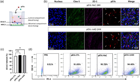
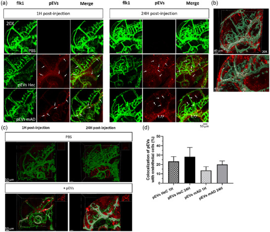
Alzheimer's disease (AD) is an age-related neurodegenerative pathology. Brain-derived extracellular vesicles (EVs) have been demonstrated to be implicated in AD pathogenesis by facilitating the propagation of Tau, amyloid-β and inflammatory cytokines. However, the impact of peripheral EVs (pEVs) in AD pathogenesis remains poorly investigated. The objective of our study was to compare the passage of pEVs from adults, cognitively healthy elderly, and AD patients through the blood-brain barrier (BBB), to evaluate their uptake in the brain and to assess their impact on the microglia activity using in vitro and in vivo models. To this end, pEVs were enriched, characterized, and fluorescently labelled. The passage of pEVs through the endothelial bEnd.3 cells was studied in a Transwell device with either neuronal or microglia cells seeded at the bottom of the well. Following the internalization of pEVs from AD patients, microglia adopted an amoeboid morphology and released a heightened level of pro-inflammatory cytokine IL-6. To further assess their in vivo transport across the BBB, pEVs were injected into the blood circulation of 2-days post-fertilization Tg(flk1:EGFP) zebrafish. The biodistribution of pEVs was monitored at 1 and 24 h post-injection using confocal microscopy. We demonstrated that pEVs traverse the BBB by transcytosis and subsequently diffuse progressively into the brain. pEVs were then internalized by neuronal and radial glial cells as seen in Tg(huc:EGFP) and Tg(gfap:EGFP) zebrafish, respectively. Additional experiments were performed with the intrahippocampal injection of pEVs in the mouse, indicating their spreading throughout the brain and their uptake by neuronal and glial cells. These findings contribute to novel insights into the fate of pEVs following their passage through the BBB in vitro and in vivo, and demonstrate for the first time that pEVs from AD patients affect microglia activity. This suggests a potential mechanism through which peripheral tissue cues may contribute to AD pathogenesis.
Keywords: Alzheimer's disease; blood‐brain barrier; brain; extracellular vesicles; microglia; zebrafish.
© 2025 The Author(s). Journal of Extracellular Biology published by Wiley Periodicals LLC on behalf of International Society for Extracellular Vesicles.
Conflict of interest statement
The authors have no relevant financial or non‐financial interests to disclose and declare no competing interests.
Figures


References
-
- Akiyama, H. , Barger, S. , Barnum, S. , Bradt, B. , Bauer, J. , Cole, G. M. , Cooper, N. R. , Eikelenboom, P. , Emmerling, M. , Fiebich, B. L. , Finch, C. E. , Frautschy, S. , Griffin, W. S. , Hampel, H. , Hull, M. , Landreth, G. , Lue, L. , Mrak, R. , Mackenzie, I. R. , … Wyss‐Coray, T. (2000). Inflammation and Alzheimer's disease. Neurobiology of Aging, 21(3), 383–421. 10.1016/s0197-4580(00)00124-x - DOI - PMC - PubMed
-
- Banks, W. A. , Sharma, P. , Bullock, K. M. , Hansen, K. M. , Ludwig, N. , & Whiteside, T. L. (2020). Transport of extracellular vesicles across the blood‐brain barrier: Brain pharmacokinetics and effects of inflammation. International Journal of Molecular Sciences, 21(12), 4407. 10.3390/ijms21124407 - DOI - PMC - PubMed
-
- Ben Khedher, M. R. , Haddad, M. , Laurin, D. , & Ramassamy, C. (2021). Apolipoprotein E4–driven effects on inflammatory and neurotrophic factors in peripheral extracellular vesicles from cognitively impaired, no dementia participants who converted to Alzheimer's disease. Alzheimer's & Dementia: Translational Research & Clinical Interventions, 7(1), e12124. 10.1002/trc2.12124 - DOI - PMC - PubMed
LinkOut - more resources
Full Text Sources
Molecular Biology Databases
Miscellaneous