. 2024 Dec 19;11(1):e41295.
doi: 10.1016/j.heliyon.2024.e41295. eCollection 2025 Jan 15.

Leveraging RIBOTAC technology: Fluorescent RNase L probes for live-cell imaging and function analysis

Affiliations

Leveraging RIBOTAC technology: Fluorescent RNase L probes for live-cell imaging and function analysis

Elias Khaskia et al. Heliyon. .

Abstract

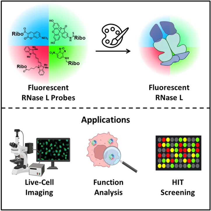
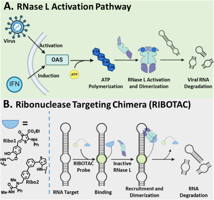
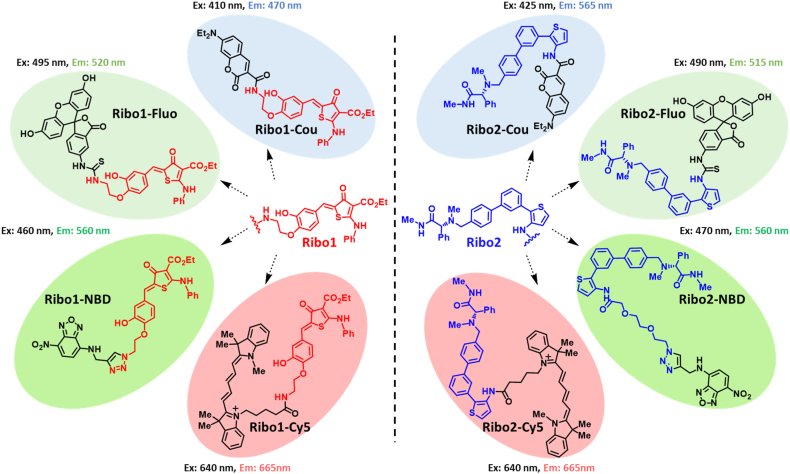
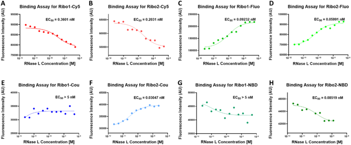
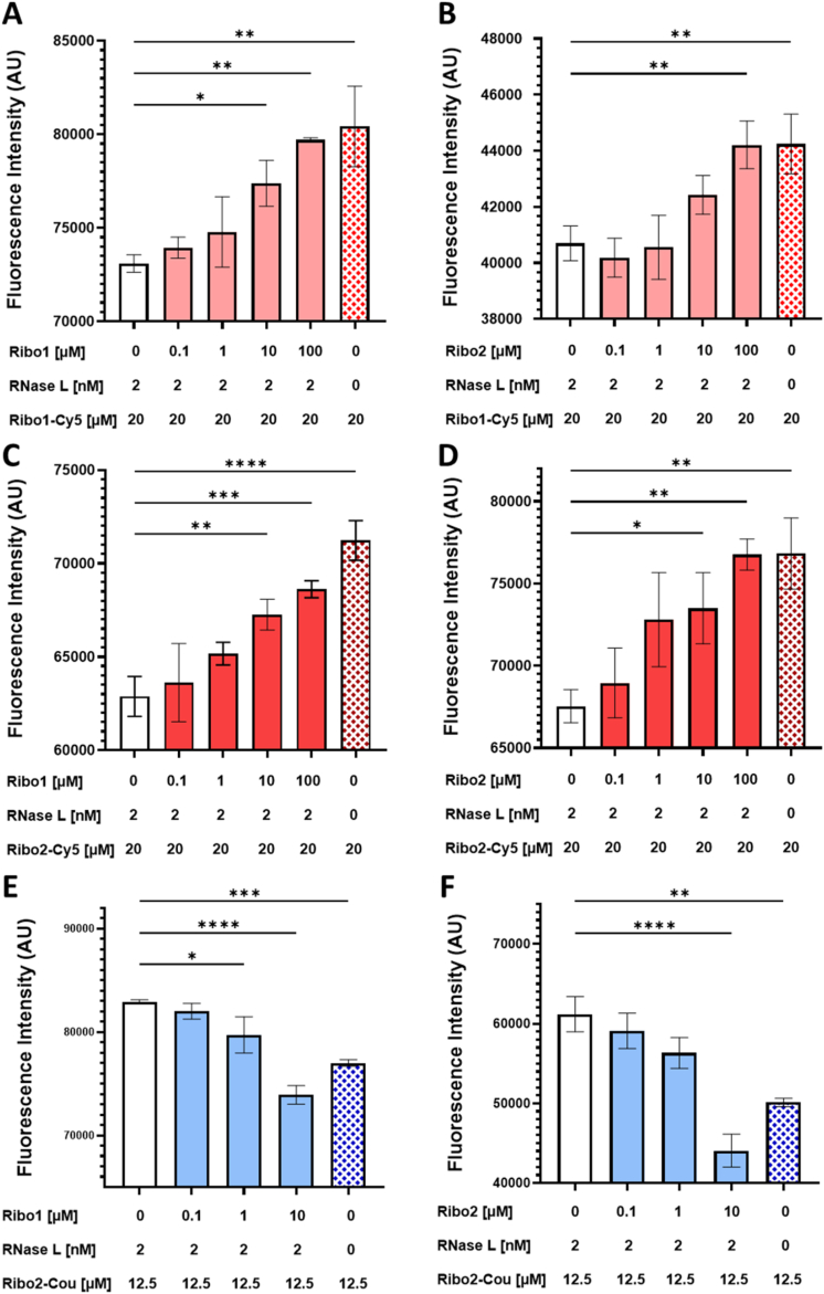
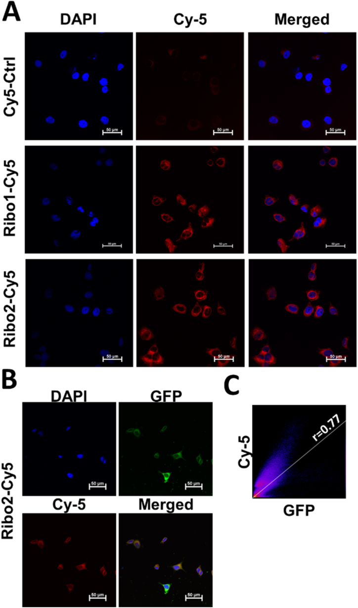
RNA-targeting small molecules, particularly RIBOnuclease TArgeting Chimeras (RIBOTACs), represent a powerful and promising therapeutic approach by selectively degrading RNAs through ribonuclease (RNase) recruitment. Despite their potential, the development of effective RNase recruitment tools is still in its early stages and remains a critical area of research. Ribonuclease L (RNase L) is a key ribonuclease targeted by RIBOTACs, yet the tools available for studying RNase L are limited. In this study, we introduce novel fluorescent ribonuclease binders that enhance the visualization and investigation of RNase L activity. Our findings provide new insights into RNase L dynamics and RNA degradation pathways, paving the way for more effective RNA-targeted degradation strategies. Furthermore, we explore the versatility of these conjugates for real-time tracking of RNase L localization, intracellular trafficking, and mechanistic studies. These fluorescent probes also enable high-throughput fluorescence-based assays to identify small molecules that bind and recruit RNase L, advancing RNA-targeted therapeutic approaches.

Keywords: Fluorescent probes; RIBOTAC; RNase L; Subcellular localization.


Conflict of interest statement

The authors declare that they have no known competing financial interests or personal relationships that could have appeared to influence the work reported in this paper.

Figures

Image 1
Graphical abstract
Fig. 1
Fig. 1
RNase L activation. A) IFN-induced RNase L activation pathway. B) RIBOnuclease TArgeting Chimera strategy for degrading target RNA.
Fig. 2
Fig. 2
Fluorescent RNase L probes. The Ribo1 binder is shown in red, and the Ribo2 binder is shown in blue. Four families of fluorophores were synthesized and are highlighted with different backgrounds: Diethylamino Coumarin (blue), Fluorescein (light green), Nitrobenzodiazole (NBD) (dark green), and Cyanine-5 (red).
Fig. 3
Fig. 3
Fluorescence-based binding assay. RNase L was titrated into a constant concentration of each derivative and was incubated at 37 °C. The EC50 values for each probe are annotated above their respective binding curves. A) Ribo1-Cy5 (20 μM). B) Ribo2-Cy5 (20 μM). C) Ribo1-Fluo (50 μM). D) Ribo2-Fluo (2 μM). E) Ribo1-Cou (12.5 μM). F) Ribo2-Cou (12.5 μM). G) Ribo1-NBD (80 μM). H) Ribo2-NBD (80 μM).
Fig. 4
Fig. 4
Fluorescent-based competition assay for target validation. RNase L was incubated with increasing concentrations of Ribo1 or Ribo2 for 10 min at 37 °C, followed by incubation with the fluorescent probes for another 2 h. A) Competition between Ribo1-Cy5 and Ribo1. B) Competition between Ribo1-Cy5 and Ribo2. C) Competition between Ribo2-Cy5 and Ribo1. D) Competition between Ribo2-Cy5 and Ribo2. E) Competition between Ribo2-Cou and Ribo1. F) Competition between Ribo2-Cou and Ribo2. Statistical analysis was performed using one-way ANOVA test. Error bars represent the SD of the mean (n = 3). ∗∗ represents P ≤ 0.01, ∗∗∗ represents P ≤ 0.001 and ∗∗∗∗ represents P ≤ 0.0001.
Fig. 5
Fig. 5
Uptake of probes by MCF-7 cells and RNase L subcellular localization. A) Representative fluorescence images showing the cellular uptake of fluorescent probes Cy5-Ctrl (1 μM), Ribo1-Cy5 (1 μM), and Ribo2-Cy5 (1 μM) (red) with nucleus marker DAPI (blue), after 2 h treatment. Scale bar = 50 μm. B) Representative fluorescence images showing RNase L subcellular localization upon 2 h treatment with Ribo2-Cy5 (0.1 μM) (red), GFP tagged RNase L (green), and nucleus marker DAPI (blue). Scale bar = 50 μm. C) Scatter plot showing the relationship between fluorescent probes and RNase L localization; the solid line represents the best-fit regression line, illustrating the positive correlation (r = 0.703 ± 0.037).
Fig. 6
Fig. 6
Fluorescent probes cellular uptake in MCF-7 and MDA-MB-231 cell lines. A) Fluorescence-activated cell sorting assay after 5 min, 2 h, and 24 h treatment (1 μM). Cyan represents untreated cells, pink represents Cy5-Ctrl, green represents Ribo1-Cy5, and orange represents Ribo2-Cy5. B) Ribo1-Cy5 and Ribo2-Cy5 cellular uptake comparison among MDA-MB-231 (orange), MCF-7 (cyan), and HEK-293 (pink) after 2 h treatment (1 μM).

References

    1. Silverman R.H. Viral encounters with 2′,5′-oligoadenylate synthetase and RNase L during the interferon antiviral response. J. Virol. 2007;81:12720–12729. doi: 10.1128/jvi.01471-07. - DOI - PMC - PubMed
    1. Cooper D.A., Banerjee S., Chakrabarti A., García-Sastre A., Hesselberth J.R., Silverman R.H., Barton D.J. RNase L targets distinct sites in influenza A virus RNAs. J. Virol. 2015;89:2764–2776. doi: 10.1128/jvi.02953-14. - DOI - PMC - PubMed
    1. Li Y., Banerjee S., Wang Y., Goldstein S.A., Dong B., Gaughan C., Silverman R.H., Weiss S.R. Activation of RNase L is dependent on OAS3 expression during infection with diverse human viruses. Proc. Natl. Acad. Sci. U. S. A. 2016;113:2241–2246. doi: 10.1073/pnas.1519657113. - DOI - PMC - PubMed
    1. Li Y., Renner D.M., Comar C.E., Whelan J.N., Reyes H.M., Cardenas-Diaz F.L., Truitt R., Tan L.H., Dong B., Alysandratos K.D., Huang J., Palmer J.N., Adappa N.D., Kohanski M.A., Kotton D.N., Silverman R.H., Yang W., Morrisey E.E., Cohen N.A., Weiss S.R. Sars-cov-2 induces double-stranded rna-mediated innate immune responses in respiratory epithelial-derived cells and cardiomyocytes. Proc. Natl. Acad. Sci. U. S. A. 2021;118 doi: 10.1073/pnas.2022643118. - DOI - PMC - PubMed
    1. Dong B., Stlvermanz R.H. 2-5A-dependent RNase molecules dimerize during activation by 2-5A. J. Biol. Chem. 1995;270:4133–4137. doi: 10.1074/jbc.270.8.4133. - DOI - PubMed

LinkOut - more resources