Review
. 2024 Dec 31;16(12):8782-8795.
doi: 10.21037/jtd-24-1125. Epub 2024 Dec 27.

The blind spots on chest computed tomography: what do we miss

Affiliations
Review

The blind spots on chest computed tomography: what do we miss

Li Zhang et al. J Thorac Dis. .

Abstract

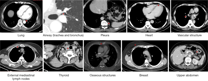
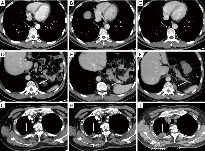
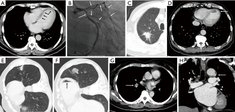
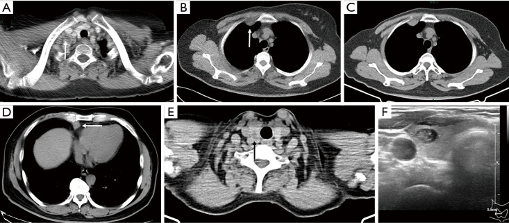
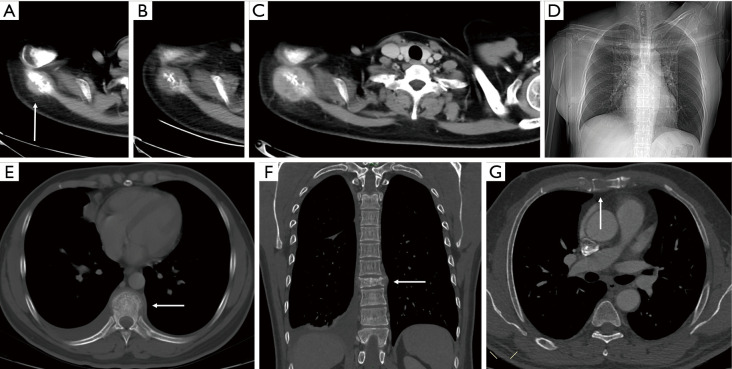
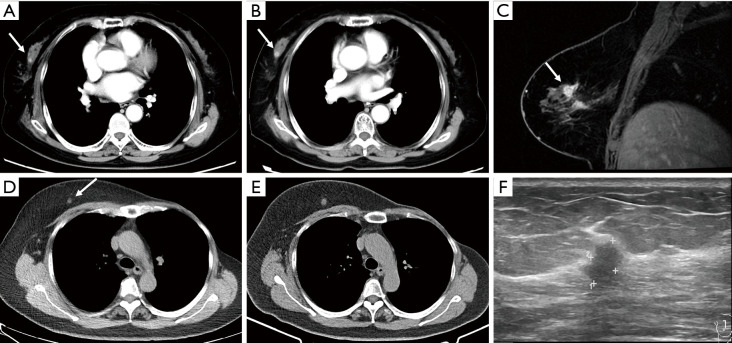
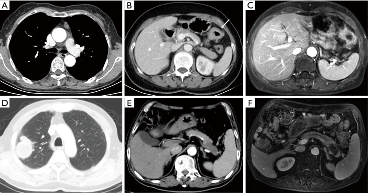
Chest computed tomography (CT) is the most frequently performed imaging examination worldwide. Compared with chest radiography, chest CT greatly improves the detection rate and diagnostic accuracy of chest lesions because of the absence of overlapping structures and is the best imaging technique for the observation of chest lesions. However, there are still frequently missed diagnoses during the interpretation process, especially in certain areas or "blind spots", which may possibly be overlooked by radiologists. Awareness of these blind spots is of great significance to avoid false negative results and potential adverse consequences for patients. In this review, we summarize the common blind spots identified in actual clinical practice, encompassing the central areas within the pulmonary parenchyma (including the perihilar regions, paramediastinal regions, and operative area after surgery), trachea and bronchus, pleura, heart, vascular structure, external mediastinal lymph nodes, thyroid, osseous structures, breast, and upper abdomen. In addition to careful review, clinicians can employ several techniques to mitigate or minimize errors arising from these blind spots in film interpretation and reporting. In this review, we also propose technical methods to reduce missed diagnoses, including advanced imaging post-processing techniques such as multiplanar reconstruction (MPR), maximum intensity projection (MIP), artificial intelligence (AI) and structured reporting which can significantly enhance the detection of lesions and improve diagnostic accuracy.

Keywords: Diagnostic imaging; blind spots; chest computed tomography (chest CT).


Conflict of interest statement

Conflicts of Interest: All authors have completed the ICMJE uniform disclosure form (available at https://jtd.amegroups.com/article/view/10.21037/jtd-24-1125/coif). The authors have no conflicts of interest to declare.

Figures

Figure 1
Figure 1
Summary of blind spots on chest CT (red star indicates the areas of common blind spots on chest CT). CT, computed tomography.
Figure 2
Figure 2
Example of lung lesions that are easily missed on chest CT. (A,B) A 52-year-old male with adenocarcinoma of the left lower lung lobe. (A) The radiologist reported the main lesion in the left lung but failed to identify multiple small pulmonary nodules, which are difficult to distinguish from cross-sections of blood vessels. (B) The use of MIP reconstruction enhanced the clarity of the visualization of small nodules (black arrows). Subsequently, follow-up of the pulmonary nodules confirmed metastasis. As there is contralateral metastasis, according to the 8th edition Lung Cancer Staging classification (13), it is classified as M1. CT, computed tomography; MIP, maximum intensity projection.
Figure 3
Figure 3
Blind spots of lung: the para-aortic, paramediastinal, pericardiac and para-diaphragm areas. (A-C) A 66-year-old female with adenocarcinoma of the right upper lung lobe. The radiologist overlooked the ground-glass opacity near the aortic arch in the left upper lobe (arrows) (B,C) because of the more noticeable lesion in the right upper lobe. (D-F) A 69-year-old female with a missed solid nodule in the first two CT images near the poster mediastinum in the left lower lobe (arrows) that gradually grew in size during follow-up. (G-I) A 63-year-old female after resection of the left lung adenocarcinoma. A new small nodule near the left pericardium (arrows) was missed in the first two CT exam, showing gradual enlargement during the follow-up period, and subsequent confirmed adenocarcinoma by biopsy. (J-L) A 52-year-old female after resection of the right upper lung lobe adenocarcinoma. (J,K) A new nodule near the diaphragm had been growing in size (arrows), raising concerns about it being a metastasis. Due to its hidden location and interference from a nearby shadow, there is a risk of overlooking this lesion. CT, computed tomography.
Figure 4
Figure 4
Blind spots on chest CT: trachea and bronchus. (A-E) A 65-year-old male with right upper lung lobe cancer. (B-D) Radiologists reported the main lesion in the right lung, but a small nodule in the bronchus of the left upper lobe was overlooked on CT scan (arrows). (E) Bronchoscopy revealed the neoplasm obstructing the lumen of the left upper lung lobe bronchus, which was pathologically confirmed to be a squamous cell carcinoma. (F) The trachea or bronchus tumor should be distinguished from intraluminal secretion which is more common seen as shown in this case (arrow). CT, computed tomography.
Figure 5
Figure 5
Blind spot on chest CT: pleura. (A-C) A 57-year-old male with lung metastases after right upper lobe adenocarcinoma resection. There was initially missed gradual thickening of the right paravertebral pleura (arrows), which was diagnosed as pleural metastasis. (D-F) A 69-year-old man with a neuroendocrine tumor in the anterior mediastinum. A initially missed spindle-shaped nodular shadow was observed in the left diaphragmatic pleura (arrows). Follow-up chest CT revealed enlargement of this nodule, suggesting a metastatic lesion. (G-I) A 71-year-old male with right lower lobe lung cancer. Enhanced CT revealed missed nodular thickening of the right pleura (arrows). The final biopsy confirmed aggressive fibromatosis. These lesions were all missed in the first two scans. CT, computed tomography.
Figure 6
Figure 6
Blind spots on chest CT: heart and vascular structure. (A,B) A 45-year-old male with nasal NK/T-cell lymphoma and coronary heart disease. (A) The radiologist failed to identify subtle linear low-density shadows in the left ventricular wall (arrows), which are consistent with myocardial infarction. (B) Subsequent coronary angiography showed multiple stenoses (arrows) in the left anterior descending and circumflex coronary arteries. (C,D) A 70-year-old male with right lung cancer and a history of prior myocardial infarction. (D) Chest CT image revealing a low-density thrombus in the left ventricle, along with linear low-density shadows within the myocardium (arrow), indicating infarction. (E,F) A 72-year-old male with lung nodules in the left lower lobe underwent CT-guided biopsy. The immediate follow-up CT image after biopsy showed air within the left ventricle (black arrow), which is easily missed and lead to fatal result (F). (G) A 61-year-old female with metastases after left lung cancer surgery. The filling defects in the right middle and lower pulmonary artery (arrow), suggesting pulmonary artery embolism, was missed in the report. (H) A 66-year-old male with a missed low-density niche in the aortic arch on CT image (arrow), suggesting a penetrating ulcer. CT, computed tomography; NK, natural killer.
Figure 7
Figure 7
Blind spots on chest CT: external mediastinal lymph nodes and thyroid. Lymph nodes located outside the mediastinal area on chest CT, such as (A) those above the right clavicle (arrow) and (B,C) those in the internal mammary region (arrow), may be easily missed. (D) A 65-year-old male after surgery for a thymoma. During the follow-up period, a tumor nodule in the right cardiophrenic angle region (arrow) was missed and later confirmed as a thymoma recurrence (type AB). (E,F) A 64-year-old female with an incidentally detected low-density nodule in the right thyroid lobe (arrow) during low-dose CT screening; this nodule was identified as a nodular thyroid goiter through ultrasound. CT, computed tomography.
Figure 8
Figure 8
Blind spot on chest CT: bone. (A-D) A 44-year-old female with thymic squamous carcinoma. Metastasis to the right scapula (arrow) was neglected on CT. Retrospective observation revealed progressive bone destruction and enlargement of the soft tissue mass. Despite a lack of relevant clinical information on the requisition form, the patient’s inability to elevate her right arm (D, CT scout image) emerged as a crucial indicator of right shoulder disease. (E,F) A 33-year-old male with adenocarcinoma of the right lung and multiple metastases. Radiologists ignored vertebral metastasis with surrounding soft tissue swelling (arrow). A clearer depiction was obtained with the MPR of the coronal plane, revealing vertebral bone destruction and compression fractures (arrow). (G) Radiologists failed to identify sternal metastasis on chest CT (arrow). CT, computed tomography; MPR, multiplanar reconstruction.
Figure 9
Figure 9
Blind spot on chest CT: breast. (A-C) A 69-year-old female after surgery for left lower lobe. A growing nodule in the right breast was ignored during the follow-up period (arrows) (A,B). Later, an irregular and abnormal enhanced mass was seen on contrast-enhanced MRI (arrow) (C), combined with its signals in different sequences, it was confirmed to be breast cancer. (D-F) A 56-year-old female after left breast cancer resection. Because of its small size, radiologists have repeatedly overlooked a small nodule in the right breast upon multiple postoperative low-dose CT exams (arrow). A retrospective review revealed that there was progressive enlargement of this right breast nodule, and ultrasound confirmed the presence of breast cancer. CT, computed tomography; MRI, magnetic resonance imaging.
Figure 10
Figure 10
Blind spot on chest CT: the upper abdomen. (A-C) A 54-year-old female after resection of a thymic cyst in the anterior mediastinum. (B) The thickening of the colonic splenic flexure was missed on chest CT (arrow). During the follow-up period, she was diagnosed with colon cancer through colonoscopy. Subsequent MRI also revealed thickening of the colonic splenic flexure wall. (D-F) A 71-year-old male with lung cancer in the right upper lung lobe. (E) CT image showing an ill-defined mass in the pancreatic tail, which was initially missed. (F) MRI images revealing the same mass with an abnormal signal; the degree of enhancement on the enhanced scan was lower than that of the normal pancreas, and the splenic artery and vein were involved, suggesting pancreatic cancer. CT, computed tomography; MRI, magnetic resonance imaging.

Similar articles

References

    1. Gefter WB, Post BA, Hatabu H. Commonly Missed Findings on Chest Radiographs: Causes and Consequences. Chest 2023;163:650-61. 10.1016/j.chest.2022.10.039 - DOI - PMC - PubMed
    1. Gefter WB, Hatabu H. Reducing Errors Resulting From Commonly Missed Chest Radiography Findings. Chest 2023;163:634-49. 10.1016/j.chest.2022.12.003 - DOI - PubMed
    1. National Lung Screening Trial Research Team ; Aberle DR, Adams AM, et al. Reduced lung-cancer mortality with low-dose computed tomographic screening. N Engl J Med 2011;365:395-409. 10.1056/NEJMoa1102873 - DOI - PMC - PubMed
    1. Wang RC, Miglioretti DL, Marlow EC, et al. Trends in Imaging for Suspected Pulmonary Embolism Across US Health Care Systems, 2004 to 2016. JAMA Netw Open 2020;3:e2026930. 10.1001/jamanetworkopen.2020.26930 - DOI - PMC - PubMed
    1. Inui S, Gonoi W, Kurokawa R, et al. The role of chest imaging in the diagnosis, management, and monitoring of coronavirus disease 2019 (COVID-19). Insights Imaging 2021;12:155. 10.1186/s13244-021-01096-1 - DOI - PMC - PubMed

LinkOut - more resources