. 2025 Dec;16(1):2453818.
doi: 10.1080/21505594.2025.2453818. Epub 2025 Jan 20.

Immunogenicity and vaccine efficacy of Actinobacillus pleuropneumoniae-derived extracellular vesicles as a novel vaccine candidate

Affiliations

Immunogenicity and vaccine efficacy of Actinobacillus pleuropneumoniae-derived extracellular vesicles as a novel vaccine candidate

Su Hyun Park et al. Virulence. 2025 Dec.

Abstract

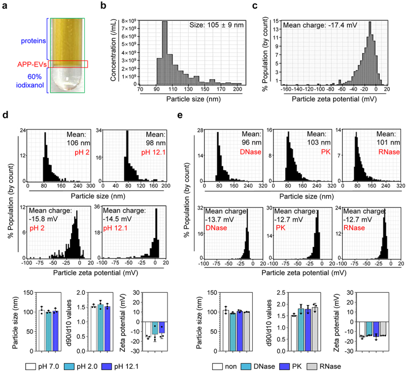
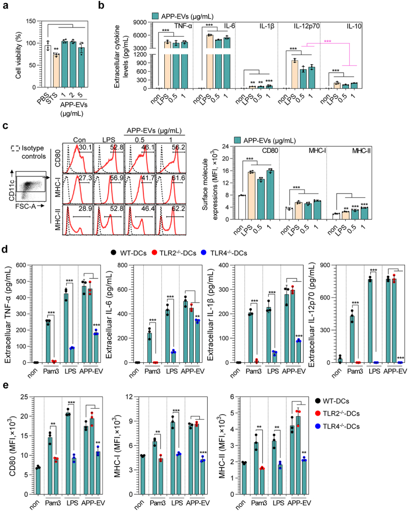
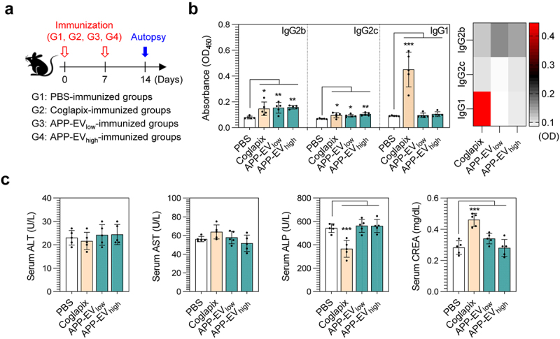
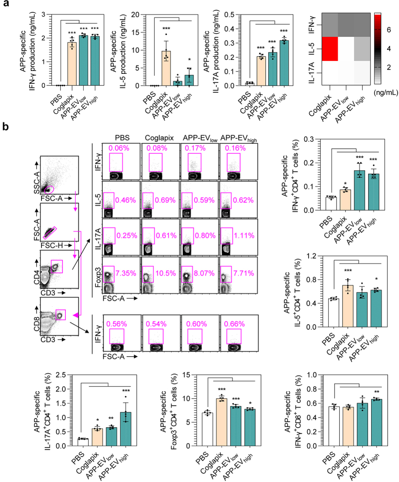
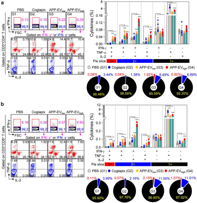
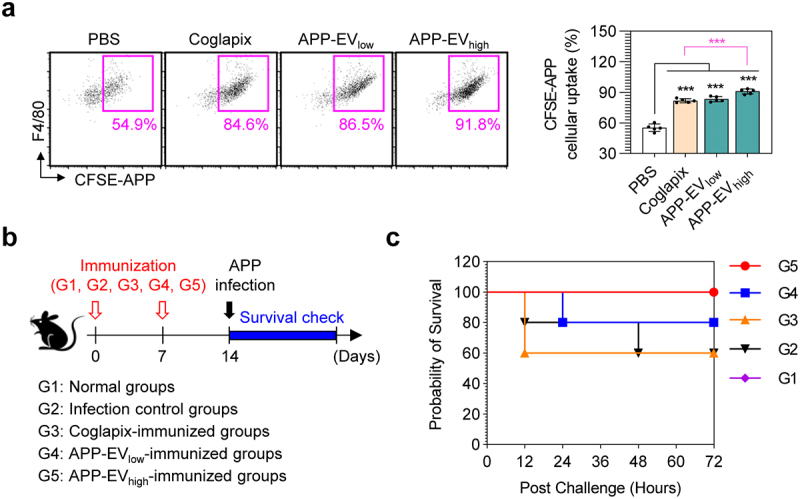
Actinobacillus pleuropneumoniae (APP) is a significant pathogen in the swine industry, leading to substantial economic losses and highlighting the need for effective vaccines. This study evaluates the potential of APP-derived extracellular vesicles (APP-EVs) as a vaccine candidate compared to the commercial Coglapix vaccine. APP-EVs, isolated using tangential flow filtration (TFF) and cushioned ultracentrifugation, exhibited an average size of 105 nm and a zeta potential of -17.4 mV. These EVs demonstrated stability under external stressors, such as pH changes and enzymatic exposure and were found to contain 86 major metabolites. Additionally, APP-EVs induced dendritic cell (DC) maturation in a Toll-like receptor 4 (TLR4)-dependent manner without cytotoxicity. APP-EVs predominantly elicited Th1-mediated IgG responses in immunized mice without significant liver and kidney toxicity. Contrarily, unlike Coglapix, which induced stronger Th2-mediated responses and notable toxicity. In addition, APP-EVs triggered APP-specific Th1, Th17, and cytotoxic T lymphocyte (CTL) responses and promoted the activation of multifunctional T-cells. Notably, APP-EV immunization enhanced macrophage phagocytosis and improved survival rates in mice challenged with APP infection compared to those treated with Coglapix. These findings suggest that APP-EVs are promising vaccine candidates, capable of inducing potent APP-specific T-cell responses, particularly Th1, Th17, CTL, and multifunctional T-cells, thereby enhancing the protective immune response against APP infection.

Keywords: Actinobacillus pleuropneumoniae; Th1-dominant cellullar immunity; Th1-dominant humoral immunity; extracellular vesicle; immunogenicity; pre-exposure vaccine.


Conflict of interest statement

No potential conflict of interest was reported by the author(s).

Figures

Figure 1.
Figure 1.
Characterization and stability of APP-EVs. (a) APP-EVs isolated via cushioned ultracentrifugation. Size (b) and zeta potential (c) of APP-EVs were analyzed using qNano-exoid. Size, zeta potential, and size distribution of APP-EVs for pH changes (d; pH 2.0 and pH 12.1) and enzyme exposures (e; DNase, RNase, PK: proteinase K) were analyzed using qNano-exoid. The qNano-exoid measurements were conducted three times, and the histograms represent typical results.
Figure 2.
Figure 2.
Change in cell viability, cytokine production, and surface molecule expression of DCs induced by APP-EV treatment. (a, b, c) Experiments were performed on DCs differentiated from WT C57BL/6 mice. (a) Cell viability was measured using the EZ-Cytox cell viability assay kit after treating DCs (mean ± SD; n = 4 samples) with staurosporine (STS, 20 nM) and APP-EVs (1, 2, and 5 μg/mL) for 18 hours. Bar graphs show mean ± SD (n = 4 samples). (B, C) DCs (mean ± SD; n = 3 samples) were treated with APP-EVs and lipopolysaccharide (LPS; 100 ng/mL) for 18 hours. (b) Cytokine levels (TNF-α, IL-6, IL-1β, IL-12p70, and IL-10) in the culture supernatants were measured using specific ELISA kits. (c) Surface molecule expression levels (anti-CD11c, -CD80, -MHC-I, and -MHC-II antibodies) were analyzed by flow cytometry, showing percentages and mean fluorescence intensity (MFI). Histograms represent typical results from three independent experiments. Statistical analysis of significant differences was performed using one-way ANOVA followed by Dunnett’s multiple comparison test for comparisons with the non-treated cell group. Statistical significance was denoted as **p < 0.01 and ***p < 0.001. (d, e) Experiments were conducted on DCs (mean ± SD; n = 3 samples) differentiated from WT, TLR2−/−, and TLR4−/− mice (WT-DCs, TLR2−/−-DCs, TLR4−/−-DCs, respectively) treated with APP-EVs (1 μg/mL) for 18 hours. (d) Cytokine production levels (TNF-α, IL-6, IL-1β, and IL-12p70) in the culture supernatants were measured. (e) Surface molecule expression levels were analyzed by flow cytometry. All data represent results from two independent experiments, with representative results shown. Statistical analysis of differences between two groups was conducted using an unpaired Student’s t-test, while differences among more than two groups were evaluated using one-way ANOVA followed by Dunnett’s multiple comparison test. Statistical significance was indicated as **p < 0.01 and ***p < 0.001.
Figure 3.
Figure 3.
Analysis in app-specific IgG responses and toxic indicators in APP-EVs-immunized mice. (a) Immunization scheme for mice (mean ± SD; n = 5 mice) with Coglapix, APP-EV low dose (APP-EVlow; 50 μg/mouse), and APP-EV high dose (APP-EVhigh; 200 μg/mouse). (b) Two weeks after the final immunization, BL-APP protein-specific IgG2b, IgG2c, and IgG1 responses in the serum of immunized mice were measured as described in the materials and methods section. (c) ALT, AST, ALP, and CREA levels in the serum of immunized mice were measured using a biochemical analyzer. All data represent results from two independent experiments, with representative results shown. Statistical analysis of significant differences was performed using one-way ANOVA followed by Dunnett’s multiple comparison test for comparisons with the pbs-immunized group. Statistical significance was denoted as *p < 0.05, **p < 0.01, ***p < 0.001.
Figure 4.
Figure 4.
Analysis in APP-specific CD4+ and CD8+ T-cell responses in APP-EVs-immunized mice. (a) Two weeks after the final immunization, splenocytes from each mouse (mean ± SD; n = 5 mice) were stimulated with BL-APP protein (5 μg/mL) for 24 hours. ifn-γ, IL-5, and IL-17A levels in the culture supernatants were analyzed using cytokine-specific ELISA kits. (b) Splenocytes from each mouse were stimulated with BL-APP protein (5 μg/mL) and transport inhibitors for 12 hours. After stimulation, cells were stained with T-cell surface-specific antibodies and intracellular cytokine staining antibodies, followed by flow cytometry analysis to measure BL-APP protein-specific IFN-γ+CD3+CD4+ T-cells (Th1 cells), IL-5+CD3+CD4+ T-cells (Th2 cells), IL-17A+CD3+CD4+ T-cells (Th17 cells), Foxp3+CD3+CD4+ T-cells (regulatory T-cells), IFN-γ+CD3+CD8+ T-cells (CTL). All data represent results from two independent experiments, with representative results shown. Statistical analysis of significant differences was performed using one-way ANOVA followed by Dunnett’s multiple comparison test for comparisons with the pbs-immunized group. Statistical significance was denoted as *p < 0.05, **p < 0.01, ***p < 0.001.
Figure 5.
Figure 5.
Analysis in APP-specific multifunctional CD4+ and CD8+ T-cell responses in APP-EVs-immunized mice. (a) Two weeks after the final immunization, splenocytes were stimulated with BL-APP protein (5 μg/mL) and transport inhibitors for 12 hours. After stimulation, cells were stained with T-cell surface-specific antibodies and intracellular Th1 cytokine staining antibodies. Flow cytometry gating strategy for analyzing multifunctional CD4+ (a; left panels) and CD8+ (b; left panels) T-cells producing IFN-γ, TNF-α , and IL-2 within CD3+CD4+ and CD3+CD8+ T-cell populations. Bar graphs and pie slices indicate the frequency of single-, double-, and triple-positive CD4+ or CD8+ T-cells for IFN-γ, IL-2, and TNF-α. All data represent results from two independent experiments, with representative results shown. Statistical analysis of significant differences was performed using one-way ANOVA followed by Dunnett’s multiple comparison test for comparisons with the pbs-immunized group. Statistical significance was denoted as *p < 0.05, ***p < 0.001.
Figure 6.
Figure 6.
Opsonophagocytic activity and protective efficacy against APP infection induced by APP-EVs immunization. (a) Two weeks after the final immunization, cfse-labelled APP (CFSE-APP) was incubated with serum from each group of immunized mice (PBS, coglapix, APP-EVlow, APP-EVhigh) at 37°C for 30 minutes as described in the materials and methods section. After incubation, CFSE-APP was collected and co-incubated with the raw 264.7 macrophage cell line for 1 hour. Uptake levels of CFSE-APP by raw 264.7 cells were analyzed by flow cytometry after staining raw 264.7 cells with the macrophage-specific antibody F4/80 to detect F4/80+CFSE-APP+ cells. Statistical analysis of significant differences was performed using one-way ANOVA followed by Dunnett’s multiple comparison test for comparisons with the pbs-immunized group. Statistical significance was denoted as *p < 0.05, ***p < 0.001. (b) Schematic of the experimental design for evaluating the protective efficacy of PBS, coglapix, APP-EVlow, and APP-EVhigh immunization against APP infection. (c) Survival data were collected timely to assess the protective efficacy of each immunization group against APP infection. All data represent results from two independent experiments, with representative results shown.

Similar articles

Cited by

References

    1. Munoz-Echeverri LM, Benavides-Lopez S, Geiger O, et al. Bacterial extracellular vesicles: biotechnological perspective for enhanced productivity. World J Microbiol Biotechnol. 2024;40(6):174. doi: 10.1007/s11274-024-03963-7 - DOI - PMC - PubMed
    1. Liu C, Yazdani N, Moran CS, et al. Unveiling clinical applications of bacterial extracellular vesicles as natural nanomaterials in disease diagnosis and therapeutics. Acta Biomater. 2024;180:18–16. doi: 10.1016/j.actbio.2024.04.022 - DOI - PubMed
    1. Aytar Celik P, Derkus B, Erdogan K, et al. Bacterial membrane vesicle functions, laboratory methods, and applications. Biotechnol Adv. 2022;54:107869. doi: 10.1016/j.biotechadv.2021.107869 - DOI - PubMed
    1. Tong Q, Li K, Huang F, et al. Extracellular vesicles hybrid plasmid-loaded lipid nanovesicles for synergistic cancer immunotherapy. Mater Today Bio. 2023;23:100845. doi: 10.1016/j.mtbio.2023.100845 - DOI - PMC - PubMed
    1. Peng Y, Yin S, Wang M.. Extracellular vesicles of bacteria as potential targets for immune interventions. Hum Vaccin Immunother. 2021;17(3):897–903. doi: 10.1080/21645515.2020.1799667 - DOI - PMC - PubMed

Publication types

MeSH terms

LinkOut - more resources