. 2025 Mar 4;13(3):e0214024.
doi: 10.1128/spectrum.02140-24. Epub 2025 Jan 20.

Protective immunity induced by a novel P1 adhesin C-terminal anchored mRNA vaccine against Mycoplasma pneumoniae infection in BALB/c mice

Affiliations

Protective immunity induced by a novel P1 adhesin C-terminal anchored mRNA vaccine against Mycoplasma pneumoniae infection in BALB/c mice

Qilin Zeng et al. Microbiol Spectr. .

Abstract

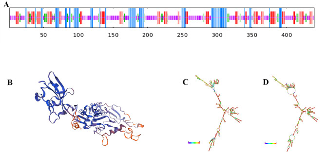
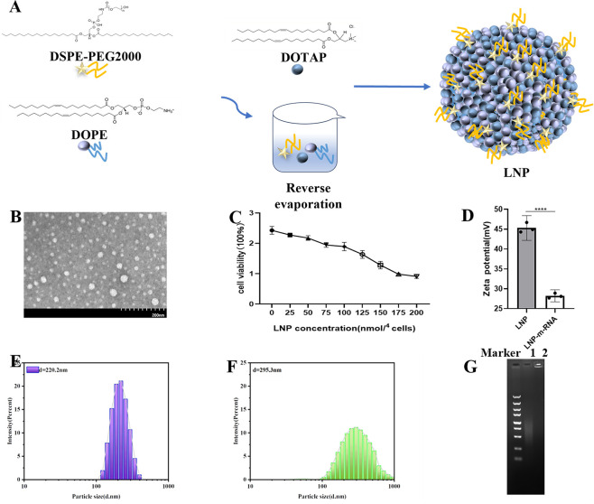
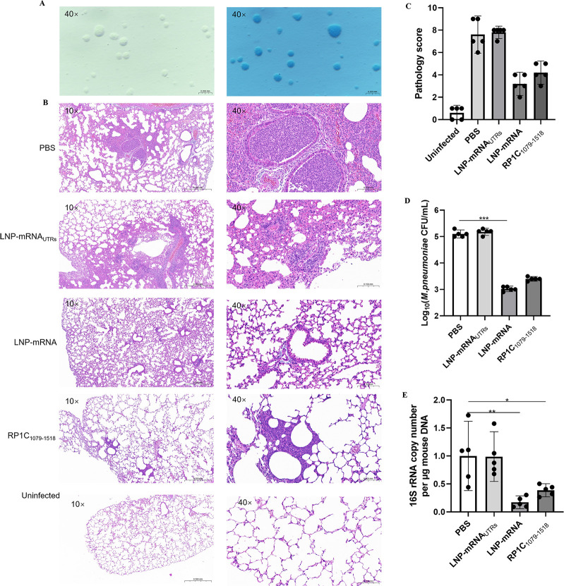
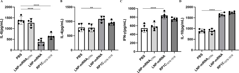
Mycoplasma pneumoniae (Mp), a unique pathogen devoid of a cell wall, is naturally impervious to penicillin antibiotics. This bacterium is the causative agent of M. pneumoniae pneumonia, an acute pulmonary affliction marked by interstitial lung damage. Non-macrolide medications may have potential adverse effects on the developmental trajectory of children, thereby establishing macrolides as the preferred treatment for M. pneumoniae in pediatric patients. However, the emergence of macrolide-resistant and multidrug-resistant strains of M. pneumoniae presents significant challenges to clinical management and public health. Vaccines, particularly those based on mRNA technology, are regarded as a promising avenue for preventing and controlling M. pneumoniae infections due to their inherent safety, immunogenicity, and adaptability. Our research delves into the P1 adhesin of M. pneumoniae, a protein that binds to host cell receptors with its immunodominant epitopes located at the carboxyl terminus, known to provoke robust immune responses and pulmonary inflammation. We have developed an mRNA vaccine harnessing this dominant antigenic epitope and assessed its protective immunity in BALB/c mice against M. pneumoniae infection. The vaccine elicited potent humoral and cellular immune responses, effectively diminishing inflammation. It notably decreased IL-6 levels in the lungs of infected mice and concurrently elevated IL-4, IL-10, and IFN-γ levels post-immunization. The vaccine also reduced pathological changes in the lungs and the M. pneumoniae DNA copy numbers in the infected animals. Collectively, these findings underscore the mRNA vaccine's remarkable immunogenicity and protective potential against M. pneumoniae infections, offering valuable insights for the development of mRNA vaccines targeting mycoplasma infections.IMPORTANCEM. pneumoniae, a bacteria without a cell wall, is known for causing pneumonia and is resistant to penicillin. The increasing prevalence of macrolide-resistant strains has complicated treatment options, emphasizing the need for new strategies. Our research explores an mRNA vaccine candidate that targets the P1 adhesin of M. pneumoniae, a protein critical for the bacteria's interaction with host cells. In a mouse model, this vaccine has shown potential by inducing immune responses and suggesting a possible reduction in inflammation, as indicated by changes in cytokine levels and lung pathology. While further research is required, the vaccine's preliminary results hint at a potential new direction in managing mycoplasma infections, offering a promising avenue for future therapeutic development. This study contributes to the ongoing search for effective preventive measures against M. pneumoniae.

Keywords: Mycoplasma pneumoniae; P1 adhesin; dominant antigenic epitope; lipid nanoparticle; mRNA vaccine; protective immunity.


Conflict of interest statement

The authors declare no conflict of interest.

Figures

Fig 1
Fig 1
Bioinformatics Analysis of M. pneumoniae P1C1079–1518. (A) Prediction of the secondary structure features of P1C1079–1518. (B) Prediction of the tertiary structure of P1C1079–1518. (C) Minimum free energy (MFE) secondary structure of mRNA encoding P1C1079–1518. (D) Centroid secondary structure of mRNA encoding P1C1079–1518.
Fig 2
Fig 2
Construction, expression, purification, and identification of pET-28a(+)/RP1C1079–1518. (A) 0.8% agarose gel electrophoresis: Lane 1 shows the double digestion electrophoresis, and Lane 2 shows the original plasmid electrophoresis. (B) Coomassie brilliant blue analysis of RP1C1079–1518 separated by 12.5% SDS-PAGE: Lanes 1–9 represent RP1C1079–1518 eluted with 80 mM imidazole wash buffer, with an expected size of approximately 47 kDa. (C) Western blot analysis of M. pneumoniae RP1C1079–1518 using anti-M. pneumoniae antibody. (D). Western blot analysis of M. pneumoniae RP1C1079–1518 using anti-His antibody.
Fig 3
Fig 3
Construction and linearization of pGEM-3zf(+)/RP1C1079–1518. (A) Schematic representation of the pGEM-3zf(+)/RP1C1079–1518 cloning vector. (B) 0.8% agarose gel electrophoresis: Lane 1 shows the original plasmid electrophoresis, and Lane 2 shows the double digestion electrophoresis. (C) 0.8% agarose gel electrophoresis: Lanes 1–5 show the single digestion electrophoresis. (D) Sequence composition of mRNA vaccine.
Fig 4
Fig 4
Preparation and characterization of LNP/mRNA. (A) Schematic diagram of LNP synthesis. (B) Transmission electron microscopy image showing the morphology of LNP particles. (C) Cytotoxicity assay of LNP. (D) Zeta potential measurement of LNP and LNP-mRNA. (E) Dispersion and particle size of LNP. (F) Dispersion and particle size of LNP-mRNA. (G) Gel retardation assay results: Lane 1 shows the mRNA group; Lane 2 shows the LNP-mRNA group.
Fig 5
Fig 5
Western blot analysis of mRNA and LNP-mRNA expression in transfected A549 Cells. (A) Western blot analysis of mRNA transfection: Lane 1 shows the mRNA UTRs transfection group; Lane 2 shows the mRNA RP1C1079–1518 transfection group. (B) Western blot analysis of mRNA and LNP-mRNA transfection: Lane 1 shows the LNP-mRNA transfection group; Lane 2 shows the mRNA transfection group. (C) Densitometric analysis of Western blot results for mRNA and LNP-mRNA transfection. Data represent three independent experiments (n = 3). *P < 0.05, **P < 0.01, ***P < 0.001, ****P < 0.0001.
Fig 6
Fig 6
Mice immunization and vaccine biosafety. (A) Schematic diagram of the mice immunization schedule. (B) Schematic representation of blood biochemical test indicators in mice. (C) Body weight of mice before each immunization. (D) Blood biochemical test results of mice. In each case, the results are expressed as the average ±SD of the five replicate samples.*P < 0.05, **P < 0.01, ***P < 0.001, ****P < 0.0001.
Fig 7
Fig 7
Vaccine-induced specific antibody response and splenic lymphocyte proliferation in BALB/c mice. (A) The level of Ags-specific IgG in mice serum from different groups post-immunization. (B) Stimulation index of splenic lymphocyte proliferation in mice. (C) Levels of IFN-γ in the mice spleen. Data are presented as mean ± 95% CI. Statistical significance tested by one-way ANOVA test (*P < 0.05; **P < 0.01; ***P < 0.001; ****P < 0.0001).
Fig 8
Fig 8
Flow cytometry analysis of T-lymphocyte subsets in spleen tissue. (A) Flow cytometry results of spleen tissue T-lymphocyte subsets. (B) Analysis of CD3+CD4+ and CD3+CD8+ T-lymphocyte levels. Data are presented as mean ± 95% CI. Statistical significance tested by one-way ANOVA test (*P < 0.05; **P < 0.01; ***P < 0.001; ****P < 0.0001).
Fig 9
Fig 9
Detection of lung tissue changes in mice on the 7th day post M. pneumoniae challenge. (A) Unstained and Dienes-Stained M. pneumoniae on PPLO Agar. (B) H&E-stained lung tissue sections of mice from different groups post M. pneumoniae challenge. (C) Pathology score of lung tissue sections from mice. (D) M. pneumoniae DNA load in lung tissue homogenates of mice. (E) M. pneumoniae DNA copy numbers. Data are presented as mean ± 95% CI. Statistical significance tested by one-way ANOVA test (*P < 0.05; **P < 0.01; ***P < 0.001; ****P < 0.0001).
Fig 10
Fig 10
Cytokine detection in lung tissue homogenate supernatants post-M. pneumoniae challenge. (A) Detection of IL-6 in lung tissue homogenate supernatants. (B) Detection of IL-4 in lung tissue homogenate supernatants. (C) Detection of IFN-γ in lung tissue homogenate supernatants. (D) Detection of IL-10 in lung tissue homogenate supernatants. Data are presented as mean ± 95% CI. Statistical significance tested by one-way ANOVA test (*P < 0.05; **P < 0.01; ***P < 0.001; ****P < 0.0001).

Similar articles

Cited by

References

    1. Rottem S. 2003. Interaction of mycoplasmas with host cells. Physiol Rev 83:417–432. doi:10.1152/physrev.00030.2002 - DOI - PubMed
    1. Feng M, Schaff AC, Cuadra Aruguete SA, Riggs HE, Distelhorst SL, Balish MF. 2018. Development of Mycoplasma pneumoniae biofilms in vitro and the limited role of motility. Int J Med Microbiol 308:324–334. doi:10.1016/j.ijmm.2018.01.007 - DOI - PubMed
    1. Feng M, Burgess AC, Cuellar RR, Schwab NR, Balish MF. 2021. Modelling persistent Mycoplasma pneumoniae biofilm infections in a submerged BEAS-2B bronchial epithelial tissue culture model. J Med Microbiol 70. doi:10.1099/jmm.0.001266 - DOI - PubMed
    1. Kornspan JD, Tarshis M, Rottem S. 2011. Adhesion and biofilm formation of Mycoplasma pneumoniae on an abiotic surface. Arch Microbiol 193:833–836. doi:10.1007/s00203-011-0749-y - DOI - PubMed
    1. Simmons WL, Dybvig K. 2015. Catalase enhances growth and biofilm production of Mycoplasma pneumoniae. Curr Microbiol 71:190–194. doi:10.1007/s00284-015-0822-x - DOI - PMC - PubMed

MeSH terms

LinkOut - more resources