. 2025 Jan 20;124(1):9.
doi: 10.1007/s00436-025-08454-8.

Activation of the COX-2/mPGES-1/PGE-2 cascade through the NLRP3 inflammasome contributes to Angiostrongylus cantonensis-induced eosinophilic meningoencephalitis

Affiliations

Activation of the COX-2/mPGES-1/PGE-2 cascade through the NLRP3 inflammasome contributes to Angiostrongylus cantonensis-induced eosinophilic meningoencephalitis

Ke-Min Chen et al. Parasitol Res. .

Abstract

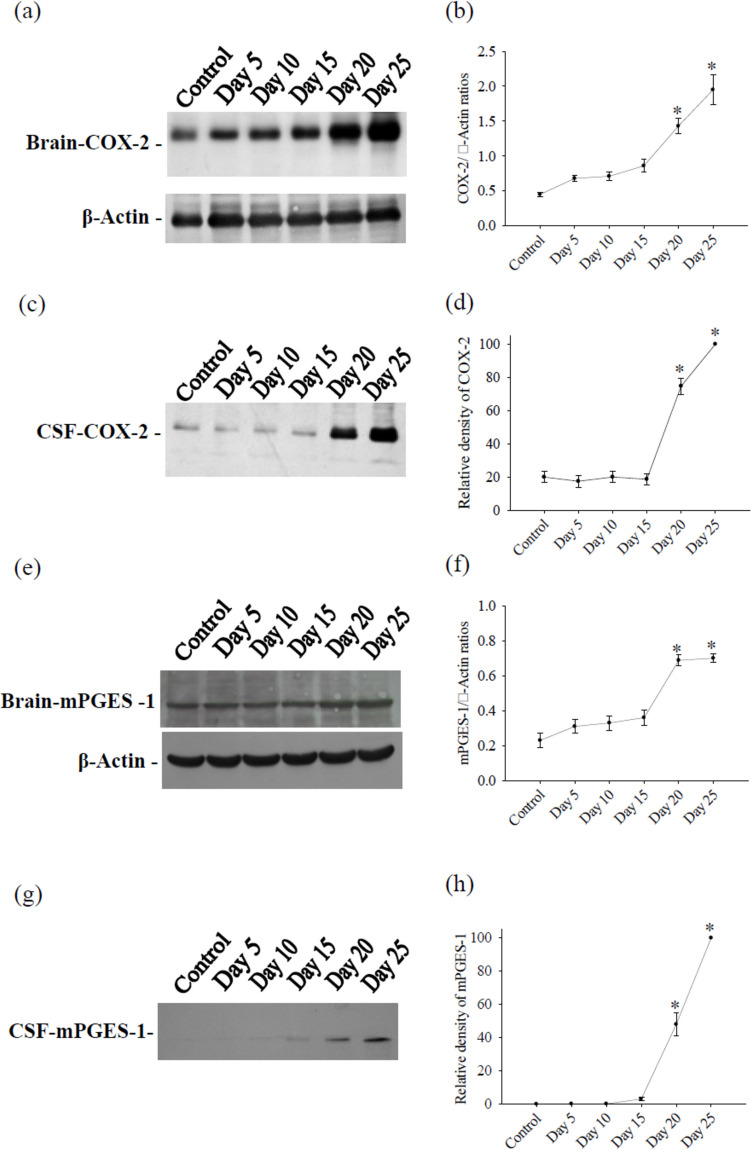
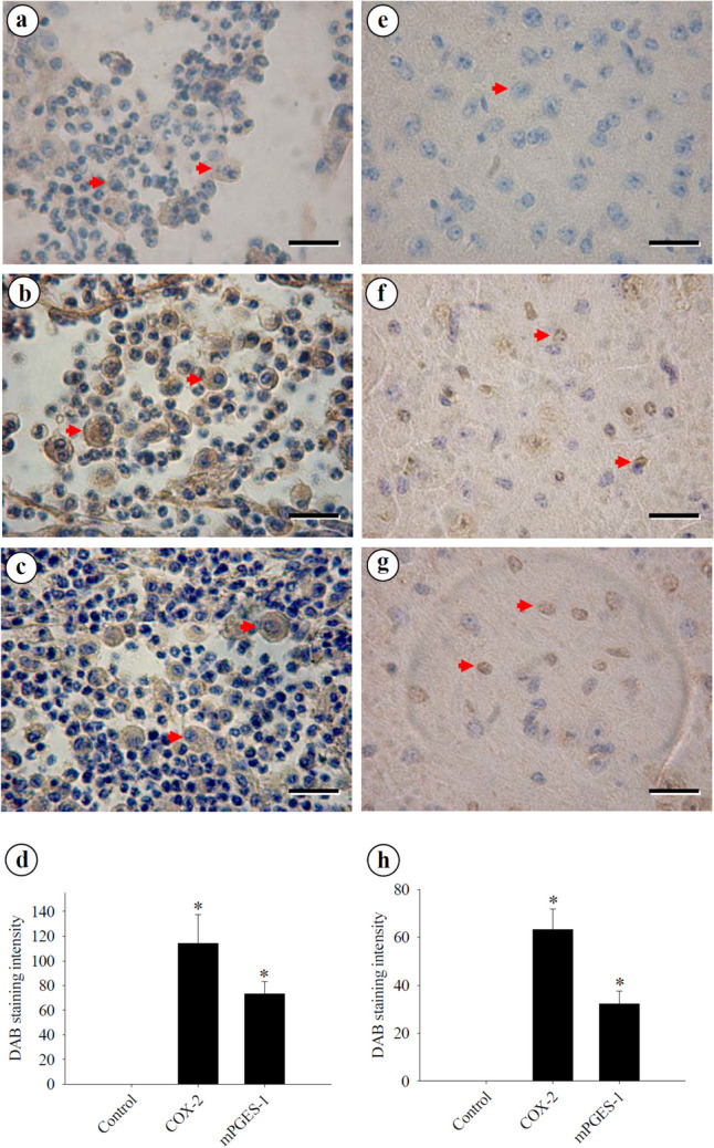
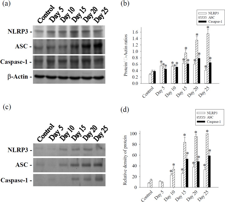
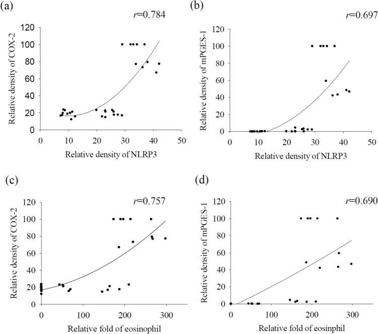
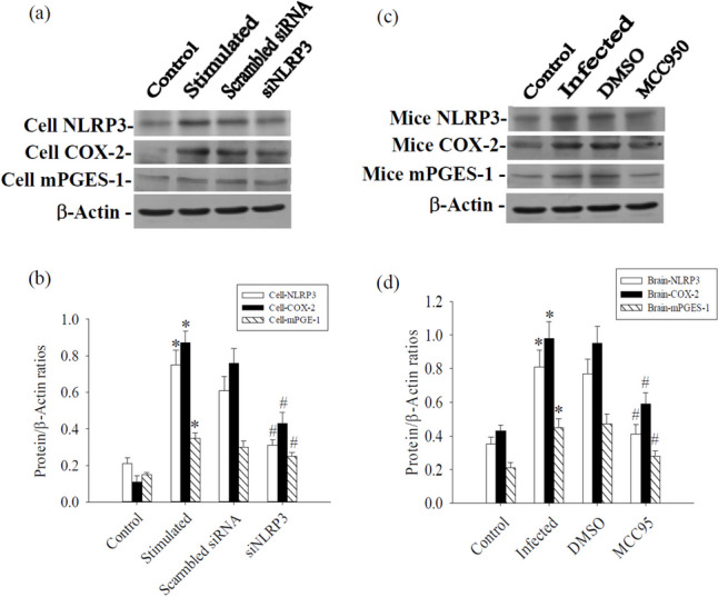
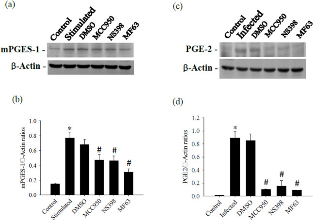
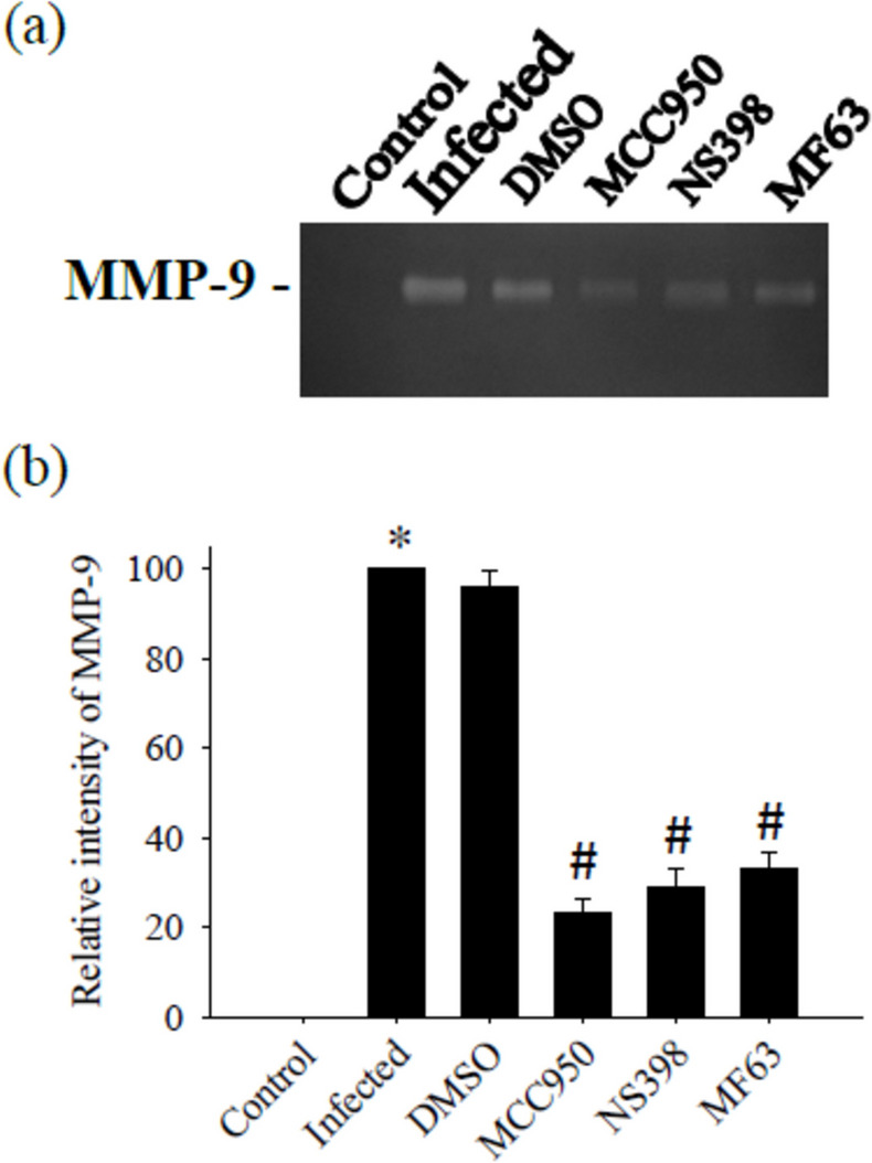
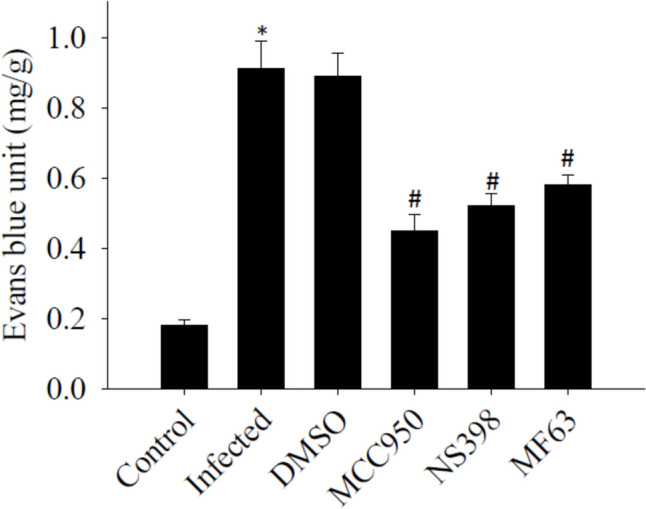
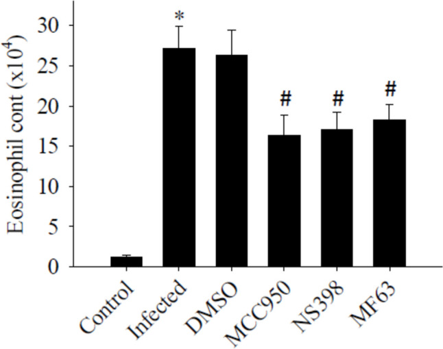
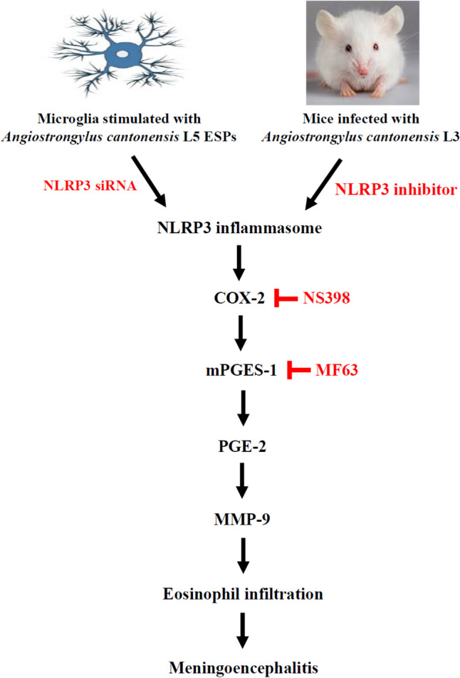
Prostaglandin E2 (PGE-2) is synthesised by cyclooxygenase-2 (COX-2) and microsomal prostaglandin E synthase 1 (mPGES-1). PGE-2 exhibits pro-inflammatory properties in inflammatory conditions. However, there remains limited understanding of the COX-2/mPGES-1/PGE-2 pathway in Angiostrongylus cantonensis-induced meningoencephalitis. This study revealed several key findings regarding the activation of the COX-2/mPGES-1/PGE-2 pathway and its correlation with eosinophilic meningoencephalitis induced by A. cantonensis infection. Immunostaining revealed an increase in the expression of COX-2 and mPGES-1 in the subarachnoid space and glial cells compared to control subjects. Inhibition of the NLRP3 inflammasome by small interfering RNA (siRNA) blocked extracellular secretory proteins (ESPs) stimulated COX-2, mPGES-1 and PGE-2 in microglia. MCC950, an NLRP3 inhibitor, inhibited the levels of the COX-2, mPGES-1, and PGE-2 proteins induced by A. cantonensis in mice. Treatment of mice infected with A. cantonensis with the COX-2 inhibitor NS398 significantly reduced the levels of mPGES-1, PGE-2, and matrix metalloproteinase-9 (MMP-9) levels. Similarly, the mPGES-1 inhibitor MF63 significantly reduced PGE-2 and MMP-9 levels in A. cantonensis-infected mice. Administration of MCC950, NS398, or MF63 resulted in marked attenuation of blood-brain barrier (BBB) permeability and eosinophil counts in A. cantonensis-infected mice. These findings highlight the critical role of the COX-2/mPGES-1/PGE-2 pathway and its regulation by the NLRP3 inflammasome in the pathogenesis of eosinophilic meningoencephalitis induced by A. cantonensis infection. Furthermore, pharmacological interventions targeting this pathway, such as MCC950, NS398, and MF63, show promising therapeutic potential in mitigating associated inflammatory responses and disruption of the BBB. The results indicate that blocking NLRP3 using pharmacological (MCC950) and gene silencing (siNLRP3) methods emphasised the crucial involvement of NLRP3 in the COX-2/mPGES-1/PGE-2 pathway. This suggests that the activation of the COX-2/mPGES-1/PGE-2 axis in response to A. cantonensis infection may be mediated through a mechanism involving the NLRP3 inflammasome.

Keywords: Angiostrongylus cantonensis; Cyclooxygenase-2; Inflammasome; Meningoencephalitis; Prostaglandin E2.


Conflict of interest statement

Declarations. Ethical approval: This study received approval from the Institutional Animal Care and Use Committee of Chung-Shan Medical University, and all procedures were conducted following institutional guidelines for animal experiments. The manuscript does not include any clinical or patient data. Consent to participate: Not applicable. Consent for publication: Not applicable. Competing interests: The authors declare no competing interests.

Figures

Fig. 1
Fig. 1
Kinetics of COX-2 and mPGES-1 from mouse cerebrum and CSF. Protein bands were detected using specific antibodies for cerebrum COX-2 a, CSF COX-2 c, cerebrum mPGES-1 e and CSF mPGES-1 g. Protein levels was performed using computer-assisted imaging densitometry b, d, f, h, with β-actin serving as loading control in cerebrum. All values are the average of at least three independent experiments ± standard error of mean (SD), with a technical duplicate in each experiment. Statistically significant differences were indicated by *
Fig. 2
Fig. 2
Immunohistochemical distribution of COX-2 and mPGES-1 in the mouse cerebrum. Immunohistochemistry staining is a powerful technique used to detect COX-2 and mPGES-1 antigen expression in cells within brain tissue sections. a Weak signals were detected in infiltrating macrophages (arrowheads) within the subarachnoid space when normal serum was used. b Strong positive signals for COX-2 were observed in infiltrating macrophages (arrowheads) within the subarachnoid space. c Strong positive signals for mPGES-1 were detected in infiltrating macrophages (arrowheads) within the subarachnoid space. Macrophages are large cells, typically round or oval in shape, with a large, kidney-shaped or oval nucleus. d Quantification of COX-2 b and PGES-1 c in leukocytes within the subarachnoid space. e No signal was detected in brain cells (arrowhead) of the mouse when normal serum was used. f Strong positive signals for COX-2 were observed in glial cells (arrowheads). g Strong positive signals for mPGES-1 were detected in glial cells (arrowheads). h Quantification of COX-2 e and PGES-1 f in glial cells in the brain tissue. Th images were analyzed using Image J 1.54 g software. All values are the average of at least three independent experiments ± standard error of mean (SD), with a technical duplicate in each experiment. Bar scales = 40 μm
Fig. 3
Fig. 3
Kinetics of NLRP3, apoptosis-associated speck-like protein containing a CARD (ASC) and caspase-1 from mouse cerebrum and CSF. a Protein bands were detected using specific antibodies for NLRP3, ASC and caspase-1 from cerebrum. β-actin serving as loading control. c Protein bands of NLRP3, ASC and caspase-1 were detected from CSF. Protein levels were performed using computer-assisted imaging densitometry b, d. All values are the average of at least three independent experiments ± standard error of mean (SD), with a technical duplicate in each experiment. Statistically significant differences were indicated by *
Fig. 4
Fig. 4
Correlation of NLRP3 and eosinophil count with COX-2 and mPGES-1. NLRP3 in the CSF showed significant correlations with the densities of COX-2 a and mPGES-1 b. The eosinophil count in the CSF showed significant correlations with the densities of COX-2 c and mPGES-1 d, determined by Spearman’s rank correlation test. All values are the average of at least three independent experiments ± standard error of mean (SD), with a technical duplicate in each experiment
Fig. 5
Fig. 5
Influence of NLRP3, COX-2 and mPGES-1 after NLRP3 silencing in microglia and MCC950 treated mice cerebrum. a NLRP3 silencing inhibited ESPs stimulated NLRP3, COX-2 and mPGES-1 in microglia. c MCC950 inhibited Angiostrongylus cantonensis-induced NLRP3, COX-2 and mPGES-1 in the cerebrum of mice. b and d Quantification of protein levels was determined using computer-assisted imaging densitometry. β-actin served as a loading control. All values are the average of at least three independent experiments ± standard error of mean (SD), with a technical duplicate in each experiment. Statistically significant differences were indicated by * for a significant increase in mice infected with A. cantonensis compared to the control and # for a significant decrease in treated mice compared to infected mice with A. cantonensis
Fig. 6
Fig. 6
Effects of NS398 and MF63 on mPGES-1 and PGE-2. The test groups were: uninfected mice (control); Angiostrongylus cantonensis-infected untreated mice (infected); mice treated with NS398; mice treated with MF63. All values are the average of at least three independent experiments ± standard error of mean (SD), with a technical duplicate in each experiment. Statistically significant differences were denoted by * to indicate a significant increase in mice infected with A. cantonensis compared to control, and # to indicate a significant decrease in treated mice compared to infected mice with A. cantonensis
Fig. 7
Fig. 7
Influence of MMP-9 activity by treatment with MCC950, NS398 and MF63. The test groups were: uninfected mice (control); Angiostrongylus cantonensis-infected untreated mice (infected); mice treated with MCC950; mice treated with NS398; mice treated with MF63. All values are the average of at least three independent experiments ± standard error of mean (SD), with a technical duplicate in each experiment. Statistically significant differences were denoted by * to indicate a significant increase in mice infected with A. cantonensis compared to control, and # to indicate a significant decrease in treated mice compared to infected mice with A. cantonensis
Fig. 8
Fig. 8
Influence of blood–brain barrier (BBB) permeability by treatment with MCC950, NS398 and MF63. The test groups were: uninfected mice (control); Angiostrongylus cantonensis-infected untreated mice (infected); mice treated with MCC950; mice treated with NS398; mice treated with MF63. All values are the average of at least three independent experiments ± standard error of mean (SD), with a technical duplicate in each experiment. The notation * signifies a statistically significant increase in mice infected with A. cantonensis compared to the control, while # indicates a statistically significant decrease in treated mice compared to infected mice with A. cantonensis
Fig. 9
Fig. 9
Influence of eosinophil counts by treatment with MCC950, NS398, and MF63. The test groups were: uninfected mice (control); Angiostrongylus cantonensis-infected untreated mice (infected); mice treated with MCC950; mice treated with NS398; mice treated with MF63. All values are the average of at least three independent experiments ± standard error of mean (SD), with a technical duplicate in each experiment. * denotes a statistically significant increase in mice infected with A. cantonensis compared to the control group, while # indicates a statistically significant decrease in treated mice compared to infected mice with A. cantonensis
Fig. 10
Fig. 10
A schematic diagram illustrating the proposed signaling pathway involved in Angiostrongylus cantonensis-induced COX-2, mPGES-1 and PGE2: COX-2 is an inducible enzyme that converts arachidonic acid into PGH2, which is further transformed into prostaglandins, including PGE-2. Inflammatory PGE2 is produced from PGH2 by an enzyme called microsomal prostaglandin E synthase-1 (mPGES-1). In a murine model of angiostrongyliasis meningoencephalitis, inhibition of NLRP3 led to decreased expression of COX-2 and mPGES-1. Additionally, treatment with MCC950, NS398 or MF63 suppressed eosinophil count by reducing MMP-9 activities by inhibiting the COX-2/mPGES-1/PGE2 pathway. These findings reveal the involvement of COX-2/mPGES-1/PGE2-mediated signaling pathways in the regulation of MMP-9 activity and subsequent eosinophilic meningoencephalitis

Similar articles

References

    1. Aid S, Silva AC, Candelario-Jalil E, Choi SH, Rosenberg GA, Bosetti F (2010) Cyclooxygenase-1 and -2 differentially modulate lipopolysaccharide-induced blood-brain barrier disruption through matrix metalloproteinase activity. J Cereb Blood Flow Metab 30:370–380. 10.1038/jcbfm.2009.223 - PMC - PubMed
    1. Andreasson K (2010) Emerging roles of PGE2 receptors in models of neurological disease. Prostaglandins Other Lipid Mediat 91:104–112 - PMC - PubMed
    1. Blais V, Turrin NP, Rivest S (2005) Cyclooxygenase 2 (COX-2) inhibition increases the inflammatory response in the brain during systemic immune stimuli. J Neurochem 95:1563–1574. 10.1111/j.1471-4159.2005.03480.x - PubMed
    1. Candelario-Jalil E, Gonzalez-Falcon A, Garcia-Cabrera M, Leon OS, Fiebich BL (2007) Post-ischaemic treatment with the cyclooxygenase-2 inhibitor nimesulide reduces blood-brain barrier disruption and leukocyte infiltration following transient focal cerebral ischaemia in rats. J Neurochem 100:1108–1120 - PubMed
    1. Chen AC, Shyu LY, Hsin YL, Chen KM, Lai SC (2017) Resveratrol relieves Angiostrongylus cantonensis - induced meningoencephalitis by activating sirtuin-1. Acta Trop 173:76–84. 10.1016/j.actatropica.2017.05.023 - PubMed

MeSH terms

LinkOut - more resources