. 2025 Jan 28;101(2):fiaf011.
doi: 10.1093/femsec/fiaf011.

Importance, structure, cultivability, and resilience of the bacterial microbiota during infection of laboratory-grown Haematococcus spp. by the blastocladialean pathogen Paraphysoderma sedebokerense: evidence for a domesticated microbiota and its potential for biocontrol

Affiliations

Importance, structure, cultivability, and resilience of the bacterial microbiota during infection of laboratory-grown Haematococcus spp. by the blastocladialean pathogen Paraphysoderma sedebokerense: evidence for a domesticated microbiota and its potential for biocontrol

Jeanne Miebach et al. FEMS Microbiol Ecol. .

Abstract

Industrial production of the unicellular green alga Haematococcus lacustris is compromised by outbreaks of the fungal pathogen Paraphysoderma sedebokerense (Blastocladiomycota). Here, using axenic algal and fungal cultures and antibiotic treatments, we show that the bacterial microbiota of H. lacustris is necessary for the infection by P. sedebokerense and that its modulation affects the outcome of the interaction. We combined metagenomics and laboratory cultivation to investigate the diversity of the bacterial microbiota associated to three Haematococcus species and monitor its change upon P. sedebokerense infection. We unveil three types of distinct, reduced bacterial communities, which likely correspond to keystone taxa in the natural Haematococcus spp. microbiota. Remarkably, the taxonomic composition and functionality of these communities remained stable during infection. The major bacterial taxa identified in this study have been cultivated by us or others, paving the way to developing synthetic communities to experimentally explore interactions within this tripartite system. We discuss our results in the light of emerging evidence concerning the structuring and domestication of plant and animal microbiota, thus providing novel experimental tools and a new conceptual framework necessary to enable the engineering of Haematococcus spp. microbiota toward the biocontrol of P. sedebokerense.

Keywords: Chlorophyta; domestication; fungal pathogen; green alga; metagenomics; microbiome.


Conflict of interest statement

None declared.

Figures

Figure 1.
Figure 1.
Effect of antibiotic treatments on the infection of H. pluvialis strain Haemc1 by P. sedebokerense PS1. Ampicillin was used at 150 mg l−1 and the combination of ampicillin and kanamycin was used at a concentration of 3.75 mg l−1. Cell counts were performed at 8 and 29 days after inoculation. The bars show the mean, and the error bars the standard deviation across four replicates.
Figure 2.
Figure 2.
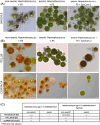
Outcome of the infection by P. sedebokerense FD61 of axenic and nonaxenic Haematococcus strains, in presence or absence of a bacterial synthetic community (SynCom). (A and B) Microscopy observation. (A) Haematococcus pluvialis SAG192.8, 14 days after inoculation. Left: non axenic strain; middle: axenic strain; right: axenic strain supplemented with SynCom 1, a mixture of 10 bacteria from the genera Mesorhizobium, Aeromicrobium, Microbacterium, Brevundimonas, and Variovorax. (B) Halictus rubicundus IT01_06 and H. pluvialis CCAP34/14, 9, and 13 days after inoculation, respectively. Arrows: collapsed algal cells; arrowheads: P. sedebokerense cysts at the surface of algal cells. Scale bar 10 μm. (C) Qualitative symptom-scoring of the above-described samples, based on a double-blind rating under the microscope: +++: heavily infected algal culture, ++: visible infection symptoms, +: less than 1 < % of infected algal cells, and −: no infection visible. PS: P. sedebokerense. The experiment with SynCom 2 was repeated twice independently with identical results.
Figure 3.
Figure 3.
Diversity and phylogenetic relationships of bacterial genera associated to Haematococcus based on 16S rRNA sequences. Representative sequences were retrieved from metagenomic reads of the 21 Haematococcus strains and from bacteria of the cultivable microbiota from the 44 Haematococcus strains. The three 'microbiota types' refer to Fig. 4. Pool: bacteria identified in the pooled DNA of the set of 21 Haematococcus strains. RAA complex: Rhizobium/Agrobacterium/Allorhizobium complex. Tree branches bearing a * sign represent genera JACVCJ01 (unclassified Bacteroidia) and JAFKFH01 (unclassified Ferrovibrionaceae) as identified with the GTDB-Tk database. In the phylogenetic tree (Fig. 3), some bacterial genera are grouped together because they cannot be distinguished based on 16S (e.g. Rhizobium, Agrobacterium, and Allorhizobium were grouped into what we called the ‘RAA’ complex). This explains why the tree has 28 branches and not 30.
Figure 4.
Figure 4.
Heatmap of the bacterial genera identified by metagenomics across the 21 Haematococcus strains from two species (see legend in the figure) when inoculated with the pathogen P. sedebokerense (PS) or in the control condition (C). The heatmap is based on bin abundance per sample normalized by the size of each library, i.e. the total number of reads in each sample. The colour scale corresponds to the abundance matrix normalized using Hellinger transformation. Dissimilarity matrix used for hierarchical cluster analysis was calculated from Euclidean distances. Pooled samples were not included in the clustering but the colour scale was the same as for the individually sequenced samples. 1: Type 1 microbiota, 2: Type 2 microbiota, and 3: Type 3 microbiota. *: low susceptibility, **: intermediate susceptibility, and ***: high susceptibility based on Allewaert et al. (2018) (Fig. S4).
Figure 5.
Figure 5.
Shannon index calculated at the bacterial genus level for the six individually analyzed Haematococcus spp. strains for control (C) and infected samples (PS). The Shannon index was calculated using the abundance of each MAG obtained from the MetaWRAP:: quant_bin module.
Figure 6.
Figure 6.
Microscopy images of the Haematococcus cultures inoculated with P. sedebokerense for the metagenomics experiment showing visible signs of infection. Paraphysoderma sedebokerense cells are visible at the algal surface (arrowheads). Haematococcus strain CCAP34/14 (A) and (B) calcofluor white staining highlights the presence of the fungal pathogen, (C) Haematococcus strain CZ01_06. Cultures were fixed in PFA. Scale bar 10 μm.
Figure 7.
Figure 7.
Results of the PCA based on the matrix with the number of reads mapping on annotated features (KOs), normalized to 1 kB and per million read of annotated KOs within each library (sample) and then summed per KO for all MAGs within a library (sample). Projection on the first and second components.
Figure 8.
Figure 8.
Maximum module completion ratio for each KEGG module within a microbiota type based on KEMET output (Palù et al. 2022). Modules encircled: ‘Nucleotide sugar biosynthesis, galactose ⇒ UDP-galactose’ (M00554), ‘Pyridoxal-P biosynthesis’ (M00916), ‘Thiosulfate oxidation by SOX complex’ (M00595), ‘ADP-l-glycero-d-manno-heptose biosynthesis’ (M00064), and ‘Phthalate, Terephthalate and Salicylate degradation’ (M00623, M00624, and M00638). Abbreviations used: AA: amino acids, BCAA: branched chain amino acids, Histi.: histidine, and P: photosynthesis.
Figure 9.
Figure 9.
Differences of functional potential between the three microbiota types. The barplot shows the differentially abundant KEGG pathways between microbiota types, based on LDA (P < .05 for Kruskal–Wallis test). Only KEGG pathways with LDA score >3 are shown here. The analysis is based on the number of reads mapping on annotated KOs, normalized to 1 kB and 1 million read of annotated KOs within each library, summed per KEGG pathway and then per sample. Here, classes were ‘Microbiota Type’. Control and infected samples were considered as replicates.

Similar articles

References

    1. Abdul Malik SA, Bedoux G, Garcia Maldonado JQ et al. Defence on surface: macroalgae and their surface-associated microbiome. In: Advances in Botanical Research. Amsterdam: Elsevier, 2020, 327–68. 10.1016/bs.abr.2019.11.009. - DOI
    1. Ajani PA, Kahlke T, Siboni N et al. The microbiome of the cosmopolitan diatom Leptocylindrus reveals significant spatial and temporal variability. Front Microbiol. 2018;9:2758. 10.3389/fmicb.2018.02758. - DOI - PMC - PubMed
    1. Allewaert CC, Hiegle N, Strittmatter M et al. Life history determinants of the susceptibility of the blood alga Haematococcus to infection by Paraphysoderma sedebokerense (Blastocladiomycota). Algal Res. 2018;31:282–90. 10.1016/j.algal.2018.02.015. - DOI
    1. Allewaert CC, Vanormelingen P, Pröschold T et al. Species diversity in European Haematococcus pluvialis (Chlorophyceae, Volvocales). Phycologia. 2015;54:583–98. 10.2216/15-55.1. - DOI
    1. Alneberg J, Bjarnason BS, De Bruijn I et al. Binning metagenomic contigs by coverage and composition. Nat Methods. 2014;11:1144–6. 10.1038/nmeth.3103. - DOI - PubMed

MeSH terms