. 2025 Jan 20;16(1):838.
doi: 10.1038/s41467-025-56114-3.

A single residue switch mediates the broad neutralization of Rotaviruses

Affiliations

A single residue switch mediates the broad neutralization of Rotaviruses

Yang Huang et al. Nat Commun. .

Abstract

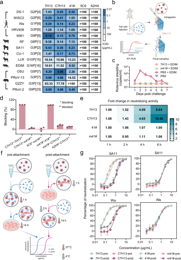
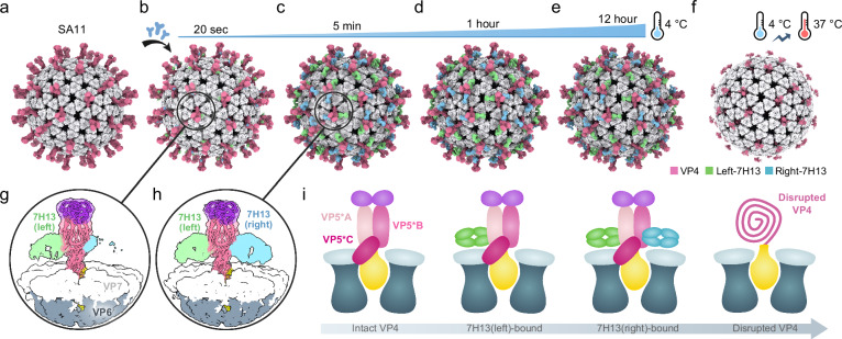
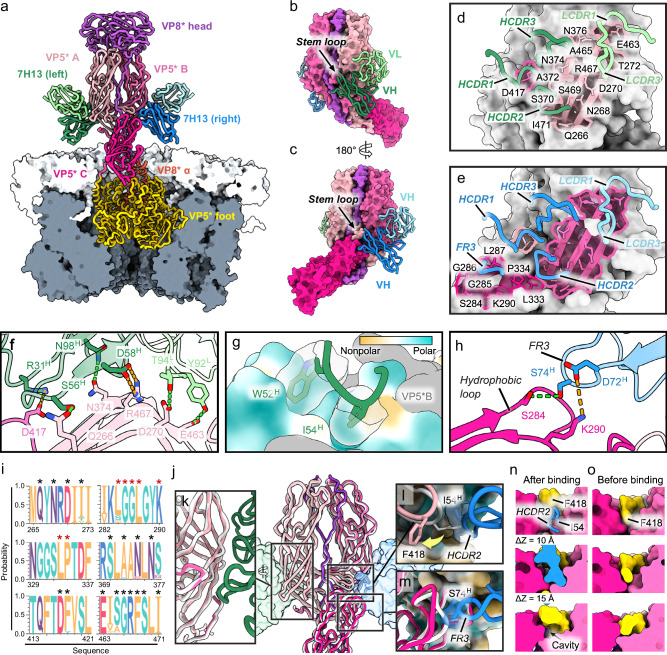
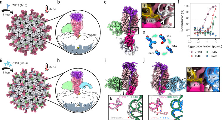
Broadly neutralizing antibodies (bNAbs) could offer escape-tolerant and lasting protection against viral infections and therefore guide development of broad-spectrum vaccines. The increasing challenge posed by viral evolution and immune evasion intensifies the importance of the discovery of bNAbs and their underlying neutralization mechanism. Here, focusing on the pivotal viral protein VP4 of rotavirus (RV), we identify a potent bNAb, 7H13, exhibiting broad-spectrum neutralization across diverse RV genotypes and demonstrating strong prevention of virus infection in female mice. A combination of time-resolved cryo-electron microscopy (cryo-EM) and in situ cryo-electron tomography (cryo-ET) analysis reveals a counterintuitive dynamic process of virus inactivation, in which 7H13 asymmetrically binds to a conserved epitope in the capsid-proximal aspect of VP4, triggers a conformational switch in a critical residue-F418-thereby disrupts the meta-stable conformation of VP4 essential for normal viral infection. Structure-guided mutagenesis corroborates the essential role of the 7H13 heavy chain I54 in activating F418 switch and destabilizing VP4. These findings define an atypical NAbs' neutralization mechanism and reveal a potential type of virus vulnerable site for universal vaccine and therapeutics design.


Conflict of interest statement

Competing interests: The authors declare that they have no conflicts of interest.

Figures

Fig. 1
Fig. 1. Characterization of NAb 7H13 in vivo and in vitro.
a Neutralization profile of 7H13 (murine Fc) and C7H13 (human Fc) against a panel of RV strains. 41# (human Fc) serves as positive control, while 5C6 (human Fc) and A2H4 (murine Fc) are negative controls. Refer to Figure S1 for detailed datasets. Cartoon illustrations were created in BioRender. Yang, H. (2024) https://BioRender.com/c98a036. b Schematic of the experimental design to assess the prophylactic effect of NAbs against RV infection and fecal shedding in a murine model. Created in BioRender. Yang, H. (2024) https://BioRender.com/s21c678. c Protective efficacy of 7H13 and m41# against fecal virus shedding in adult mice (n = 5 per group) following RV EDIM challenge. Data are log10 transformed and presented as mean ± SEM. d Cross-inhibition between C7H13 and m41# as evaluated by blocking ELISA. Blocking mAbs (100 μg/mL) were pre-incubated with microplates before the addition of blocked mAbs, and the binding of blocked mAbs was detected by HRP-conjugated secondary antibodies. Data are shown as mean ± SEM of three technical replicates. e Fold change in neutralizing activity of NAbs over time. Fold change represents the variation in IC50 across time points (1, 2, 4 and 6 h), calculated as the ratio of IC50 at 1 h relative to those at subsequent time points. Refer to Figure S3 for detailed datasets. f Schematic of the experimental setup for pre- and post-attachment neutralization assays. Created in BioRender. Yang, H. (2024) https://BioRender.com/j09b061. g Neutralizing activities of NAbs against SA11 and Wa strains in pre- and post-attachment assays. Data are shown as mean ± SEM of three technical replicates. Source data are provided as a Source Data file.
Fig. 2
Fig. 2. The unusual binding mode of 7H13 and binding-induced VP4 destabilization.
a Radially colored surface representation of reconstructed RV strain SA11, showing external and sectional views of the intact virion. Symmetry axes are annotated with geometric markers. b Ribbon diagram of VP4 model, color-coded by domain: VP8* α in orange, VP8* lectin and its linker to VP8* α in purple, VP5* β-barrel (VP5*A, *B and *C) in shades of pink, VP5* C-terminal foot in yellow. c Focused 3D classification of SA11, with VP4 densities color-coded by domain. VP6 and VP7 are depicted in deep and light gray, respectively, and clipped to visualize the VP4 foot. d SA11:7H13 complex incubated at 37 °C for 5 minutes. e, f Focused 3D classifications of SA11-7H13 (37 °C-5min). e shows two 7H13 bound to opposite sides of the VP4, differentiated by green and blue. f shows disrupted VP4, characterized by noise-dominated densities in pink. g SA11:7H13 complex incubated at 37 °C for 1 h. h Focused 3D classification of SA11-7H13 (37 °C-1h), highlighting extensive VP4 disruption. i Schematic representation of disrupted VP4. A semi-transparent surface representation of VP4 density segmented from SA11:7H13 (37 °C-1h) in h is overlaid with atomic models. Disrupted VP4 segments and their connectors to the spike base are illustrated with patches and dotted lines, respectively, colored to match their domains. j, k Linear diagrams of VP4 domain organization before (j) and after (k) 7H13 binding, with amino acid positions numbered. Disrupted domains are shaded in gray in (k). l SDS-PAGE analysis of SA11 following incubation with 7H13-Fab at two time points. M: molecular mass protein marker; C: SA11 control.
Fig. 3
Fig. 3. Cryo-ET reveals the disordered VP4 on virion upon 7H13 binding.
a Representative tomographic slice of the native SA11 virion. b Magnified view of VP4 from a, showing intact VP4 morphology. c Segmented SA11 volume with radial coloring to highlight the core-to-periphery gradient. Blue and pink arrows indicate areas detailed in (d and e) respectively. d, e Close-up views of VP4 from the native SA11 virion. The virus is shown as semi-transparent surfaces overlaid with atomic models of RRV strain (PDB: 4V7Q). f Representative tomographic slice of the SA11:7H13 complex after 1-h incubation at 37 °C, showing altered VP4 morphology. g Magnified view of VP4 from (f) showing disrupted VP4 in the presence of 7H13. h Segmented SA11:7H13 volume with radial coloring, highlighting disordered regions of VP4. Blue and pink arrows indicate areas detailed in i and j, respectively. i, j Close-up views of disordered VP4 densities from the SA11:7H13 complex. The complex is shown as semi-transparent surfaces overlaid with composite SA11:7H13 model, generated by superimposing VP4:7H13 structure onto the virus model (PDB: 4V7Q). Scale bars: 50 nm in (a and f); 10 nm in (b and g).
Fig. 4
Fig. 4. Cryo-EM analysis reveals temperature and reacting time are dependent for the destabilization of VP4 by 7H13.
af Comparative structural analyzes of the native SA11 virion (a) and SA11:7H13 complexes following incubations under varying conditions: at 4 °C for 20 seconds (b), 5 minutes (c), 1 h (d), 12 h (e), and 4 °C for 1 h followed by 37 °C for another 1 h (f). VP7, VP4, left-7H13, and right-7H13 are colored light gray, pink, green, and blue, respectively. g, h 3D class averages of the SA11:7H13 complex for 20-second (g) and 5-minute (h) incubations, with VP4 represented as semi-transparent surfaces overlaid with ribbon diagrams, color-coded by domain: head in purple, body and stack in pink, and foot in yellow. i Schematic illustration of the hypothesized sequential VP4 disruption process. The color scheme corresponds to that used in h, with VP5* β-barrel domains shown in shades of pink.
Fig. 5
Fig. 5. High-resolution structure reveals an asymmetric mode of 7H13 binding triggered localized conformational alteration of VP4.
a Composite model of SA11:7H13 complex, with VP6 and VP7 surfaces clipped for clarity. VP8* α is shown in orange, VP8* lectin and its linker to VP8* α in purple, VP5* β-barrel in shades of pink, VP5* foot in gold, VP6 and VP7 in deep and light gray, respectively. Left- and right-7H13 are depicted in green and blue, respectively. b, c Atomic model of VP4:7H13, with VP4 depicted as a surface and 7H13 as ribbons. c represents a 180° rotated view relative to b. d, e Interactions of left- (d) and right-7H13 (e) with VP4. Epitopes are represented as semi-transparent surfaces with outlined boundaries, color-coded according to the corresponding VP5* β-barrel domains. f-h Detailed interactions between VP4 and 7H13. f Interactions between left-7H13 and the VP5* projection, with key residues shown in stick representation. Hydrogen bonds and salt bridges are indicated by green and orange dashed lines. g Close-up view of W52 and I54 from left-7H13 heavy chain, depicted in stick and semi-transparent surface representations, inserting into the pocket of VP5*A, visualized as hydrophobic (yellow) and charged (turquoise) surfaces. h Interactions between right-7H13 FR3 and the VP5*C hydrophobic loop. i Sequence conservation of 7H13 epitopes. Black asterisks mark residues recognized by both left- and right-7H13, while red asterisks indicate those unique to right-7H13. jm Structural rearrangements induced by 7H13 binding. j Superimposition of unbound VP4 (white ribbon) and VP4:7H13 complex (colored ribbon and surface). k Close-up view of the VP4:left-7H13 binding interface. l Rearrangements of the VP5*A stem-loop F418 induced by right-7H13 HCDR2, highlighted by a yellow arrow. m Rearrangements of VP5*C hydrophobic loop induced by right-7H13 FR3. In l and m, the residues in 7H13 that dominantly contribute to inter-VP4-right-7H13 collisions are shown in stick and semi-transparent surface. n, o Cross-sectional comparison of VP5*A stem-loop after (n) and before (o) right-7H13 binding. The stem-loop is rendered in yellow, with other domain elements colored as in a.
Fig. 6
Fig. 6. Structural validation of the biased binding of 7H13 and binding-induced F418 switch.
a, b Cryo-EM reconstruction (a) and focused classification (b) of SA11:7H13 complex prepared at 1/10 antibody concentration and incubated at 37 °C for 1 h, showing VP4 (pink) bound exclusively with left-7H13 (green). c Localized reconstruction of SA11:7H13−1/10 complex incubated at 4 °C for 1 h. d Close-up view of VP5*A stem-loop from (c) depicted as a semi-transparent yellow surface overlaid with atomic model. The right-7H13 model (white) is superimposed to highlight the absence of collisions between its I54 residue and VP5*A F418 (both in stick). e Structural representations of I54 residue variants (I54A, I54S, and I54G) illustrate mutation-induced variations in side-chain size. f Neutralization activities of 7H13 and its mutants. Data are shown as mean ± SEM of three technical replicates. g, h Cryo-EM reconstruction (g) and focused classification (h) of SA11:7H13-I54G complex incubated at 37 °C for 1 h, showing VP4 bound by mutated left-7H13 (green) and right-7H13 (blue). i, j Localized reconstructions of the SA11:7H13-I54G complex incubated at 4 °C for 1-h, targeted to left-7H13 (i) and right-7H13 (j). k, l Superimpositions of VP4:left-7H13-I54G onto VP4:7H13 (k) and VP4:right-7H13-I54G (l), showing the mutation-induced alterations in binding postures. m Close-up view of VP5*A stem-loop from (j) with right-7H13-I54G depicted as a semi-transparent blue surface overlaid with the corresponding model, demonstrating that the 7H13-I54G mutant fails to induce conformational changes in VP5*A F418. Source data are provided as a Source Data file.

Similar articles

Cited by

References

    1. Chen, Y. J. et al. Broadly neutralizing antibodies to SARS-CoV-2 and other human coronaviruses. Nat. Rev. Immunol.23, 189–199 (2023). - PMC - PubMed
    1. Zhang, L. et al. A proof of concept for neutralizing antibody-guided vaccine design against SARS-CoV-2. Natl. Sci. Rev.8, 10.1093/nsr/nwab053 (2021). - PMC - PubMed
    1. Britto, C. & Alter, G. The next frontier in vaccine design: blending immune correlates of protection into rational vaccine design. Curr. Opin. Immunol.78, 10.1016/j.coi.2022.102234 (2022). - PMC - PubMed
    1. Thadani, N. et al. Learning from prepandemic data to forecast viral escape. Nature622, 1–8 (2023). - PMC - PubMed
    1. Burton, D. R., Poignard, P., Stanfield, R. L. & Wilson, I. A. Broadly neutralizing antibodies present new prospects to counter highly antigenically diverse viruses. Science337, 183–186 (2012). - PMC - PubMed

MeSH terms