. 2025 Jan 20;11(1):17.
doi: 10.1038/s41420-025-02291-5.

Exocyst complex component 1 (Exoc1) loss in dormant oocyte disrupts c-KIT and growth differentiation factor (GDF9) subcellular localization and causes female infertility in mice

Affiliations

Exocyst complex component 1 (Exoc1) loss in dormant oocyte disrupts c-KIT and growth differentiation factor (GDF9) subcellular localization and causes female infertility in mice

Chi Lieu Kim Nguyen et al. Cell Death Discov. .

Abstract

A limited number of female germ cells support reproduction in many mammals. The follicle, composed of oocytes and supporting granulosa cells, forms the basis of oogenesis. Crosstalk between oocytes and granulosa cells is essential for the formation, dormancy, re-awakening, and maturation of oocytes. The oocyte expresses c-KIT and growth differentiation factor-9 (GDF-9), which are major factors in this crosstalk. The downstream signalling pathways of c-KIT and GDF-9 have been well-documented; however, their intra-oocyte trafficking pathway remains unclear. Our study reveals that the exocyst complex, a heterotetrameric protein complex important for tethering in vesicular transport, is important for proper intra-oocyte trafficking of c-KIT and GDF9 in mice. We found that depletion of oocyte-specific EXOC1, a component of the exocyst complex, impaired oocyte re-awakening and cyst breakdown, and inhibited granulosa cell proliferation during follicle growth. The c-KIT receptor is localised on the oocyte plasma membrane. The oocyte-specific Kit conditional knockout mice were reported to exhibit impaired oocyte re-awakening and reduced oocyte cyst breakdown. GDF9 is a protein secreted extracellularly in the oocyte. Previous studies have shown that Gdf9 knockout mice impaired proliferation and granulosa cell multilayering in growing follicles. We found that both c-KIT and GDF9 abnormally stuck in the EXOC1-depleted oocyte cytoplasm. These abnormal phenotypes were also observed in oocytes depleted of exocyst complex members EXOC3 and EXOC7. These results clearly show that the exocyst complex is essential for proper intra-oocyte trafficking of c-KIT and GDF9. Inhibition of this complex causes complete loss of female fertility in mice. Our findings build a platform for research related to trafficking mechanisms of vital crosstalk factors for oogenesis.


Conflict of interest statement

Competing interests: The authors declare no competing interests. Ethics: Animal experimentation: All animal experiments were conducted in a humane manner with approval from the Institutional Animal Experiment Committee of the University of Tsukuba according to the Regulations for Animal Experiments of the University of Tsukuba and the Fundamental Guidelines for Proper Conduct of Animal Experiments and Related Activities in Academic Research Institutions under the jurisdiction of the Ministry of Education, Culture, Sports, Science, and Technology of Japan.

Figures

Fig. 1
Fig. 1. Follicle growth in Exoc1-G-cKO mice.
Follicle growth in the ovaries of 4, 8, 10, and 12-week-old Exoc1-G-cKO (Exoc1flox/flox::Gdf9+/Cre) and control Exoc1-G-ctrl (Exoc1+/flox::Gdf9+/Cre) mice. A Representative Haematoxylin and Eosin staining images of Exoc1-G-cKO mice ovaries. No antral follicles were observed in Exoc1-G-cKO mice ovaries that were eight weeks old. No follicles were observed at any stage in 28-week-old Exoc1-G-cKO mice. Scale bar = 500 μm. B Representative Haematoxylin and Eosin staining images of primordial, primary, secondary and antral follicle in 4-week-old Exoc1-G-cKO ovary. Scale bar = 20 μm. C Follicle count in ovaries in Exoc1-G-cKO mice. No secondary follicles were observed in the Exoc1-G-cKO mice that were 10 and 12 weeks old. n = 3, Student’s t test.
Fig. 2
Fig. 2. Ovarian transplantation of Exoc1-G-cKO ovaries into ROSAGRR/GRR ovaries.
Images of harvested ovaries nine weeks after ovary transplantation from three-week-old Exoc1-G-cKO (Exoc1flox/flox::Gdf9+/Cre) and Exoc1-G-ctrl (Exoc1+/flox::Gdf9+/Cre) mice into three-week-old ROSAGRR/GRR mice. A Representative immunofluorescence images for green fluorescence protein and Haematoxylin and Eosin staining. Dashed circles: transplanted donor ovarian regions a, c: Oocytes in donor ovaries. b, d: Oocytes in recipient ovaries. Scale bar = 500 µm (at low magnification), scale bar = 50 µm (high magnification). B Follicle growth in donor ovaries. Primordial and primary follicles, but not secondary follicles, were found in the Exoc1-G-cKO donor ovaries. C Comparison of transplanted and non-transplanted Exoc1-G-cKO mice ovaries. The number of primordial oocytes was significantly higher in transplanted Exoc1-G-cKO ovaries than in non-transplanted Exoc1-G-cKO mice ovaries (9 + 3 and 12 weeks). Three ovarian regions per mouse were measured, and the number of primordial oocytes in nine regions, 3 regions ×3 mice, was plotted. One-way analysis of variance.
Fig. 3
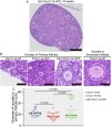
Fig. 3. Diameter of the oocytes in primary follicles in Exoc1-G-cKO mice.
A Morphology of oocytes in primary follicles in Exoc1-G-cKO (Exoc1flox/flox::Gdf9+/Cre) ovaries compared to that of the control (Exoc1+/flox::Gdf9+/Cre). White line: measured diameter. Scale bar = 20 μm. B Diameter of oocytes in primordial and primary follicles in Exoc1-G-cKO mice. The diameter of oocytes in primordial follicles between control and Exoc1-G-cKO mice showed no significant differences. In contrast, the oocytes in primary follicles in Exoc1-G-cKO mice showed significantly shorter diameters than those in control mice. n = 3, Student’s t test.
Fig. 4
Fig. 4. Impairment of c-KIT trafficking in Exoc1-G-cKO mice.
A Representative immunofluorescence images of 10-week-old Exoc1-G-cKO (Exoc1flox/flox::Gdf9+/Cre) and Exoc1-G-ctrl (Exoc1+/flox::Gdf9+/Cre) mice ovaries. The c-KIT signals were observed mainly on the plasma membrane in oocytes in primary follicles of control mice. In contrast, extensive c-KIT dot-like signals were detected in the oocyte cytoplasm in primary follicles of Exoc1-G-cKO mice. Scale bar = 20 μm. B Area ratio of plasma membrane c-KIT (c-KIT co-localised with wheat germ agglutinin (WGA), WGA+::KIT+) to cytoplasmic c-KIT (c-KIT not co-localised with WGA, WGA-::KIT+). n = 3, Student’s t test. C Signal intensity ratio of plasma membrane c-KIT to cytoplasmic c-KIT. n = 3, Student’s t test. D Intensity of the total KIT signal in each oocyte. n = 3, Student’s t test.
Fig. 5
Fig. 5. Rescue of oocyte re-awakening failure in Exoc1-G-cKO mice using a PTEN inhibitor.
A Macroscopic Haematoxylin and Eosin-stained images of PTEN inhibitor bpV-treated Exoc1-G-cKO (bpV-Exoc1-G-cKO) mice ovaries. Scale bar = 500 μm. B Representative primary follicles of each mouse genotype (10 weeks old). The control mice were Exoc1+/flox::Gdf9+/Cre, referred to as Exoc1-G-ctrl. Scale bar = 50 μm. C Plots of the oocyte sizes in primary follicles of each mouse genotype at 10 weeks of age. n = 3, one-way analysis of variance. Oocyte diameters in primary follicles in bpV Exoc1-G-cKO mice were significantly longer than those in Exoc1-G-cKO mice. In the bpV Exoc1-G-cKO group, enlarged oocytes appeared in primary follicles (plots circled by dashed line), which were absent in the control group.
Fig. 6
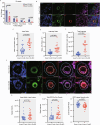
Fig. 6. Exoc1 deletion during adult stage impairs the subcellular location of growth differentiation factor-9 (GDF9).
Tamoxifen was injected into eight-week-old Exoc1-D-cKO (Exoc1flox/flox::Ddx4+/CreERT2) and Exoc1-D-ctrl (Exoc1flox/+::Ddx4+/CreERT2) mice. A Oocyte count in ovaries in 16-week-old mice. No growing follicles were observed in the Exoc1-D-cKO mice. N = 3, Student’s t test. B Representative immunofluorescence images of 16-week-old Exoc1-D-cKO and control mice ovaries. The c-KIT signals were observed mainly on the plasma membrane of oocytes in the primary follicles of control mice, whereas exclusive c-KIT signals were observed in the cytoplasm of oocytes in primary follicles of Exoc1-D-cKO mice. Scale bar = 20 μm C Area ratio of plasma membrane c-KIT (c-KIT co-localised with wheat germ agglutinin (WGA), WGA + ::KIT + ) to cytoplasmic c-KIT (c-KIT not co-localised with WGA, WGA-::KIT+). D Intensity ratio of plasma membrane to cytoplasmic c-KIT. E Intensity of the total KIT signal in each oocyte. N = 3, Student’s t test. F Representative immunofluorescence images of 16-week-old Exoc1-D-cKO and control mice ovaries. Most GDF9 signals were found in the extra-oocyte region of the Exoc1-D-ctrl mice primary follicles. However, GDF9 signals were mostly localised in ooplasm of the primary follicles of Exoc1-D-cKO mice. Blue dashes: Follicle area. White dashes: oocyte areas in primary follicles. Scale bar = 20 μm G Plot of the sum of the GDF9 signal intensities in each follicle. No significant differences were observed among the two groups. H Plot of the GDF9 signal intensity in the oocytes in primary follicles only. Oocyte GDF9 levels in Exoc1-D-cKO mice were significantly higher than those in the control. I The ratio of GDF9 signal intensity between the extra-oocyte and follicle area. GDF9 signals were found mainly in oocyte cytoplasm of Exoc1-D-cKO primary follicles. N = 3, Student’s t test.
Fig. 7
Fig. 7. Downstream pathways of c-KIT and growth differentiation factor-9 (GDF9).
A Representative immunofluorescence images of 16-week-old Exoc1-D-cKO (Exoc1flox/flox::Ddx4+/CreERT2) and Exoc1-D-ctrl (Exoc1flox/+::Ddx4+/CreERT2) primary follicles. FOXO3a signals concentrated in the nucleus of Exoc1-D-cKO oocytes in primary follicles. Scale bar = 10 µm. B The signal intensity of FOXO3a in the nucleus of oocytes in primary follicles. n = 3, Student’s t test. C Representative PCNA and FOXL2, a marker of granulosa cells, co-immunofluorescence images of 16-week-old Exoc1-D-cKO and control primary follicles. Scale bar = 10 µm. D The percentage of PCNA-positive granulosa cells in the primary follicles of Exoc1-D-cKO mice was significantly lower than that in the control. n = 3, Student’s t test.
Fig. 8
Fig. 8. Exoc1 deletion during foetal ovarian development significantly disrupts folliculogenesis.
A Intensity level of plasma membrane c-KIT. The c-KIT located in plasma membrane of Exoc1-D-cKO-E (Exoc1flox/flox::Ddx4+/CreERT2) oocytes at postnatal day 0 (P0) compared to control group Exoc1-D-ctrl-E (Exoc1flox/+::Ddx4+/CreERT2. B Representative Haematoxylin and Eosin staining images of Exoc1-D-cKO-E ovaries at P5. Dashed white line: oocytes in the cyst. Hashtag: single follicles. C Total number of oocytes in ovaries, follicles, and cysts of both groups. The number of oocytes in cyst of Exoc1-D-cKO-E ovaries was significantly higher than those in control. n = 3, Student’s t test. D Representative ovaries and Macroscopic Haematoxylin and Eosin-stained images of Exoc1-D-cKO-E and Exoc1-D-ctrl-E mice that were 10 weeks old. Dashed black line: ovary area. Asterisk: follicles observed on the ovary surface. Scale bar = 500 µm. E Ovary maximum diameter. Exoc1-D-cKO-E ovaries were smaller than those of the control group. F Oocyte counts in whole ovaries at 10 weeks of age. Secondary and antral follicles were absent in the Exoc1-D-cKO-E mice. n = 3, Student’s t test. G Oocyte area of primary follicles. Oocytes in primary follicles of Exoc1-D-cKO-E mice were significantly smaller than those in control group. n = 3, Student’s t test.

Similar articles

Cited by

References

    1. Zhang H, Liu L, Li X, Busayavalasa K, Shen Y, Hovatta O, et al. Life-long in vivo cell-lineage tracing shows that no oogenesis originates from putative germline stem cells in adult mice. Proc Natl Acad Sci USA. 2014;111:17983–8. - PMC - PubMed
    1. Pepling ME, C. Spradling A. Female mouse germ cells form synchronously dividing cysts. Development. 1998;125:3323–8. - PubMed
    1. Niu W, Spradling AC. Mouse oocytes develop in cysts with the help of nurse cells. Cell. 2022;185:2576–90. - PubMed
    1. Martinez-Marchal A, Huang Y, Guillot-Ferriols MT, Ferrer-Roda M, Guixe A, Garcia-Caldes M, et al. The DNA damage response is required for oocyte cyst breakdown and follicle formation in mice. PLoS Genet. 2020;16:e1009067. - PMC - PubMed
    1. Clarke HJ. Regulation of germ cell development by intercellular signaling in the mammalian ovarian follicle. Wiley Interdiscip Rev Dev Biol. 2018;7:e294. - PMC - PubMed

LinkOut - more resources