. 2025 Jan 20;16(1):853.
doi: 10.1038/s41467-024-55669-x.

Positively charged specificity site in cyclin B1 is essential for mitotic fidelity

Affiliations

Positively charged specificity site in cyclin B1 is essential for mitotic fidelity

Christian Heinzle et al. Nat Commun. .

Abstract

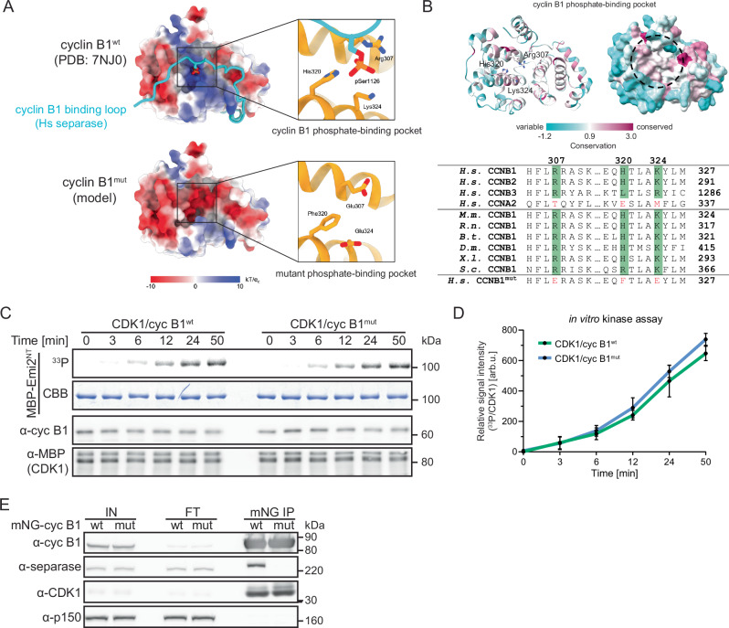
Phosphorylation of substrates by cyclin-dependent kinases (CDKs) is the driving force of cell cycle progression. Several CDK-activating cyclins are involved, yet how they contribute to substrate specificity is still poorly understood. Here, we discover that a positively charged pocket in cyclin B1, which is exclusively conserved within B-type cyclins and binds phosphorylated serine- or threonine-residues, is essential for correct execution of mitosis. HeLa cells expressing pocket mutant cyclin B1 are strongly delayed in anaphase onset due to multiple defects in mitotic spindle function and timely activation of the E3 ligase APC/C. Pocket integrity is essential for APC/C phosphorylation particularly at non-consensus CDK1 sites and full in vitro ubiquitylation activity. Our results support a model in which cyclin B1's pocket facilitates sequential substrate phosphorylations involving initial priming events that assist subsequent pocket-dependent phosphorylations even at non-consensus CDK1 motifs.


Conflict of interest statement

Competing interests: The authors declare no competing interests.

Figures

Fig. 1
Fig. 1. Cyclin B1mut is capable of activating CDK1, but deficient in separase binding.
A Electrostatic surface potentials of human cyclin B1wt (top, PDB: 7NJ0) and cyclin B1mut (bottom, modeled) are illustrated. Close-up views of the cyclin B1 phosphate-binding pocket in cyclin B1wt and in cyclin B1mut bound to phosphoserine 1126 of human separase. Mutations of three critical residues (R307E, H320F, K324E) in the binding pocket lead to a complete charge reversal. Electrostatic potentials are contoured from -10 (red) to +10 kTe-1 (blue). B Sequence conservation of cyclin B1 mapped onto the human cyclin B1 structure (PDB: 7NJ0). The cyclin B1 structure is shown as ribbon (left) or surface representation (right). Residues that form the phosphate-binding pocket are displayed as sticks in the ribbon representation, and the phosphate-binding pocket is indicated with a dashed circle on the surface representation. The AL2CO program has been used to map the conservation indices from a multiple sequence alignment of 2000 different cyclin B homologs (including yeast and human) onto the spatial structure of human cyclin B1. Variable regions are shown in cyan, conserved regions in maroon. Bottom, sequence alignment of the pocket region for different cyclins. Sequence comparison of human B-type cyclins with cyclin A2 (top four rows). Sequence conservation of the binding pocket in B1-cyclins across different species (middle six rows). Last row shows the sequence of the designed cyclin B1mut pocket. Residue numbers for each species are labeled on the right. H.s. (Homo sapiens), M.m. (Mus musculus), R.n. (Rattus norvegicus), B.t. (Bos taurus), D.m. (Drosophila melanogaster), X.l. (Xenopus laevis), S.c. (Saccharomyces cerevisiae). C Radioactive in vitro kinase assay using MBP-CDK1/cyclin B1wt/mut-His and as substrate MBP-Emi2NT (aa 1-350) containing a single CDK1 consensus site. At indicated timepoints after reaction start, samples were immunoblotted for MBP and cyclin B1 (α-cyc B1) and analyzed by autoradiography (33P). CBB (Coomassie brilliant blue). D Quantification of n = 3 independent in vitro kinase assays. Shown are mean values with standard deviations. E Immunoblot analyses of anti-mNG immunoprecipitation (IP) samples of HeLa cells expressing mNG-cyclin B1wt/mut. Doxycycline treated cells were enriched in mitosis by treatment with 333 nM nocodazole for 20 h. Shown are representative input (IN), flow through (FT) and IP samples. p150 served as IP control.
Fig. 2
Fig. 2. Cyclin B1’s phosphate-binding pocket is critical for mitotic fidelity.
A Experimental outline to synchronize HeLa cells transfected with control (ctrl) or cyclin B1 and B2 siRNAs (siB1&B2) in S-phase via double thymidine block and to induce expression of mNG-cyclin B1wt/mut by doxycycline (dox) treatment. B Immunoblot assessing depletion of endogenous cyclin B1 and B2 and expression of mNG-cyclin B1wt/mut. p150 served as loading control. C Quantification of live-cell imaging. The mean of n = 3 or n = 4 independent experiments is shown with standard deviation. Unpaired t-test. **p = 0.002, *p = 0.0149. D Quantification shown in (C) with different y-axis scale for reversine conditions. Unpaired t-test**p = 0.002. E Representative pictures of mitotic mNG-cyclin B1wt/mut (green) HeLa cells treated with either DMSO or nocodazole and stained for tubulin (red) and DNA (blue). Scale bar = 10 µm. F Timecourse to synchronize cells for STLC washout experiments. Images of representative cells processed at indicated timepoints, 1 to 3 h after STLC washout. Tubulin (green), pericentrin (PCNT) (cyan), CREST (red), and DNA (blue) are shown. Scale bar = 10 µm. G Representative images of siB1&B2 cells expressing mNG-cyclin B1wt/mut imaged 3.5 h post STLC release. Shown are examples of either lagging chromosomes or chromosome bridges with DNA and CREST in green and red, respectively. Right panel shows the quantification of both phenotypes. The mean with standard deviation of n = 3 independent experiments is shown. Unpaired t-test: **p = 0.0023.
Fig. 3
Fig. 3. Cyclin B1’s phosphate-binding pocket is important for efficient APC/C activation.
A Heat-map visualizing the hierarchical clustering of the 14 significantly enriched proteins identified by quantitative DIA mass spectrometry in the Flag-cyclin B1wt over Flag-cyclin B1mut IP after ANOVA (s0 = 0.25, FDR = 0.05) and post-hoc Tukey HSD (FDR = 0.05). Technical replicates of n = 3 biological replicates are averaged and log2 abundances are z-score normalized. Euclidean distances are not shown, for details see methods. B Experimental outline of cell synchronization procedure. Immunoblot assessing α-mNG IP samples of mitotically arrested siB1&B2 HeLa cells expressing mNG-cyclin B1wt/mut. p150 served as IP control. Right panels show the quantification of the APC3 signal intensity in the input and co-IP samples from n = 3 independent repetitions and plotted against the distance on the blot. C Schematic of experimental design of α-Flag-APC4 IP from mitotic siB1&B2 HeLa cells expressing mNG-cyclin B1wt/mut and treated for 30 min with 1 µM reversine for SAC silencing. Immunoblot assessing α-APC4 IP samples. D Fluorescent readout (488 nm) of in vitro ubiquitylation assay using immunoprecipitated APC/C from (C), recombinant ubiquitin, UBA1, UBE2C, where indicated UBE2S and as substrate fluorescein-labeled cyclin B11–70 (cyc B11–70). E Schematic of in vitro ubiquitylation assay using APC/C immunoprecipitated from interphasic Xenopus egg extract. To prevent cyclin B resynthesis following its calcium-induced degradation, extract was co-treated with the translation inhibitor cycloheximide (CHX). Phosphorylation by recombinant PLK1 and CDK1/CKS1/cyclin B1wt/mut. In vitro kinase reactions were performed in the presence of DMSO or the CDK and PLK1 inhibitors flavopiridol (FL) and BI2536 (BI), respectively. Immunoblot assessing samples taken at indicated steps shown in experimental outline. F Fluorescent readout (488 nm) of in vitro ubiquitylation assay using APC/C purified according to (E) and supplemented with recombinant UBA1, UBE2C, ubiquitin and as substrate fluorescein-labeled cyclin B11–70. Immunoblot assessing phosphorylation state of APC3 under different experimental conditions.
Fig. 4
Fig. 4. Cyclin B1’s phosphate-binding pocket is important for APC/C phosphorylation at non-consensus CDK1 sites.
A In vitro kinase assay using recombinant CCCwt/mut to phosphorylate recombinant strep-tagged apo-APC/C and MBP-Emi2NT, both present in the same kinase reaction mixture. Shown are western blots for all relevant proteins involved in the reaction. As readout for phosphorylation efficiency, pT97 (MBP-Emi2NT) and APC3 (apoAPC/C) phosphorylation pattern were used. APC3 and APC1 blots were conducted on PhosTag™ gels. In vitro kinase reactions were performed at 30 °C, and at indicated timepoints samples were taken and analyzed by WB. B Pie charts and discrete heatmaps of APC/C phosphosites identified after in vitro phosphorylation by CCCwt/mut. Dephosphorylated and purified APC/C has been subjected to an in vitro phosphorylation assay using either cyclin B1wt or cyclin B1mut and phosphosites have been identified by MS. The phosphosites were stringently filtered (see Fig. S4 for details). Then, the obtained phosphosites from CCCwt were divided in either minimal CDK1 (S/T)P consensus sites (blue) or non-consensus sites (orange). The upper pie chart shows the percentage and number of identified consensus or other phosphosites in the CCCwt samples. Using these phosphosites as basis, the CCCmut data set was analyzed and classified in sites that were equally well detected in CCCmut samples (left) and sites that were either rarely (at least 2 fold reduced in intensity and in half or less than half of the raw files compared to wt; middle) or not at all detected (right). Below the pie charts heatmaps of the identified phosphosites are shown (red: present; light red: reduced at least 2 fold and in half or less than half of the rawfiles compared to CCCwt; gray: absent. C Bar plot of the fold changes of APC/C subunits, CDC20, cyclin B1 and UBE2S of the immunopurified APC/C of cells overexpressing either cyclin B1wt or B1mut. APC3 was immunopurified from siB1&B2 cells expressing either cyclin B1wt or B1mut and interaction partners were quantified by MS. For proteins of interest, the ratio of the quantified protein intensities between cyclin B1wt and cyclin B1mut was calculated (log2 intensities cyclin B1wt – log2 intensities B1mut) and normalized on the ratio of the bait protein APC3. The x-axis shows the normalized log2 changes of relevant indicated proteins (y-axis). The experiment was conducted in n = 3 independent biological replicates, each dot represents a single measurement. Data are represented as mean values ±SD. D Model of pocket-dependent multisite substrate phosphorylations. Step 1: Priming phosphorylation carried out by any kinase creates a docking site for cyclin B1’s phosphate-binding pocket. Step 2: Upon pocket-dependent recruitment, CCC catalyzes the phosphorylation of an additional site. Step 3: This phosphosite can serve as docking site for CCC itself using cyclin B1 or CKS1 as phosphate adaptor or other kinases with phosphate adaptor properties such as casein kinase 1 (CK1) or PLK1. For illustration, CCC (PDB: 7NJ0) is shown in blue (CDK1), orange (CKS1), and green (cyclin B1), PLK1 (AlphaFold2 prediction) in red, and CK1 (PDB: 6GZD) in purple.

References

    1. Hochegger, H., Takeda, S. & Hunt, T. Cyclin-dependent kinases and cell-cycle transitions: does one fit all? Nat. Rev. Mol. Cell Biol.9, 910–916 (2008). - PubMed
    1. Crncec, A. & Hochegger, H. Triggering mitosis. FEBS Lett.593, 2868–2888 (2019). - PubMed
    1. Kamenz, J. & Ferrell, J. E. The temporal ordering of cell-cycle phosphorylation. Mol. Cell65, 371–373 (2017). - PubMed
    1. Enserink, J. M. & Kolodner, R. D. An overview of Cdk1-controlled targets and processes. Cell Div.5, 11 (2010). - PMC - PubMed
    1. Holt, L. J. et al. Global analysis of Cdk1 substrate phosphorylation sites provides insights into evolution. Science325, 1682–1686 (2009). - PMC - PubMed

LinkOut - more resources