Enhanced multiscale human brain imaging by semi-supervised digital staining and serial sectioning optical coherence tomography
- PMID: 39833166
- PMCID: PMC11746934
- DOI: 10.1038/s41377-024-01658-0
Enhanced multiscale human brain imaging by semi-supervised digital staining and serial sectioning optical coherence tomography
Abstract
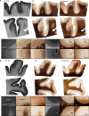
A major challenge in neuroscience is visualizing the structure of the human brain at different scales. Traditional histology reveals micro- and meso-scale brain features but suffers from staining variability, tissue damage, and distortion, which impedes accurate 3D reconstructions. The emerging label-free serial sectioning optical coherence tomography (S-OCT) technique offers uniform 3D imaging capability across samples but has poor histological interpretability despite its sensitivity to cortical features. Here, we present a novel 3D imaging framework that combines S-OCT with a deep-learning digital staining (DS) model. This enhanced imaging modality integrates high-throughput 3D imaging, low sample variability and high interpretability, making it suitable for 3D histology studies. We develop a novel semi-supervised learning technique to facilitate DS model training on weakly paired images for translating S-OCT to Gallyas silver staining. We demonstrate DS on various human cerebral cortex samples, achieving consistent staining quality and enhancing contrast across cortical layer boundaries. Additionally, we show that DS preserves geometry in 3D on cubic-centimeter tissue blocks, allowing for visualization of meso-scale vessel networks in the white matter. We believe that our technique has the potential for high-throughput, multiscale imaging of brain tissues and may facilitate studies of brain structures.
© 2025. The Author(s).
Conflict of interest statement
Conflict of interest: The authors declare no competing interests.
Figures


Update of
-
Enhanced Multiscale Human Brain Imaging by Semi-supervised Digital Staining and Serial Sectioning Optical Coherence Tomography.Res Sq [Preprint]. 2024 Mar 21:rs.3.rs-4014687. doi: 10.21203/rs.3.rs-4014687/v1. Res Sq. 2024. Update in: Light Sci Appl. 2025 Jan 20;14(1):57. doi: 10.1038/s41377-024-01658-0. PMID: 38562721 Free PMC article. Updated. Preprint.
References
-
- Yushkevich, P. A. et al. 3D mouse brain reconstruction from histology using a coarse-to-fine approach. Proc. 3rd Biomedical Image Registration (Utrecht: Springer, 2006).
Grants and funding
LinkOut - more resources
Full Text Sources

