. 2025 Jan 20;15(1):2509.
doi: 10.1038/s41598-024-85072-x.

Enhanced effect of the immunosuppressive soluble HLA-G2 homodimer by site-specific PEGylation

Affiliations

Enhanced effect of the immunosuppressive soluble HLA-G2 homodimer by site-specific PEGylation

Chisato Yamada et al. Sci Rep. .

Abstract

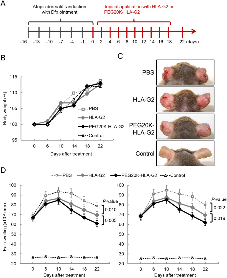
Human leukocyte antigen (HLA)-G is a nonclassical HLA class I molecule that has an immunosuppressive effect mediated by binding to immune inhibitory leukocyte immunoglobulin-like receptors (LILR) B1 and LILRB2. A conventional HLA-G isoform, HLA-G1, forms a heterotrimeric complex composed of a heavy chain (α1-α3 domains), β2-microglobulin (β2m) and a cognate peptide. One of the other isoforms, HLA-G2, lacks a α2 domain or β2m to form a nondisulfide-linked homodimer, and its ectodomain specifically binds to LILRB2 expressed in human monocytes, macrophages, and dendritic cells. The administration of the ectodomain of HLA-G2, designated the soluble HLA-G2 homodimer, showed significant immunosuppressive effects in mouse models of rheumatoid arthritis and systemic lupus erythematosus, presumably by binding to a mouse ortholog of LILRB2, paired immunoglobulin-like receptor B. However, the refolded soluble HLA-G2 homodimer used in these studies tends to aggregate and degrade; thus, its stability for clinical use has been a concern. In the present study, we improved the stability of the refolded soluble HLA-G2 homodimer via a site-directed PEGylation method. PEGylation at an original free cysteine residue, Cys42, resulted in increased lyophilization and thermal and serum stability. Furthermore, the PEGylated soluble HLA-G2 homodimer could better suppress atopic symptoms in mice than the non-PEGylated homodimer. These results suggest that PEGylated soluble HLA-G2 homodimers could be candidates for immunosuppressive biologics that specifically target LILRB2-positive myelomonocytic antigen-presenting cells.

Keywords: HLA-G2; Immune checkpoint; Immunosuppressive; LILR; PEGylation.


Conflict of interest statement

Declarations. Competing interests: The authors declare no competing interests. Consent for publication: All the authors read and approved the final manuscript.

Figures

Fig. 1
Fig. 1
Randomly PEGylated soluble HLA-G2 homodimer protein in lysine residues. (A) SPR analysis of HLA-G2 (6.2 μM, left) or PEGylated HLA-G2 in lysine residues (randomPEG-HLA-G2, 5.3 μM, right) to the immobilized PIR-B. Each soluble HLA-G2 homodimer protein was injected over the immobilized PIR-B (3000 RU, black line) and negative control BSA (2000 RU, gray line). The response of the SPR results to BSA was due to buffer mismatch and nonspecific interactions between the analytes and BSA. (B) Schematic diagram of the domain structure (left) and the model structure (right) of the soluble HLA-G2 homodimer. The original Cys42 (green circle) and lysine residues (blue circle) are shown in the schematic structures (left). Three lysine residues are shown in the blue stick model (right). K183, which is not visible in the crystal structure of the soluble HLA-G1 monomer (PDB ID: 2DYP), is shown in the blue circle (right).
Fig. 2
Fig. 2
The positions of the targeted residues on the soluble HLA-G2 homodimer for PEGylation. (A) Schematic diagram of the domain structures of soluble HLA-G2 homodimers and their mutants. The cysteine residues used for PEG attachment are shown in green circles (right). In HLA-G2 N86C and HLA-G2 CG, C42 was substituted for serine (C42S). (B) Model structure of soluble HLA-G2 homodimers. The positions of the target residues for PEGylation are shown. All targeted residues are exposed on the surface of the soluble HLA-G2 homodimer. The lysine residues in the blue stick models targeted for random PEGylation (Fig. 1) are also shown.
Fig. 3
Fig. 3
PEGylation of soluble HLA-G2 homodimers by four different molecular weights of PEG-maleimide molecules. (A) SDS‒PAGE analysis of the PEGylated soluble HLA-G2 homodimer mixture with each molecular size of PEG under nonreducing conditions followed by CBB (left) and BaI2 (right) staining. The PEGylated proteins were detected as brown bands in BaI2 staining. The non-PEGylated soluble HLA-G2 monomer separated in the SDS‒PAGE gel (21 kDa) is indicated by a white arrow, and the black arrows indicate the PEGylated soluble HLA-G2 homodimer. Multimerization of the non-PEGylated soluble HLA-G2 homodimer resulted in the existence of a 42 kDa band due to disulfide bonding via Cys42. (B) Purification of the PEGylated soluble HLA-G2 homodimer proteins by SEC and SDS‒PAGE analysis under nonreducing conditions followed by BaI2 staining of the SEC fractions indicated by black lines in the chromatograms. The band derived from the PEGylated soluble HLA-G2 homodimer is indicated by black arrows. A schematic diagram of the domain structure of each soluble HLA-G2 homodimer is shown. A representative chromatogram of five (5 kDa) or six (10 kDa and 20 kDa) experiments is shown. The 40 kDa PEG experiment was performed once. See Supplementary Figs. 2 and 3 for the uncropped gel images.
Fig. 4
Fig. 4
PEGylation of soluble HLA-G2 homodimers at different positions by 20 kDa PEG. (A) Purification of soluble HLA-G2 homodimer proteins (left) and SDS‒PAGE analysis of the peak fraction indicated by a black bar in each chromatogram (HLA-G2) and soluble HLA-G2 homodimers after the PEGylation reaction (PEGylated) under nonreducing conditions followed by CBB and BaI2 staining. The black arrows indicate the PEGylated soluble HLA-G2 homodimer. The non-PEGylated soluble HLA-G2 homodimer (21 kDa via SDS‒PAGE) is indicated by a white arrow. Multimerization of the non-PEGylated HLA-G2 resulted in the existence of a 42 kDa band in the gel due to disulfide bonds via Cys42. (B) Purification of the PEGylated soluble HLA-G2 homodimer mutants with 20 kDa PEG (upper) and SDS‒PAGE analysis under nonreducing conditions followed by BaI2 staining of the fractions shown by black bars in each chromatogram (lower). The black arrows indicate PEGylated HLA-G2, and non-PEGylated HLA-G2 (21 kDa) and multimerized HLA-G2-derived 42 kDa bands were observed. A schematic diagram of the domain structure of each soluble HLA-G2 homodimer is shown. Representative data from three independent experiments are shown. See Supplementary Fig. 4 for the uncropped gel images.
Fig. 5
Fig. 5
SPR analysis of the ability of sHLA-G2 and PEG20K-HLA-G2 to immobilize LILRB2 on the sensor chip. (A) Kinetic analysis of sHLA-G2 (left) and PEG20K-HLA-G2 (right) at the indicated concentrations. The binding response curves at each concentration of sHLA-G2 or PEG20K-HLA-G2 are shown by subtracting the response measured in the control flow cell (BSA) from the response in the sample flow cells (LILRB2). The response curves (black lines) were fitted locally with the Langmuir binding mode (gray lines), and apparent KD values were calculated. LILRB2 was immobilized at approximately 500 RU for sHLA-G2 and approximately 270 RU for PEG20K-HLA-G2. BSA was immobilized at the same level for each experiment. This experiment was independently performed twice. (B) Kinetic parameters of the binding between sHLA-G2 or PEG20K-HLA-G2 and immobilized LILRB2 calculated by local fitting. The mean value and standard deviation (SD) were determined via six fitting data points (three concentrations in two independent experiments).
Fig. 6
Fig. 6
In vitro stability analyses of sHLA-G2 and PEG20K-HLA-G2. (A) Heat stability was analyzed by SDS‒PAGE under nonreducing conditions. The black arrow shows the band derived from each soluble HLA-G2 homodimer protein in the gel. The soluble HLA-G2 homodimer was partially multimerized during incubation as a disulfide-bonded 42 kDa band. Representative data from four independent experiments are shown. (B) Serum stability was analyzed by western blotting using MEM-G1 (the first antibody) and anti-mouse IgG-HRP (the second antibody). Left top: The intensity of the sHLA-G2 band after zero-hour incubation with FBS/without FBS was defined as 100%. Left bottom: Two bands of PEG20K-HLA-G2 were used for the quantification analysis, as PEG20K-HLA-G2 without serum was detected as two bands in this WB analysis. Right: The graph shows the residual ratio of sHLA-G2 (gray) and PEG20K-HLA-G2 (black) proteins in the WB. Representative data from four independent experiments are shown. (C) Lyophilization stability was analyzed by SDS‒PAGE to compare degradation and aggregation under nonreducing conditions, followed by CBB staining. Representative data from two independent experiments are shown. (D) LILRB2 binding activity of sHLA-G2 and PEG20K-HLA-G2 (0.6 μM) before (dotted line) and after lyophilization (solid line) to the immobilized LILRB2. The sensorgrams were obtained by subtracting the responses measured in the BSA-immobilized flow cell (control) from those measured in the LILRB2-immobilized flow cell. (E) Kinetic analysis of lyophilized sHLA-G2 (left) and PEG20K-HLA-G2 (right) to immobilize LILRB2. The concentrations of the injected samples are shown on the right of the sensorgrams. This experiment was independently performed twice. The mean value and standard deviation (SD) were determined via six fitting data points (three concentrations in two independent experiments). See Supplementary Fig. 5 for the uncropped gel images of A and C, and Supplementary Fig. 6 for the full blot images and another result of B.
Fig. 7
Fig. 7
Effect of PEG20K-HLA-G2 on Dfb ointment-induced atopic dermatitis in NC/Nga mice. NC/Nga mice without dermatitis induced by Dfb ointment were used as the control group. (A) Timeline of the experiments. Atopic dermatitis was induced with Dfb ointment treatment 6 times. The mice were treated with 5 μg of soluble HLA-G1 monomer, sHLA-G2, PEG20K-HLA-G2 or PBS every other day, and the thickness of the ear was monitored every 4 days, as indicated by underlines. (B) The average body weight changes in NC/Nga mice treated with soluble HLA-G2 homodimer proteins or PBS were monitored twice a week. (C) Macroscopic features of atopic dermatitis-like skin lesions in NC/Nga mice on Day 18. Representative data from four mice/group are shown. (D) Thickness of the left (left) and right (right) ears. The means ± standard error of the means (SEMs) (n = 4) and statistically significant differences are shown as P values (*P < 0.05, **P < 0.01, ***P < 0.001).

References

    1. Kamishikiryo, J. & Maenaka, K. HLA-G molecule. Curr. Pharm. Des.15, 3318–3324. 10.2174/138161209789105153 (2009). - PubMed
    1. Kuroki, K. & Maenaka, K. Immune modulation of HLA-G dimer in maternal-fetal interface. Eur. J. Immunol.37, 1727–1729. 10.1002/eji.200737515 (2007). - PMC - PubMed
    1. Kuroki, K. et al. Cutting edge: Class II-like structural features and strong receptor binding of the nonclassical HLA-G2 isoform homodimer. J. Immunol.198, 3399–3403. 10.4049/jimmunol.1601296 (2017). - PubMed
    1. Bahri, R. et al. Soluble HLA-G inhibits cell cycle progression in human alloreactive T lymphocytes. J. Immunol.176, 1331–1339. 10.4049/jimmunol.176.3.1331 (2006). - PubMed
    1. Gonen-Gross, T. et al. Complexes of HLA-G protein on the cell surface are important for leukocyte Ig-like receptor-1 function. J. Immunol.171, 1343–1351. 10.4049/jimmunol.171.3.1343 (2003). - PubMed

Publication types

MeSH terms

LinkOut - more resources