

Dehydrocorydaline maintains the vascular smooth muscle cell contractile phenotype by upregulating Spta1
- PMID: 39833304
- PMCID: PMC12032006
- DOI: 10.1038/s41401-024-01464-9
Dehydrocorydaline maintains the vascular smooth muscle cell contractile phenotype by upregulating Spta1
Abstract
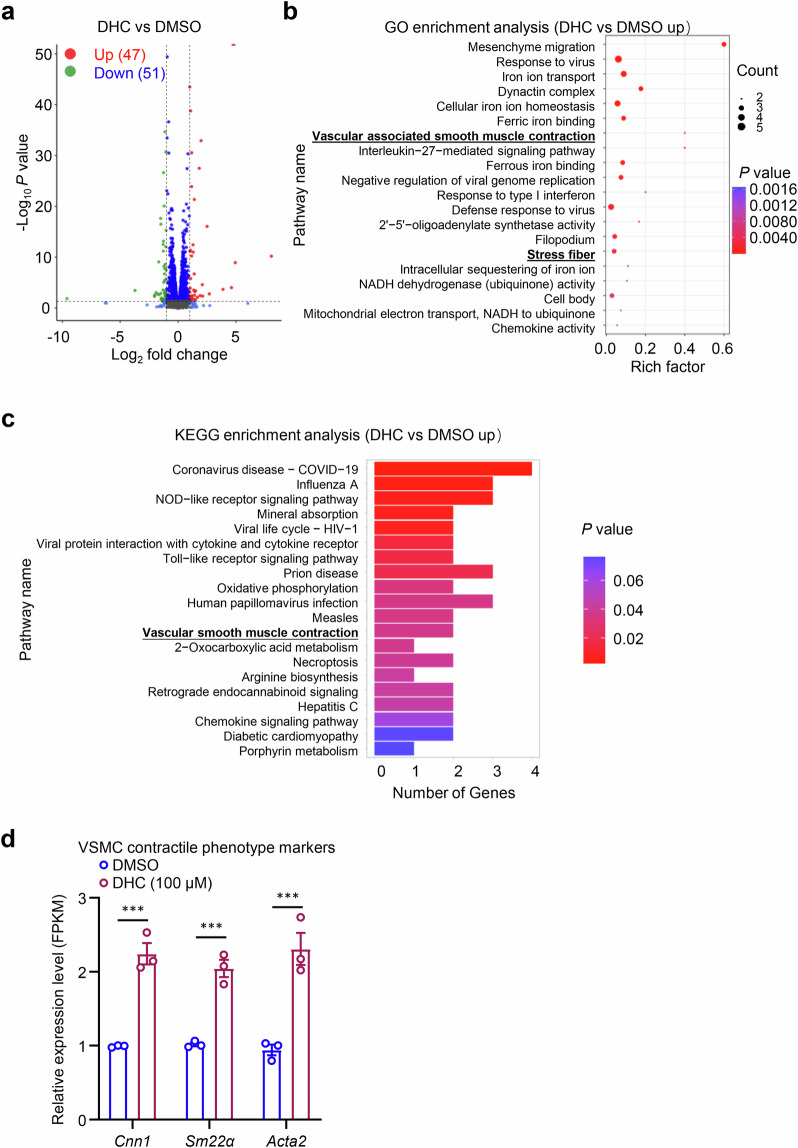
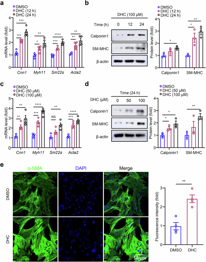
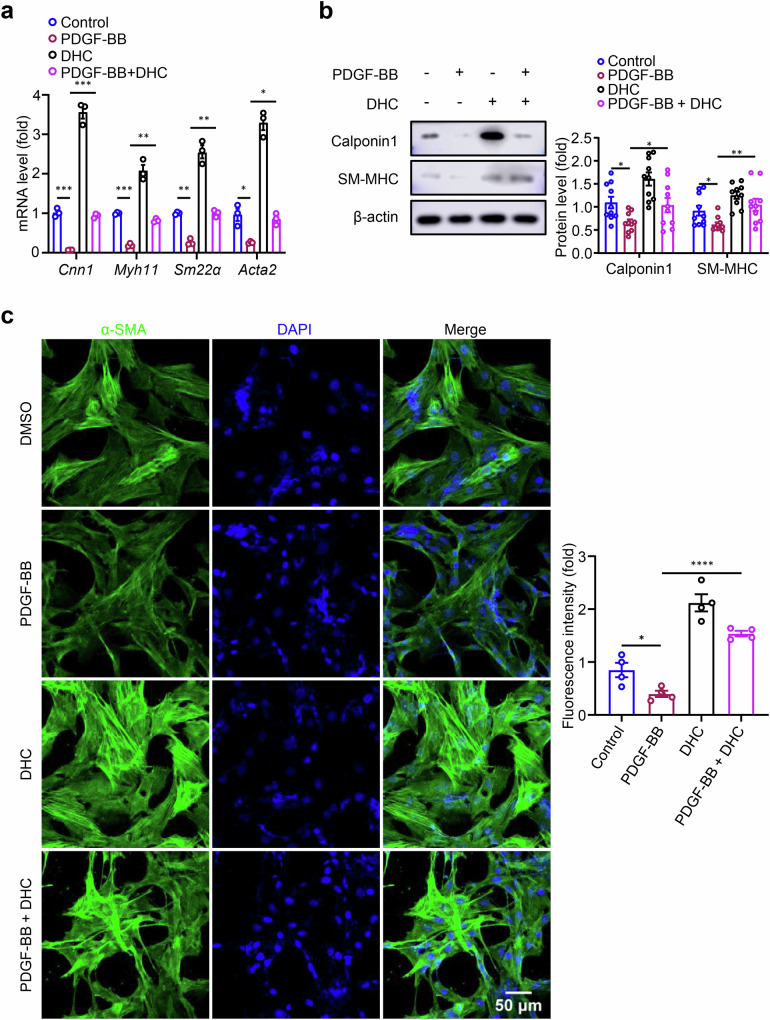
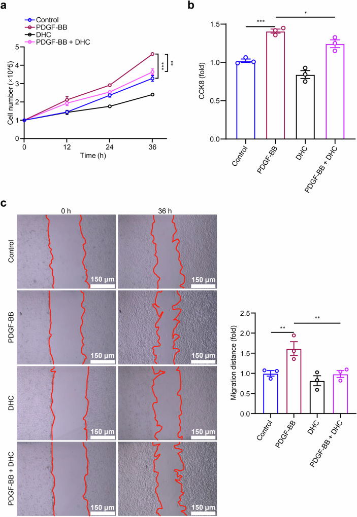
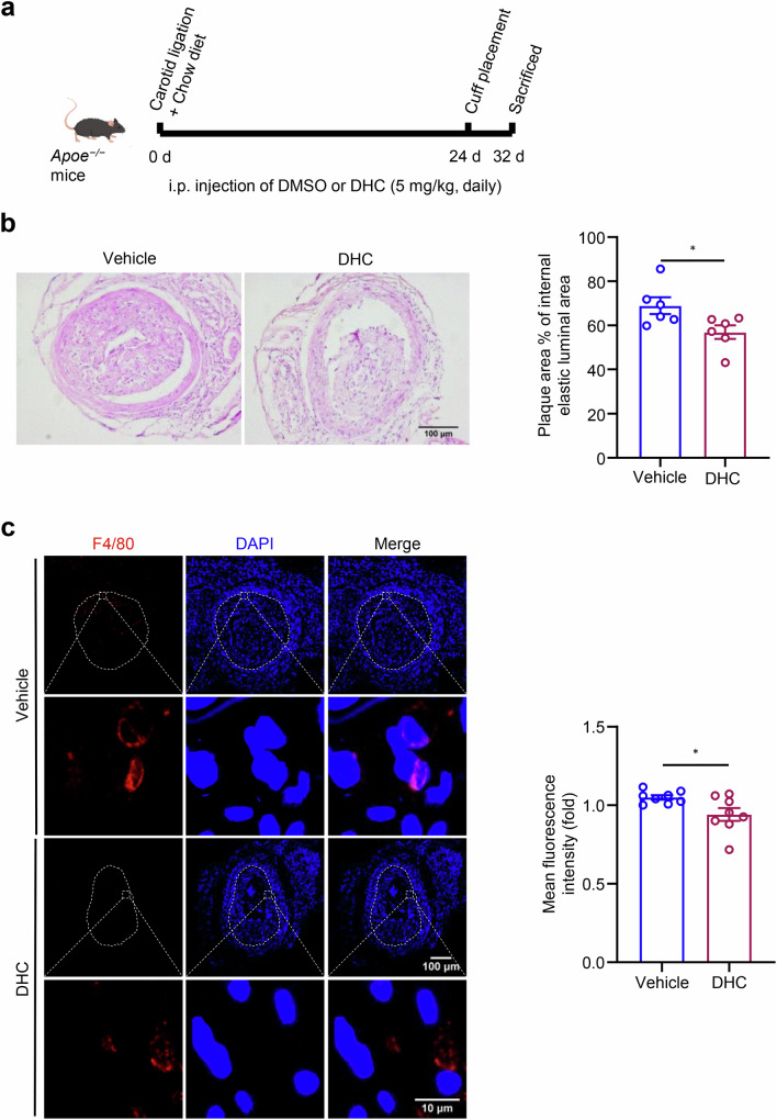
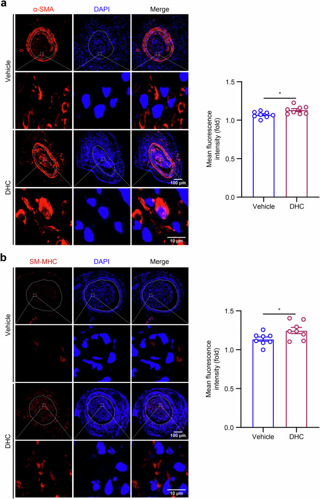
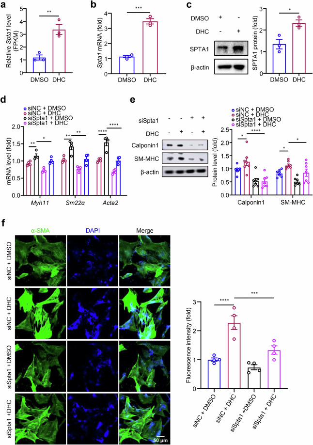
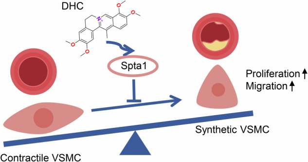
Vascular smooth muscle cell (VSMC) phenotypic switching plays a crucial role in the initiation and progression of atherosclerosis. Dehydrocorydaline (DHC), a major active component of the traditional Chinese herbal medicine Rhizoma Corydalis, exhibits diverse pharmacological effects. However, its impact on VSMCs remains largely unknown. This study aims to investigate the effects and underlying mechanisms of DHC in phenotypic switching of VSMCs. Our study revealed that DHC increased the mRNA and protein levels of rat VSMC contractile phenotype markers, such as calponin 1 (Cnn1), myosin heavy chain (Myh11, SM-MHC), smooth muscle 22α (Sm22α), and alpha-smooth muscle actin (Acta2, α-SMA) in a time- and dose-dependent manner. Additionally, DHC inhibited platelet-derived growth factor-BB-induced VSMC proliferation and migration. In Apoe-/- mice, DHC treatment resulted in reduced carotid plaque areas and macrophage infiltration, along with increased contractile phenotype marker expression. RNA sequencing analysis revealed a significant upregulation of spectrin alpha, erythrocytic 1 (Spta1) in DHC-treated rat VSMCs. Strikingly, Spta1 knockdown effectively negated the increase in contractile phenotype marker expression in VSMCs that was initially prompted by DHC. Therefore, DHC preserves the VSMC contractile phenotype through Spta1, thereby attenuating carotid artery atherosclerotic plaques in Apoe-/- mice. This study provides evidence supporting the potential use of Chinese herbal medicines, particularly those containing DHC such as Rhizoma Corydalis, in the treatment of atherosclerotic cardiovascular disease, thus expanding the clinical application of such herbal remedies.
Keywords: atherosclerosis; dehydrocorydaline; phenotypic switching; vascular smooth muscle cell.
© 2025. The Author(s).
Conflict of interest statement
Competing interests: The authors declare no competing interests.
Figures
Similar articles
-
AMP-Activated Protein Kinase Alpha 2 Deletion Induces VSMC Phenotypic Switching and Reduces Features of Atherosclerotic Plaque Stability.Circ Res. 2016 Sep 2;119(6):718-30. doi: 10.1161/CIRCRESAHA.116.308689. Epub 2016 Jul 20. Circ Res. 2016. PMID: 27439892 Free PMC article.
-
M2 Macrophage-Derived Exosomes Inhibit Atherosclerosis Progression by Regulating the Proliferation, Migration, and Phenotypic Transformation of Smooth Muscle Cells.Front Biosci (Landmark Ed). 2024 Aug 19;29(8):288. doi: 10.31083/j.fbl2908288. Front Biosci (Landmark Ed). 2024. PMID: 39206919
-
The protective effect of HOXA5 on carotid atherosclerosis occurs by modulating the vascular smooth muscle cell phenotype.Mol Cell Endocrinol. 2021 Aug 20;534:111366. doi: 10.1016/j.mce.2021.111366. Epub 2021 Jun 11. Mol Cell Endocrinol. 2021. PMID: 34126188
-
The Effect of Retinoids in Vascular Smooth Muscle Cells: From Phenotyping Switching to Proliferation and Migration.Int J Mol Sci. 2024 Sep 25;25(19):10303. doi: 10.3390/ijms251910303. Int J Mol Sci. 2024. PMID: 39408632 Free PMC article. Review.
-
Vascular Smooth Muscle Cells in Atherosclerosis.Circ Res. 2016 Feb 19;118(4):692-702. doi: 10.1161/CIRCRESAHA.115.306361. Circ Res. 2016. PMID: 26892967 Free PMC article. Review.
Cited by
-
The NLRP3 inflammasome: a therapeutic target of phytochemicals in treating atherosclerosis (a systematic review).Front Immunol. 2025 May 15;16:1568722. doi: 10.3389/fimmu.2025.1568722. eCollection 2025. Front Immunol. 2025. PMID: 40443656 Free PMC article.
References
-
- Joseph P, Leong D, Mckee M, Anand SS, Schwalm JD, Teo K, et al. Reducing the global burden of cardiovascular disease, Part 1: the epidemiology and risk factors. Circ Res. 2017;121:677–94. - PubMed
-
- Ridker PM, Everett BM, Thuren T, Macfadyen JG, Chang WH, Ballantyne C, et al. Antiinflammatory therapy with Canakinumab for atherosclerotic disease. N Engl J Med. 2017;377:1119–31. - PubMed
-
- Tardif JC, Kouz S, Waters DD, Bertrand OF, Diaz R, Maggioni AP, et al. Efficacy and safety of low-dose colchicine after myocardial infarction. N Engl J Med. 2019;381:2497–505. - PubMed
-
- Yin C, Ge Z, Yuan J, Chen Y, Tang Y, Xiang Y, et al. NEAT1 regulates VSMC differentiation and calcification in as long noncoding RNA NEAT1 enhances phenotypic and osteogenic switching of vascular smooth muscle cells in atherosclerosis via scaffolding EZH2. Am J Physiol Cell Physiol. 2024;326:C1721–c1734. - PMC - PubMed
MeSH terms
Substances
LinkOut - more resources
Full Text Sources
Research Materials
Miscellaneous
