. 2025 Jan 20;8(1):89.
doi: 10.1038/s42003-025-07545-7.

Positively charged cytoplasmic residues in corin prevent signal peptidase cleavage and endoplasmic reticulum retention

Affiliations

Positively charged cytoplasmic residues in corin prevent signal peptidase cleavage and endoplasmic reticulum retention

Hui Li et al. Commun Biol. .

Abstract

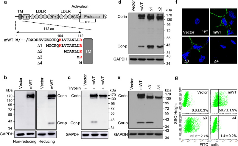
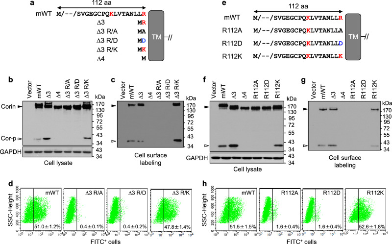
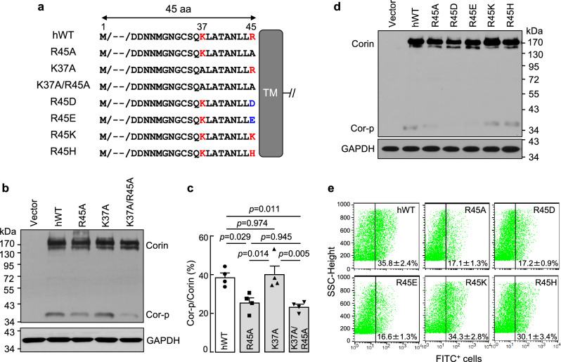
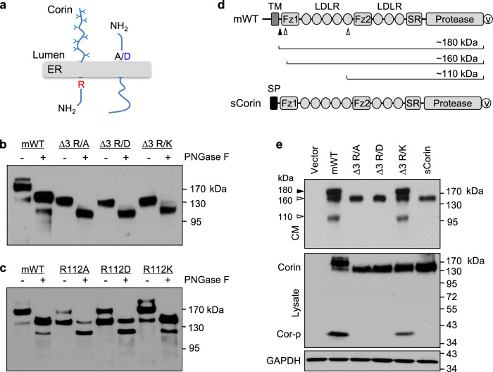
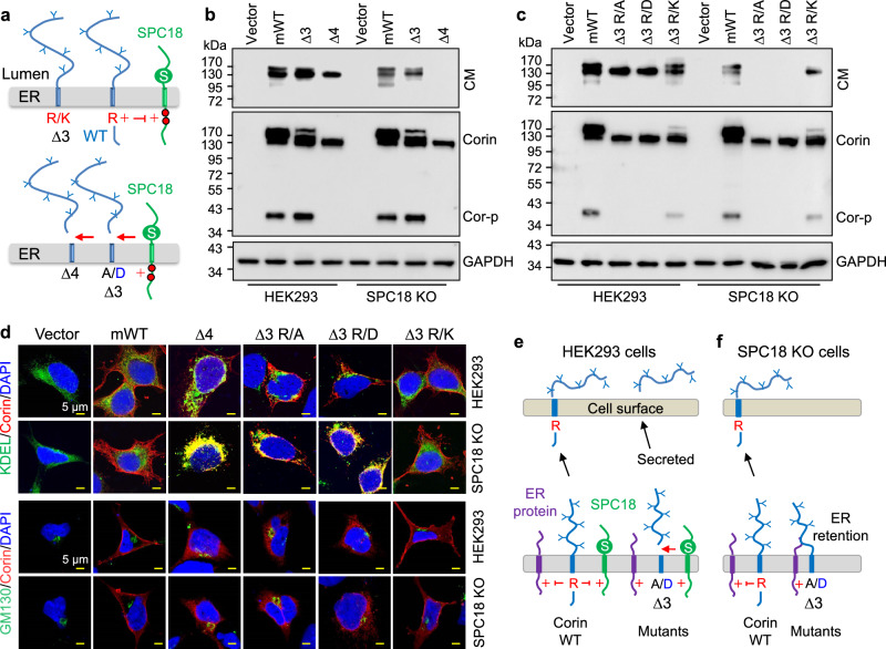
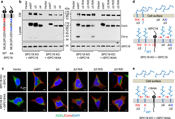
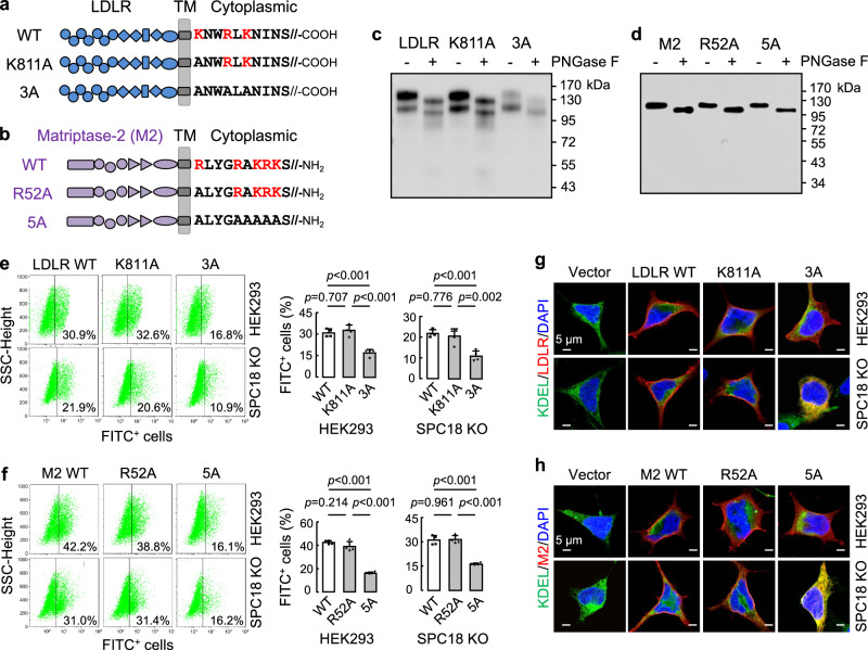
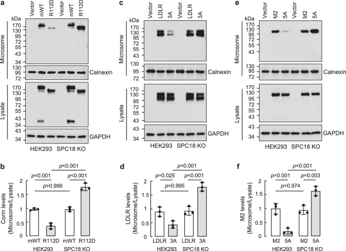
Positively charged residues are commonly located near the cytoplasm-membrane interface, which is known as the positive-inside rule in membrane topology. The mechanism underlying the function of these charged residues remains poorly understood. Herein, we studied the function of cytoplasmic residues in corin, a type II transmembrane serine protease in cardiovascular biology. We found that the positively charged residue at the cytoplasm-membrane interface of corin was not a primary determinant in membrane topology but probably served as a charge-repulsion mechanism in the endoplasmic reticulum (ER) to prevent interactions with proteins in the ER, including the signal peptidase. Substitution of the positively charged residue with a neutral or acidic residue resulted in corin secretion likely due to signal peptidase cleavage. In signal peptidase-deficient cells, the mutant corin proteins were not secreted but retained in the ER. Similar results were found in the low-density lipoprotein receptor and matriptase-2 that have positively charged residues at and near the cytoplasm-membrane interface. These results provide important insights into the role of the positively charged cytoplasmic residues in mammalian single-pass transmembrane proteins.


Conflict of interest statement

Competing interests: The authors declare no competing interests.

Figures

Fig. 1
Fig. 1. Activation cleavage and cell surface expression of mouse corin mutants with shortened cytoplasmic tails.
a Corin protein domains and plasmids expressing mouse WT corin (mWT) with a 112-amino-acid cytoplasmic tail and mutants with shortened cytoplasmic tails (Δ1-Δ4). TM, transmembrane; Fz, frizzled; LDLR, LDL receptor, SR, scavenger receptor. The activation cleavage site (arrow), a disulfide bond (s-s) between the stem region and the protease domain, and a C-terminal V5 (v) tag are indicated. b Western blotting of mWT in HEK293 cell lysates under non-reducing (left) and reducing (right) conditions. The full-length corin (Corin) and the activation cleaved corin protease domain (Cor-p) are indicated. GAPDH was a control. Data are representative of four experiments. c Western blotting of mWT in transfected cells without (−) or with (+) trypsin digestion. Western blotting was under reducing conditions. Data are representative of three experiments. d, e Western blotting of mWT and corin mutants from transfected cells under reducing conditions. Data are representative of five experiments. f Immunostaining of corin proteins on the surface of non-membrane permeabilized cells. Scale bars, 5 μm. Data are representative of five experiments. g Flow cytometric analysis of corin protein on the cell surface. Percentages of corin-positive cells are indicated. n = 5 per group.
Fig. 2
Fig. 2. Activation cleavage and cell surface expression of mouse corin mutants with substituted residues at the cytoplasm-membrane interface.
a Mouse corin mutants with altered cytoplasmic sequences. Corin extracellular domains are not shown. TM, transmembrane. b Western blotting of corin proteins from transfected cells under reducing conditions. Data are representative of five experiments. c Western blotting of biotin-labeled cell surface corin proteins under reducing conditions. Data are representative of three experiments. d Flow cytometric analysis of corin on the cell surface. Percentages of corin-positive cells are indicated. n = 6 per group. e Mouse corin mutants with a full-length cytoplasmic tail. Western blotting of corin proteins in lysates (f) and biotin-labeled cell surface proteins (g) from transfected cells. Western blotting was done under reducing conditions. Data are representative of three experiments. h Flow cytometric analysis of corin proteins on the cell surface. Percentages of corin-positive cells are indicated. n = 6 per group.
Fig. 3
Fig. 3. Activation cleavage and cell surface expression of full-length human corin mutants with substituted residues at the cytoplasm-membrane interface.
a Full-length human corin mutants with altered cytoplasmic residues. Corin extracellular domains are not shown. TM, transmembrane. b Western blotting of human corin proteins from transfected cells under reducing conditions. Data are representative of four experiments. c Quantitative data of corin activation cleavage, as measured by the ratio of Cor-p vs. Corin bands from densitometric analysis of western blots. n = 4. P values were analyzed by one-way ANOVA. d Western blotting of human corin mutants from transfected cells under reducing conditions. Data are representative of three experiments. e Flow cytometric analysis of corin proteins on the cell surface. Percentages of corin-positive cells are indicated. n = 4 per group.
Fig. 4
Fig. 4. N-glycosidase digestion and detection of corin proteins in the conditioned media.
a Alternative corin membrane orientations in the ER. WT corin with the cytoplasmic Arg (R) (left) and the mutants, in which the Arg was replaced by Ala (A) or Asp (D) (right). NH2, N-terminus. Y shaped symbols indicate N-glycans. b, c Western blotting of corin proteins from transfected cells without (−) or with (+) PNGase F treatment. Western blotting was done under reducing conditions. Data are representative of three experiments. d Illustration of cleaved corin ectodomain fragments. The ADAM10 cleavage site (filled arrowhead) and corin autocleavage sites (open arrowheads) and corresponding cleaved fragments are indicated. A soluble corin (sCorin), in which the cytoplasmic tail and the transmembrane domain were replaced with a signal peptide (SP), was a control. e Western blotting of corin proteins in the conditioned media (CM) (top panel) and cell lysates (middle panel) from transfected cells. In the top panel, ADAM10-cleaved and corin self-cleaved fragments in mWT are indicated by filled and open arrowheads, respectively. GAPDH in cell lysates was a control (bottom panel). Data are representative of three experiments.
Fig. 5
Fig. 5. Analysis of corin proteins in HEK293 and SPC18 KO cells.
a A proposed model. WT corin and the mutants Δ3 and Δ3 R/K had a positively charged Arg (R) or Lys (K) at the cytoplasm-membrane interface, preventing cleavage by SPC18 with the active Ser (S) and positively charged cytoplasmic residues (red dots). The mutants Δ4, Δ3 R/A, and Δ3 R/D lacking the positively charged cytoplasmic residue were cleaved by SPC18 (arrows). Western blotting of corin fragments in the conditioned media (CM) (top panels) and lysates (bottom panels) from HEK293 and SPC18 KO cells expressing mWT and the mutants Δ3 and Δ4 (b), or Δ3 R/A, Δ3 R/D, and Δ3 R/K (c). Data are representative of at least four experiments. d Co-straining of KDEL or GM130 and corin in HEK293 and SPC18 KO cells expressing mWT and the corin mutants. Nuclei were stained with DAPI. Scale bars: 5 μm. Data are representative of at least six experiments. e, f A proposed model. In HEK293 cells, the cytoplasmic Arg protects WT corin from interacting with SPC18 and ER proteins with positively charged cytoplasmic residues, allowing corin expression on the cell surface. When the cytoplasmic Arg is replaced by Ala (A) or Asp (D), the mutants were cleaved by SPC18 and secreted from HEK293 cells or trapped by ER proteins in SPC18 KO cells.
Fig. 6
Fig. 6. Analysis of corin mutants in SPC18 KO cells expressing recombinant SPC18 proteins.
a Cytoplasmic sequences in SPC18 WT and the mutant SPC18/4A. b Western blotting of corin fragments in the conditioned media (CM) and lysates from SPC18 KO cells expressing recombinant SPC18 WT (rSPC18) or the mutant SPC18/4A (rSPC18/4A). rSPC18 proteins in the transfected cells were verified. Data are representative of at least five experiments. c Co-staining of KDEL and corin in SPC18 KO cells expressing rSPC18 or rSPC18/4A. Nuclei were stained with DAPI. Scale bars: 5 μm. Data are representative of at least five experiments. Proposed models. In SPC18 KO cells expressing rSPC18 (d), the charge-repulsion mechanism protected corin WT and the mutant Δ3 R/K, but not the mutants Δ4, Δ3 R/A, and Δ3 R/D, from rSPC18 cleavage. Membrane-bound, but not secreted, corin proteins were activated, as indicated by the Cor-p band in western blotting. In SPC18 KO expressing rSPC18/4A (e), substitution of the positively charged residues (red dots) with Ala residues (grey dots) at the cytoplasm-membrane interface in SPC18 abolished the protective mechanism, resulting in rSPC18/4A cleavage and secretion of corin WT and all the mutants.
Fig. 7
Fig. 7. ER retention of LDLR and matriptase-2 mutants lacking positively charged cytoplasmic residues.
Illustration of human LDLR (a) and matriptase-2 (M2) (b) domains and partial cytoplasmic sequences with positively charged residues in red. LDLR has a C-terminal cytoplasmic tail, whereas M2 has an N-terminal cytoplasmic tail. LDLR and M2 mutants with mutated cytoplasmic Lys and/or Arg residues are indicated. c, d Western blotting of LDLR (c) and M2 (d) WT and the mutants from HEK293 cell lysates without (−) or with (+) PNGase F treatment. Western blotting was under reducing conditions. Data are representative of three experiments. Flow cytometric analysis of LDLR (e) and M2 (f) WT and the mutants on the surface of HEK293 and SPC18 KO cells. Quantitative data of LDLR- and M2-positive cells from four experiments are shown in bar graphs. Co-straining of KDEL and LDLR (g) or M2 (h) proteins in HEK293 and SPC18 KO cells transfected with a vector or plasmids expressing corresponding WT and the mutants. Nuclei were stained with DAPI. Scale bars, 5 μm. Data are representative of at least five experiments.
Fig. 8
Fig. 8. Analysis of corin, LDLR, matriptase-2, and their mutants in microsomal fractions.
Mouse corin mWT and the mutant R112D (a), human LDLR and the mutant 3A (c), and human matriptase-2 (M2) and the mutant 5A (e) were expressed in HEK293 and SPC18 KO cells. Microsomal fractions were isolated from the cells. Corin, LDLR, and matriptase-2 proteins in the microsomal fractions (top panels) and cell lysates (third panels) were analyzed with western blotting under reducing conditions. Calnexin (second panels) and GAPDH (bottom panels) were loading controls for proteins in the microsomal preparations and cell lysates, respectively. Protein bands on western blots were quantified by densitometry. Levels of microsomal corin (b), LDLR (d), and matriptase-2 (f) proteins were calculated. The data were mean ± S.D. from three experiments, analyzed by one-way ANOVA.

Similar articles

References

    1. Almén, M. S., Nordström, K. J., Fredriksson, R. & Schiöth, H. B. Mapping the human membrane proteome: a majority of the human membrane proteins can be classified according to function and evolutionary origin. BMC Biol.7, 50 (2009). - PMC - PubMed
    1. Fagerberg, L., Jonasson, K., von Heijne, G., Uhlén, M. & Berglund, L. Prediction of the human membrane proteome. Proteomics10, 1141–1149 (2010). - PubMed
    1. Dobson, L., Reményi, I. & Tusnády, G. E. The human transmembrane proteome. Biol. Direct10, 31 (2015). - PMC - PubMed
    1. Hubert, P. et al. Single-spanning transmembrane domains in cell growth and cell-cell interactions: More than meets the eye? Cell Adh Migr.4, 313–324 (2010). - PMC - PubMed
    1. Lomize, A. L., Lomize, M. A., Krolicki, S. R. & Pogozheva, I. D. Membranome: a database for proteome-wide analysis of single-pass membrane proteins. Nucleic Acids Res. 45, D250–D255 (2017). - PMC - PubMed

LinkOut - more resources