. 2025 Jun;21(3):485-498.
doi: 10.1007/s11302-025-10066-x. Epub 2025 Jan 21.

Extracellular ATP regulates phagocytic activity, mitochondrial respiration, and cytokine secretion of human astrocytic cells

Affiliations

Extracellular ATP regulates phagocytic activity, mitochondrial respiration, and cytokine secretion of human astrocytic cells

Sijie Shirley Yang et al. Purinergic Signal. 2025 Jun.

Abstract

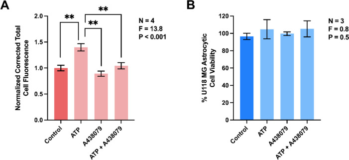
The two main glial cell types of the central nervous system (CNS), astrocytes and microglia, are responsible for neuroimmune homeostasis. Recent evidence indicates astrocytes can participate in removal of pathological structures by becoming phagocytic under conditions of neurodegenerative disease when microglia, the professional phagocytes, are impaired. We hypothesized that adenosine triphosphate (ATP), which acts as damage-associated molecular pattern (DAMP), when released at high concentrations into extracellular space, upregulates phagocytic activity of human astrocytes. This study is the first to measure changes in phagocytic activity and mitochondrial respiration of human astrocytic cells in response to extracellular ATP. We demonstrate that ATP-induced phagocytic activity of U118 MG astrocytic cells is accompanied by upregulated mitochondrial oxidative phosphorylation, which likely supports this energy-dependent process. Application of a selective antagonist A438079 provides evidence identifying astrocytic purinergic P2X7 receptor (P2X7R) as the potential regulator of their phagocytic function. We also report a rapid ATP-induced increase in intracellular calcium ([Ca2+]i), which could serve as regulator of both the phagocytic activity and mitochondrial metabolism, but this hypothesis will need to be tested in future studies. Since ATP upregulates interleukin (IL)-8 secretion by astrocytes but has no effect on their cytotoxicity towards neuronal cells, we conclude that extracellular ATP affects only specific functions of astrocytes. The selectivity of P2X7R-dependent regulation of astrocyte functions by extracellular ATP could allow targeting this receptor-ligand interaction to upregulate their phagocytic function. This could have beneficial outcomes in neurodegenerative disorders, such as Alzheimer's disease, that are characterized by reactive astrocytes and defective phagocytic processes.

Keywords: Astrocytes; Damage-associated molecular pattern (DAMP); Extracellular adenosine triphosphate (ATP); Oxidative phosphorylation; Phagocytosis; Purinergic receptor P2X7.


Conflict of interest statement

Compliance with ethical standards. Ethics approval and consent to participate: All animal experiments were conducted in compliance with the guidelines of the Animal Care Committee at the University of British Columbia, BC, Canada, under protocol A20-0105. Competing interests: The authors declare no competing interests.

Figures

Fig. 1
Fig. 1
Extracellular ATP upregulates the phagocytic activity of human U118 MG astrocytic cells (A) and murine primary astrocytes (B). Cells were exposed to ATP (0.1 mM), LPS (400 ng/ml), their combination, or vehicle solutions (Control) for 48 h, followed by addition of fluorescent latex beads for one h (A) or two h (B). Corrected total cell fluorescence values (means ± SEM) from 163–238 (A) or 152–162 (B) randomly selected cells from three to five (A) or four (B) independent experiments were measured and normalized to control values obtained in the absence of immune mediators. P and F values for the one-way ANOVA are displayed (A); * P < 0.05, ** P < 0.01, according to Tukey’s post-hoc tests (A) or the unpaired Student’s t-test (B). Representative fluorescent images demonstrating the uptake of latex beads (green) by human U118 MG astrocytic cells and murine primary astrocytes (C). U118 MG cell membranes were stained with rhodamine-labeled WGA (red), while murine astrocytes were visualized by sequential application of anti-GFAP antibodies and CY5-lableled (red) secondary antibodies. Nuclei of both cell types were stained with bisbenzimide (blue). The scale bars represent 50 μm
Fig. 2
Fig. 2
Extracellular ATP upregulates the phagocytic activity of human U118 MG astrocytic cells in a P2X7R-dependent manner. A438079 (10 µM) or its vehicle solution was added to human U118 MG astrocytic cells for 30 min before their exposure to ATP (0.1 mM) or its vehicle solution for 48 h, followed by addition of fluorescent latex beads for one h. Corrected total cell fluorescence from 154–206 randomly selected cells from four independent experiments was measured (A) and normalized to control values obtained in the absence of immune mediators. The MTT assay was used to assess cell viability in three independent experiments (B). P and F values for the one-way ANOVA are displayed; ** P < 0.01, according to Tukey’s post-hoc tests
Fig. 3
Fig. 3
Extracellular ATP induces rapid increase in intracellular calcium concentration ([Ca2+]i) in human U118 MG astrocytic cells (A, B) and murine BV-2 microglia (C, D). To detect changes in [Ca2+]i, cells were loaded with Fluo-4 AM and real-time fluorescence intensities were measured for 25 min. A: ATP (1 mM, 265 cells monitored) or its vehicle solution (deionized water, 255 cells monitored) was added 5 min after signaling stabilized. C: ATP (1 mM, 114 cells monitored) or its vehicle solution (deionized water, 110 cells monitored) was added 2 min after the fluorescence signal had stabilized. B, D: The normalized maximum [Ca2+]i responses after the addition of ATP or deionized water were quantified. Data from six to eight independent experiments performed on three different days are presented as means ± SEM. ** P < 0.01, according to the unpaired Student’s t-test
Fig. 4
Fig. 4
Extracellular ATP stimulates mitochondrial respiration in human U118 MG astrocytic cells. Mitochondrial respiration was assessed in cells stimulated for 48 h with ATP (0.1 mM) in the presence or absence of LPS (400 ng/ml). High resolution respirometry was used to measure mitochondrial respiration in the basal state (A; basal respiration), after cell permeabilization (B; leak respiration), and following addition of mitochondrial complex I substrates (2 mM malate and 5 mM pyruvate) in the presence of submaximal (C; 50 µM) and maximal (D; 5 mM) concentrations of ADP. Complex I + II supported respiration (E) was then assessed following addition of succinate (10 mM) prior to titrations with the uncoupler CCCP (0.5 µM steps) to determine maximal electron transfer capacity (F; uncoupled respiration). Data from five to six independent experiments are presented as means ± SEM. P and F values for the one-way ANOVA are displayed. * P < 0.05, ** P < 0.01, according to Tukey’s post-hoc tests
Fig. 5
Fig. 5
Extracellular ATP upregulates IL-8 secretion by human U118 MG astrocytic cells, but has no effect on their secretion of cytotoxins. Human astrocytic cells were treated with ATP (0.1 mM), LPS (400 ng/ml), their combination, or vehicle solutions for 48 h. Subsequently, an ELISA was used to measure the concentration of IL-8 in cell culture supernatants (A) and the MTT assay was used to assess the viability of astrocytic cells (C). Aliquots of U118 MG cell supernatants were also transferred to human SH-SY5Y neuronal cell cultures and their viability was measured by the MTT assay after 72 h incubation (B). Data from eight (A) or five (B, C) independent experiments are presented as means ± SEM. P and F values for the one-way randomized block ANOVA are displayed. * P < 0.05, according to Dunnett’s post-hoc tests. The limit of detection for the ELISA is shown as a dotted line (A)

Similar articles

References

    1. Konishi H, Koizumi S, Kiyama H (2022) Phagocytic astrocytes: Emerging from the shadows of microglia. Glia 70:1009–1026. 10.1002/glia.24145 - PMC - PubMed
    1. Chung WS, Clarke LE, Wang GX et al (2013) Astrocytes mediate synapse elimination through MEGF10 and MERTK pathways. Nature 504:394–400. 10.1038/nature12776 - PMC - PubMed
    1. Davis CHO, Kim KY, Bushong EA et al (2014) Transcellular degradation of axonal mitochondria. Proc Natl Acad Sci U S A 111:9633–9638. 10.1073/pnas.1404651111 - PMC - PubMed
    1. Nguyen JV, Soto I, Kim KY et al (2011) Myelination transition zone astrocytes are constitutively phagocytic and have synuclein dependent reactivity in glaucoma. Proc Natl Acad Sci U S A 108:1176–1181. 10.1073/pnas.1013965108 - PMC - PubMed
    1. Damisah EC, Hill RA, Rai A et al (2020) Astrocytes and microglia play orchestrated roles and respect phagocytic territories during neuronal corpse removal in vivo. Sci Adv 6:3239. 10.1126/sciadv.aba3239 - PMC - PubMed

MeSH terms

LinkOut - more resources