. 2025 Jan 21;82(1):51.
doi: 10.1007/s00018-024-05570-z.

Serotonin receptor 5-HT7 modulates inflammatory-associated functions of macrophages

Affiliations

Serotonin receptor 5-HT7 modulates inflammatory-associated functions of macrophages

Frauke S Bahr et al. Cell Mol Life Sci. .

Abstract

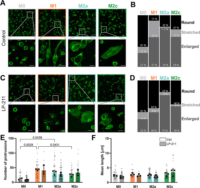
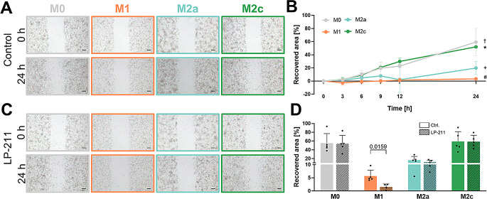
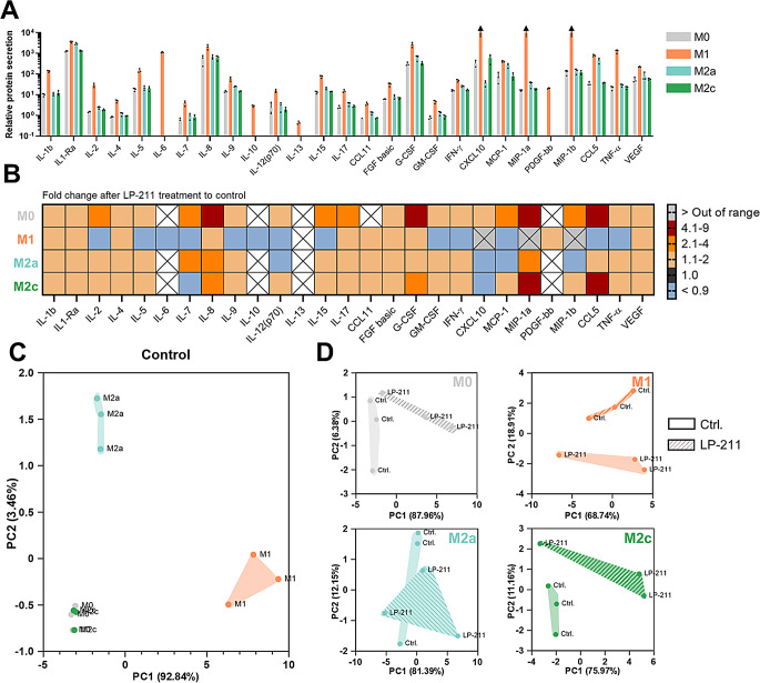
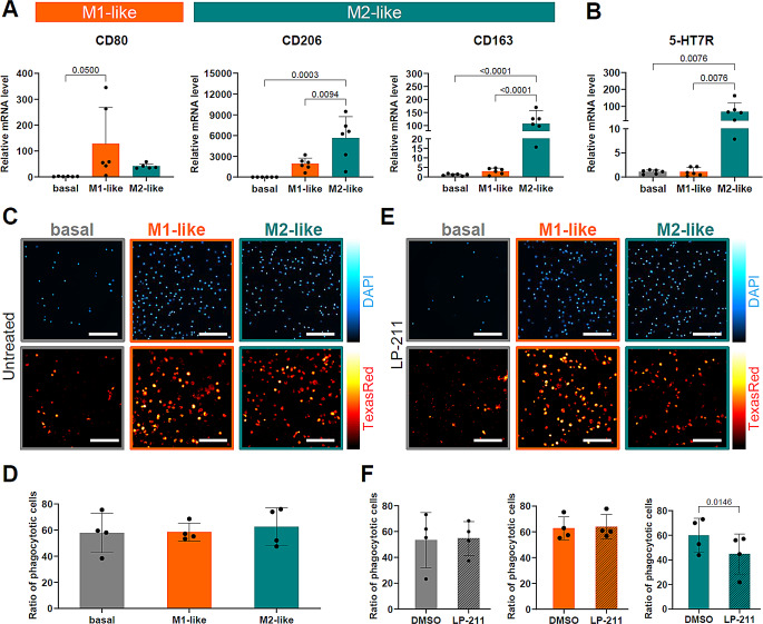
The hormone and neurotransmitter serotonin regulates numerous physiological functions within the central nervous system and in the periphery upon binding to specific receptors. In the periphery, the serotonin receptor 7 (5-HT7R) is expressed on different immune cells including monocytes and macrophages. To investigate the impact of 5-HT7R-mediated signaling on macrophage properties, we used human THP-1 cells and differentiated them into pro-inflammatory M1- and anti-inflammatory M2-like macrophages. Pharmacological 5-HT7R activation with the specific agonist LP-211 especially modulates morphology of M1-like macrophages by increasing the number of rounded cells. Furthermore, 5-HT7R stimulation results in significantly reduced phagocytic and migratory ability of M1-like macrophages. Noteworthy, LP-211 treatment leads to changes in secretory properties of all macrophage types with the highest effects obtained for M0- and M2c-like macrophages. Finally, the importance of 5-HT7R for regulation of phagocytosis was confirmed in human primary CD14+ cells. These results indicate that 5-HT7R activation selectively impairs basic functions of macrophages and might thus be a new access point for the modulation of macrophage responses in the future treatment of inflammatory diseases.

Keywords: 5-HT7 receptor; Macrophages; Phagocytosis; Serotonin; THP-1 cells.


Conflict of interest statement

Declarations. Ethical approval: Not applicable. Consent for publication: The authors hereby consent to publication of the work in Cellular and Molecular Life Sciences. Competing interests: The authors declare that they have no known competing financial interests or personal relationships that could have appeared to influence the work reported in this paper.

Figures

Fig. 1
Fig. 1
Basal characterization of THP-1-derived macrophage subtypes. A: Differentiation scheme of THP-1-monocytes into M0-, M1-, M2a-, and M2c-like macrophages. B: Representative morphology of differentiated macrophages, arrow indicates stretched cells, * highlights enlarged cells. C: mRNA expression levels of macrophage markers. Statistical significance was evaluated using Kruskal-Wallis with Dunn’s multiple comparison test (N = 5 independent differentiations). D: Quantitative analysis of surface marker expression of CD11b, HLA-DR, CD86, and MERTK for each macrophage subtype using flow cytometry. Fluorescence intensity shifts are displayed with normalized counts; u: unstained control. Fold changes of median fluorescence intensities (MFI) relative to M0-like macrophages were analyzed using ordinary one-way ANOVA with Tukey’s multiple comparisons test (CD11b, HLA-DR, CD86) and Kruskal-Wallis with Dunn’s multiple comparison test (MERTK) (N ≥ 3 independent differentiations)
Fig. 2
Fig. 2
THP-1-derived macrophages express the 5-HT7R and downstream signaling molecules. A: mRNA levels of 5-HT7R and Gαs, Gα12, and Cdc42. Statistical difference was evaluated using Kruskal-Wallis with Dunn’s multiple comparison test (N ≥ 5). B: Protein expression levels of 5-HT7R and its downstream effectors shown by western blot. Statistical analysis was performed using ordinary one-way ANOVA with Tukey’s multiple comparisons (5-HT7R, Gα12) and Kruskal-Wallis with Dunn’s multiple comparison (Gαs, Cd42) test (N = 5). C: Representative images visualizing 5-HT7R localization. Antibody specificity was confirmed with a corresponding blocking peptide (right panel; scale bars 20 μm)
Fig. 3
Fig. 3
5-HT7R activation influences macrophage morphology. A: Characterization of macrophages morphology using plasma membrane staining in living cells. Representative images of cell shape on day 5 of differentiation are shown. Scale bars 50 μm in upper panel and 20 μm in magnification. B: Relative quantification of cell shape by counting round, stretched and enlarged phenotypes after THP-1 differentiation. C: Representative images of THP-1-derived macrophage morphology following LP-211 treatment (10 µm; 48 h). Scale bars 50 μm in upper panel and 20 μm in magnification (N = 3). D: Quantification of morphology profiles after treatment with LP-211 (10 µm; 48 h). E: Quantification of protrusion numbers in differentiated macrophages without and after LP-211 (10 µm; 48 h) treatment. Statistical analysis was performed on daily mean values (N = 3) using two-way ANOVA with Tukey’s multiple comparisons test. F: Analysis of mean length of protrusions under basal conditions and after 5-HT7R stimulation with LP-211 (10 µm; 48 h). Statistical analysis was performed on daily mean values (N = 3) using two-way ANOVA with Tukey’s multiple comparisons test
Fig. 4
Fig. 4
Motility of macrophages is influenced by 5-HT7R activity. A: Representative images of scratch area at beginning (0 h) and 24 h after scratch initiation for all macrophage subtypes. Scale bars 200 μm. B: Recovered area of macrophage subtypes relative to scratch area at 0 h. Quantification of migratory ability after 24 h. Data are presented as mean with standard deviation. Adjusted p values: † M0 vs. M1 p = 0.0004, + M0 vs. M2a p = 0.0084, # M1 vs. M2c p = 0.002, and M2a vs. M2c p = 0.0398 using one-way ANOVA with Tukey’s multiple comparisons test (N = 4). C: Images of scratch area at 0 h and 24 h after LP-211 treatment (10 µm). Scale bars 200 μm. D: Quantification of macrophage migration ability after 24 h after LP-211 (10 µm) or control treatment. Statistical significance was evaluated using unpaired Mann-Whitney test between control and LP-211 conditions of each macrophage subtype (N = 5)
Fig. 5
Fig. 5
Phagocytosis rate of M1-like macrophages is reduced upon 5-HT7R activation. A: Representative images of DAPI stained nuclei (upper row) and Texas red-coupled Zymosan A particles (lower row) in control-treated macrophages. Scale bars 200 μm. B: Fraction of phagocytic macrophages showing engulfment of Texas red-coupled Zymosan A particles. Statistical differences are evaluated using ordinary one-way ANOVA with Tukey’s multiple comparison test (N = 8; each point displays the mean of 3 replicate wells). C: Representative images of macrophages treated with LP-211 (10 µm) showing DAPI and Texas red-coupled Zymosan A particle fluorescence after phagocytosis assay. Scale bars 200 μm. D: Quantification of phagocytosis rate of macrophage subtypes with control or LP-211 treatment (10 µm; 48 h). Statistical significance was determined using unpaired two-tailed t-test (N = 8; each point displays the mean of 3 replicate wells)
Fig. 6
Fig. 6
Modulation of secretion ability of macrophages upon 5-HT7R stimulation. A: Relative secretion profile in the supernatant of THP-1-derived macrophages by normalization to protein concentration (N=3). Arrows indicate concentrations above the highest value of the standard. B: Relative fold changes of secreted proteins after LP-211 (10 µm) treatment starting day 3 after seeding compared to control. C: Principal component analysis of three independent differentiations (one dot displays one differentiation) using the first two principal components (N=3). D: Principal component analysis to visualize clustering under basal conditions and after LP-211 treatment for each macrophage subtype. Each principal component was assessed in a separated analysis using Kaiser’s rule (N=3)
Fig. 7
Fig. 7
Impact of 5-HT7R signaling on primary human CD14+ macrophages. A: mRNA expression levels of differentiation markers in CD14+ cells. Statistical significance was evaluated using ordinary one-way ANOVA with Tukey’s multiple comparisons test (N = 6 independent differentiations/donors). B: mRNA expression level of 5-HT7R at day 7 of differentiation. Statistical difference was evaluated using ordinary one-way ANOVA with Tukey’s multiple comparisons test (N = 6). C: Representative images of DAPI stained nuclei (upper row) and Texas red-coupled Zymosan A particles (lower row) in CD14+-derived macrophages after 24 h phagocytosis assay under basal conditions. Scale bars 200 μm. D: Quantification of phagocytic cells showing engulfment of Texas red-coupled Zymosan A particles under basal conditions. Statistical differences were evaluated using ordinary one-way ANOVA with Tukey’s multiple comparison test (N = 4; each point displays the mean of ≥ 4 replicate wells). E: Representative images of macrophages treated with LP-211 (10 µM) showing DAPI and Texas red-coupled Zymosan A particle fluorescence after phagocytosis assay. Scale bars 200 μm. F: Quantification of phagocytosis rate of macrophage subtypes with control (DMSO) or LP-211 treatment (10 µM; 48 h). Statistical significance was determined using paired two-tailed t-test (N = 4; each point displays the mean of ≥ 4 replicate wells)

References

    1. Barnes NM, Ahern GP, Becamel C et al (2021) International Union of Basic and Clinical Pharmacology. CX. Classification of receptors for 5-hydroxytryptamine; pharmacology and function. Pharmacol Rev 73:310–520. 10.1124/pr.118.015552 - PMC - PubMed
    1. Quintero-Villegas A, Valdés-Ferrer SI (2019) Role of 5-HT7 receptors in the immune system in health and disease. Mol Med 26:2. 10.1186/s10020-019-0126-x - PMC - PubMed
    1. Rieder M, Gauchel N, Bode C, Duerschmied D (2021) Serotonin: a platelet hormone modulating cardiovascular disease. J Thromb Thrombolysis 52:42–47. 10.1007/s11239-020-02331-0 - PMC - PubMed
    1. El-Merahbi R, Löffler M, Mayer A, Sumara G (2015) The roles of peripheral serotonin in metabolic homeostasis. FEBS Lett 589:1728–1734. 10.1016/j.febslet.2015.05.054 - PubMed
    1. Sharp T, Barnes NM (2020) Central 5-HT receptors and their function; present and future. Neuropharmacology 177:108155. 10.1016/j.neuropharm.2020.108155 - PubMed

MeSH terms

Substances

LinkOut - more resources