

Three-dimensional sonographic findings of diprosopus: a case report and literature review
- PMID: 39833719
- PMCID: PMC11744991
- DOI: 10.1186/s12884-025-07168-0
Three-dimensional sonographic findings of diprosopus: a case report and literature review
Abstract
Background: Diprosopus is one of the rarest anomalies. It typically manifests as bilateral alterations and often involves anomalies within the cranial structures. In this report, we present a case of a fetus with diprosopus diagnosed prenatally. Along with reviewing relevant literature on prenatal ultrasound diagnosis of diprosopus, we aim to raise awareness of its ultrasound characteristics.
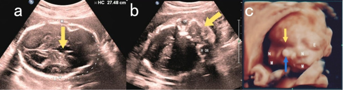
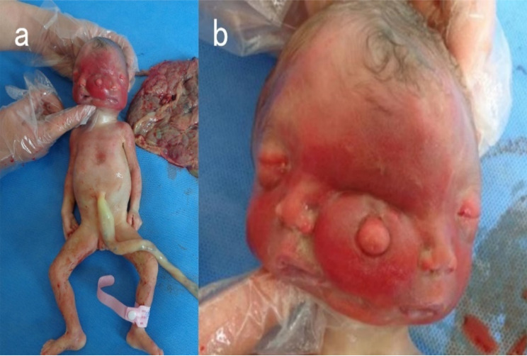
Case presentation: We report a case of craniofacial and intracranial abnormalities detected during a 26-week ultrasound examination. Two-dimensional ultrasound (2D ultrasound) demonstrates significant increases in head circumference, widening of the interocular distance, and abnormal echo patterns in the facial structure. Three-dimensional ultrasound (3D ultrasound) revealed the presence of three eye sockets (the lateral eye sockets contained eyeballs, while the central region exhibited fusion without visible eyeballs), two noses, and two mouths, with no abnormalities observed in other areas. The ultrasound findings suggested diprosopus. Following risk counseling at the prenatal diagnosis center, the pregnant woman decided to induce labor. The newborn passed away thirty minutes after delivery. The facial features of the newborn were consistent with the 3D ultrasound imaging, and the appearance of the trunk and limbs was normal. Both CT and MRI scans confirmed the diagnosis of diprosopus.
Conclusion: The prenatal 2D ultrasound revealed intracranial and facial abnormalities in the fetus. 3D ultrasound imaging clearly displayed the facial duplication anomalies, highlighting the advantages of 3D ultrasound in diagnosing diprosopus. We hope to raise awareness of this rare condition and provide insights into prenatal ultrasound diagnosis through this case.
Keywords: Case report; Diprosopus; Prenatal ultrasound examination; Three-dimensional ultrasound imaging.
© 2025. The Author(s).
Conflict of interest statement
Declarations. Ethics approval and consent to participate: We declare that this research has been reviewed by the Ethics Committee of the Second Affiliated Hospital of Dalian Medical University and adheres to relevant ethical standards and regulations (Approval No. 2023869). Consent for publication: The patient described herein provided written informed consent to publish this case report. A copy of the written consent is available for review by the editor of this journal on request. Competing interests: The authors declare no competing interests.
Figures


Similar articles
-
Diprosopus (partially duplicated head) associated with anencephaly: a case report.Pathol Res Pract. 1999;195(1):45-50; discussion 51-2. doi: 10.1016/s0344-0338(99)80094-6. Pathol Res Pract. 1999. PMID: 10048094
-
Prenatal diagnosis of parapagus diprosopus dibrachius dipus twins with spina bifida in the first trimester using two- and three-dimensional ultrasound.Taiwan J Obstet Gynecol. 2015 Dec;54(6):780-3. doi: 10.1016/j.tjog.2015.10.010. Taiwan J Obstet Gynecol. 2015. PMID: 26701003
-
Antenatal diagnosis of complete facial duplication--a case report of a rare craniofacial defect.Prenat Diagn. 1998 Jun;18(6):618-20. Prenat Diagn. 1998. PMID: 9664609
-
Prenatal ultrasound diagnosis of Tessier number 7 cleft: Case report and review of the literature.J Obstet Gynaecol. 2017 May;37(4):421-427. doi: 10.1080/01443615.2017.1285274. Epub 2017 Mar 13. J Obstet Gynaecol. 2017. PMID: 28287290 Review.
-
Prenatal 2D and 3D ultrasound diagnosis of diprosopus: case report with post-mortem magnetic resonance images (MRI) and review of the literature.Prenat Diagn. 2009 Oct;29(10):992-4. doi: 10.1002/pd.2321. Prenat Diagn. 2009. PMID: 19565608 Review. No abstract available.
References
-
- Bidondo MP, Groisman B, Tardivo A, Tomasoni F, Tejeiro V, Camacho I, Vilas M, Liascovich R, Barbero P. Diprosopus: systematic review and report of two cases. Birth Defects Res Clin Mol Teratol. 2016;106(12):993–1007. - PubMed
-
- al Muti Zaitoun A, Chang J, Booker M. Diprosopus (partially duplicated head) associated with anencephaly: a case report. Pathol Res Pract. 1999;195(1):45–52. - PubMed
-
- Bulbul Y, Drummond CL, Hillion Y, Bidat L, Ville Y. Diprosopus associated with neural tube defect and facial cleft in the first trimester. Fetal Diagn Ther. 2004;19(3):246–50. - PubMed
Publication types
MeSH terms
LinkOut - more resources
Full Text Sources
Medical
