Case Reports
. 2025 Jan 20;13(1):12.
doi: 10.1186/s40478-025-01929-w.

A novel case of glial transdifferentiation in renal medullary carcinoma brain metastasis

Affiliations
Case Reports

A novel case of glial transdifferentiation in renal medullary carcinoma brain metastasis

Maria A Gubbiotti et al. Acta Neuropathol Commun. .

Abstract

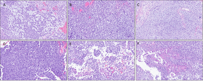
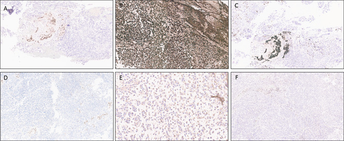
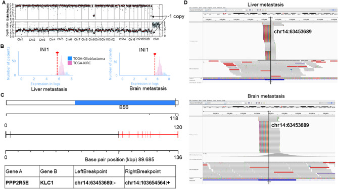
Renal medullary carcinoma is a rare undifferentiated tumor of the kidney associated with sickle cell trait and characterized by INI1 (SMARCB1) loss. Although metastasis to lungs, lymph nodes, and bone is commonly reported, distant spread to the central nervous system almost never occurs. Here we present an unusual case of a patient with renal medullary carcinoma with metastasis to the brain following treatment which included tazemetostat, an EZH2 inhibitor. The metastatic brain lesion harbored morphologic, immunohistochemical, and methylation profile supportive of a primary CNS phenotype with loss of the trimethylated lysine 27 residue of histone 3 while maintaining INI1 loss and a specific gene fusion shared with the patient's tumor prior to initiation of tazemetostat therapy. Therefore, given the common genetic signatures in the brain metastasis and the patient's prior tumor, this case represents a rare event of glial transdifferentiation in a brain metastasis of renal medullary carcinoma following the use of an epigenetic modulator. As renal medullary carcinoma has been known to cleverly utilize adaptive mechanisms for survival, we propose that such cell plasticity seen in this case may have been provoked by the use of a drug that alters the epigenetic signature of the tumor cells. Thus, careful assessment of tumor biology following novel therapeutic treatment options must be performed in order to note such unexpected consequences of treatment.

Keywords: EZH2; Glial transdifferentiation; H3K27me3; INI1; Renal medullary carcinoma; SMARCB1; Tazemetostat.


Conflict of interest statement

Declarations. Ethics approval and consent to participate: The study was conducted in accordance with the Declaration of Helsinki and was approved by the Institutional Review Board of The University of Texas MD Anderson Cancer Center (protocols #2024 − 1035 and PA11-1045). Consent for publication: A waiver of consent was granted by the institutional review board which is available upon request. Competing interests: PM has received honoraria for service on a Scientific Advisory Board for Mirati Therapeutics, Bristol Myers Squibb, and Exelixis; consulting for Axiom Healthcare Strategies; non-branded educational programs supported by DAVA Oncology, Exelixis and Pfizer; and research funding for clinical trials from Regeneron Pharmaceuticals, Takeda, Bristol Myers Squibb, Mirati Therapeutics, Gateway for Cancer Research, and the University of Texas MD Anderson Cancer Center.

Figures

Fig. 1
Fig. 1
Histomorphologic evaluation of the CT-guided liver biopsy of the metastatic renal medullary carcinoma. A, H&E stained section showing atypical epithelioid cells with abundant eosinophilic cytoplasm arranged in glands and embedded in a desmoplastic stroma. B, PAX8 shows strong, diffuse nuclear positivity within the neoplastic cells. C, INI1 shows high background staining, but demonstrates loss of nuclear expression, similar to the external control, within the tumor cells. D, A recent kidney biopsy performed confirms the absence of nuclear INI1 expression in the primary tumor cells with appropriate internal positive control E, H3K27me3 stain highlighting intact nuclear expression in both neoplastic cells and background stromal cells
Fig. 2
Fig. 2
Morphologic appearance of the cerebellar lesion following H&E staining. The undifferentiated tumor shows variable histologic features including A, adenoid structures, B, large atypical cells with prominent nucleoli and perinuclear clearing, C, loosely arranged cells in a myxoid matrix, D, densely cellular areas with high mitotic activity, E, focal clusters of epithelioid cells with abundant eosinophilic cytoplasm, and F, rare cells with abundant eosinophilic cytoplasm imparting a vaguely rhabdoid appearance
Fig. 3
Fig. 3
Immunohistochemical studies performed on the cerebellar lesion. A, PAX8 is largely negative within the undifferentiated tumor cells but shows nuclear positivity in the regions with epithelioid morphology. B, GFAP shows diffuse positivity within the tumor. C, Cam5.2 highlights the epithelioid structures. D, INI1 expression is lost within the tumor cells with internal positive control showing retained expression in background non-neoplastic tissue. E, H3K27M stain is negative. F, H3K27me3 stain shows loss of nuclear expression within the tumor cells with internal positive control demonstrating intact expression within the non-neoplastic cells
Fig. 4
Fig. 4
Molecular sequencing of the RMC liver and brain metastases. A, Detailed copy number alteration overview of the brain metastasis sample showed partial loss of one copy of chromosome 22 including the SMARCB1 gene encoding INI1. B, RNA expression of INI1 in each of the RMC liver and brain metastasis samples (denoted by the dashed red line in each panel) compared with the distribution of INI1 RNA expression in glioblastoma and clear cell renal cell carcinoma (KIRC) samples from the Cancer Genome Atlas (TCGA). C, PPP2R5E::KLC1 fusion identified in both liver and brain metastasis. Annotation was performed using the AGFusion package. D, Integrative genome viewer (IGV) showing that the PPP2R5E::KLC1 fusion fully corresponds with nucleotide precision in both the liver and brain metastasis samples
Fig. 5
Fig. 5
Principal component analysis (PCA) plots of RNA-seq expression in the liver and brain metastasis samples compared with gene expression from the Cancer Genome Atlas (TCGA) of clear cell renal cell carcinoma (KIRC), papillary renal cell carcinoma (KIRP), sarcoma, astrocytoma, glioblastoma and oligodendroglioma samples

References

    1. Bhat KPL, Balasubramaniyan V, Vaillant B, Ezhilarasan R, Hummelink K, Hollingsworth F, Wani K, Heathcock L, James JD, Goodman LD al (2013) Mesenchymal differentiation mediated by NF-kappaB promotes radiation resistance in glioblastoma. Cancer Cell 24:331–346. 10.1016/j.ccr.2013.08.001 - PMC - PubMed
    1. Capper D, Jones DTW, Sill M, Hovestadt V, Schrimpf D, Sturm D, Koelsche C, Sahm F, Chavez L, Reuss DE et al (2018) DNA methylation-based classification of central nervous system tumours. Nature 555: 469–474 10.1038/nature26000 - PMC - PubMed
    1. Cheng JX, Tretiakova M, Gong C, Mandal S, Krausz T, Taxy JB (2008) Renal medullary carcinoma: rhabdoid features and the absence of INI1 expression as markers of aggressive behavior. Mod Pathol 21:647–652. 10.1038/modpathol.2008.44 - PubMed
    1. Chi SN, Yi JS, Williams PM, Roy-Chowdhuri S, Patton DR, Coffey BD, Reid JM, Piao J, Saguilig L, Alonzo TA al (2023) Tazemetostat for tumors harboring SMARCB1/SMARCA4 or EZH2 alterations: results from NCI-COG pediatric MATCH APEC1621C. J Natl Cancer Inst 115:1355–1363. 10.1093/jnci/djad085 - PMC - PubMed
    1. Conway E, Healy E, Bracken AP (2015) PRC2 mediated H3K27 methylations in cellular identity and cancer. Curr Opin Cell Biol 37:42–48. 10.1016/j.ceb.2015.10.003 - PubMed

Publication types

MeSH terms

Substances