. 2025 Jan 20;13(1):e4744.
doi: 10.1002/fsn3.4744. eCollection 2025 Jan.

Ginsenoside RG3 Synergizes With STING Agonist to Reverse Cisplatin Resistance in Gastric Cancer

Affiliations

Ginsenoside RG3 Synergizes With STING Agonist to Reverse Cisplatin Resistance in Gastric Cancer

Zhongqi Lu et al. Food Sci Nutr. .

Abstract

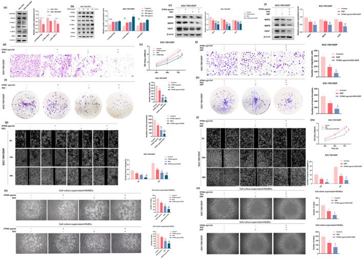
This study investigates the synergistic inhibitory effects of combining the stimulator of interferon genes (STING) agonist cyclic diadenylate monophosphate (c-di-AMP) and ginsenoside RG3 on cisplatin (DDP)-resistant gastric cancer (GC) cells. The objective is to identify novel therapeutic targets and offers insights for the clinical management of DDP resistance. Various techniques were employed, including western blot, MTT assay, colony formation assay, scratch assay, transwell assay, tubule formation assay, flow cytometry, Hoechst 33342 fluorescence staining, and in vivo experiments, to investigate the potential mechanisms and effects of the combined application of the STING agonist and ginsenoside RG3 in reversing cisplatin resistance in gastric cancer. The combination markedly suppressed key malignant behaviors, including proliferation, migration, invasion, and angiogenesis of SGC-7901/DDP cells. Additionally, this treatment inhibited the epithelial-mesenchymal transition (EMT) process and stem cell-like characteristics of SGC-7901/DDP cells, while downregulating the expression of resistance-related proteins. The STING agonist effectively suppresses the growth and proliferation of gastric cancer cells. Ginsenoside RG3, well-documented for its multifaceted properties, including antioxidant, anti-aging, and anti-cancer effects, is widely used in cancer treatment and in managing chemotherapy-related side effects. Furthermore, RG3 enhances anti-tumor immunity by regulating signal transduction. This study comprehensively evaluated the efficacy of the STING agonist and RG3 combination through in vitro and in vivo experiments, demonstrating significant inhibition of malignant progression and reversal of drug resistance in gastric cancer. These findings offer a robust theoretical foundation for clinical applications and highlight new therapeutic targets for future research.

Keywords: STING agonist; cisplatin resistance; gastric cancer; ginsenoside RG3; immunotherapy.


Conflict of interest statement

The authors declare no conflicts of interest.

Figures

FIGURE 1
FIGURE 1
STING agonist and RG3 inhibit proliferation, migration, invasion, and tube formation in SGC‐7901/DDP cells and enhance sensitivity to Cisplatin. (a) Differential expression of cGAS‐STING signaling pathway‐related proteins in SGC‐7901 and SGC‐7901/DDP cells. (b) Gastric cancer cells SGC‐7901/DDP were treated with different concentrations (0, 100, 200, and 300 μg/mL) of c‐di‐AMP for 48 h, followed by Western blot analysis to assess the expression of STING, p‐STING, TBK1, P‐TBK1, IRF3, and P‐IRF3. (c) Western blot analysis evaluated the impact of STING agonist and RG3 treatment on the expression of VEGF, MMP2, and MMP9 proteins in SGC‐7901/DDP cells. (d) Transwell assays were conducted to observe the effect of STING agonist and RG3 on the vertical migration of SGC‐7901/DDP cells. (e) MTT assay was performed to assess the influence of STING agonist and RG3 treatment on the proliferation capacity of SGC‐7901/DDP cells. (f) Impact of STING agonist and RG3 on the colony‐forming ability of SGC‐7901/DDP cells, evaluated through colony formation assays. (g) Scratch assays were employed to assess the effect of STING agonist and RG3 treatment on the wound healing ability of SGC‐7901/DDP cells. (h) Evaluation of the impact of STING agonist and RG3 on the tubulogenesis ability of gastric cancer cells. Endothelial cell tube formation assays detected microvessel formation in HUVECs (human umbilical vein endothelial cells) and HLECs (human lymphatic endothelial cells) treated with conditioned medium. (i) Western blot analysis evaluated the impact of DDP and STING agonist + RG3 + DDP treatment on the expression of VEGF, MMP2, and MMP9 proteins in SGC‐7901/DDP cells. (j) Transwell assays were conducted to observe the effect of DDP and STING agonist + RG3 + DDP on the vertical migration of SGC‐7901/DDP cells. (k) Impact of DDP and STING agonist + RG3 + DDP on the colony‐forming ability of SGC‐7901/DDP cells, evaluated through colony formation assays. (l) Scratch assays were employed to assess the effect of DDP and STING agonist + RG3 + DDP treatment on the wound healing ability of SGC‐7901/DDP cells. (m) MTT assay was performed to assess the influence of DDP and STING agonist+RG3 + DDP treatment on the proliferation capacity of SGC‐7901/DDP cells. (n) Evaluation of the impact of DDP and STING agonist + RG3 + DDP on the tubulogenesis ability of gastric cancer cells. Endothelial cell tube formation assays detected microvessel formation in HUVECs and HLECs treated with conditioned medium. (* denotes comparison with the control group. # represents comparison with the STING agonist group or DDP group. & denotes comparison with the RG3 group. *, #, & indicate statistical significance (p < 0.05). **, ##, && indicate statistical significance at p < 0.01. ***, ###, &&& indicate statistical significance at p < 0.001. ****, #### indicate statistical significance at p < 0.0001).
FIGURE 2
FIGURE 2
Impact of STING agonist and RG3 on the apoptotic function of gastric cancer cells. (a and b) Western blot analysis assessing the influence of STING agonist and RG3 on the expression of apoptotic‐related proteins in gastric cancer cells. (c, e) Flow cytometry used to evaluate the induction of apoptosis in SGC‐7901/DDP cells following treatment with STING agonist and RG3. (d, f) Hoechst 33342 staining depicting the morphological changes induced by STING agonist and RG3 treatment in apoptotic SGC‐7901/DDP cells. (g, h) Western blot analysis assessing the influence of DDP and STING agonist+RG3 + DDP on the expression of apoptotic‐related proteins in gastric cancer cells. (i, j) Flow cytometry used to evaluate the induction of apoptosis in SGC‐7901/DDP cells following treatment with DDP and STING agonist+RG3 + DDP. (k, l) Hoechst 33342 staining depicting the morphological changes induced by DDP and STING agonist + RG3 + DDP treatment in apoptotic SGC‐7901/DDP cells. (* denotes comparison with the control group. # represents comparison with the STING agonist group or DDP group. & denotes comparison with the RG3 group. *, #, & indicate statistical significance at p < 0.05. **, ##, && indicate statistical significance at p < 0.01. ***, ###, &&& indicate statistical significance at p < 0.001. ****, #### indicate statistical significance at p < 0.0001).
FIGURE 3
FIGURE 3
Impact of STING agonist and RG3 on the Epithelial‐Mesenchymal Transition (EMT) process in gastric cancer. (a and b) Western blot analysis assessing the expression of EMT‐related proteins in SGC‐7901/DDP cells following treatment with STING agonist and RG3. (c and d) Immunofluorescence evaluation of the fluorescence signal intensity of E‐cadherin and Vimentin in SGC‐7901/DDP cells after treatment with STING agonist and RG3. (* denotes comparison with the control group. # represents comparison with the STING agonist group. & denotes comparison with the RG3 group. *, #, & indicate statistical significance at p < 0.05. **, ##, && indicate statistical significance at p < 0.01. ***, ###, &&& indicate statistical significance at p < 0.001. ****, #### indicate statistical significance at p < 0.0001).
FIGURE 4
FIGURE 4
Inhibitory effect of the combined use of STING agonist and RG3 on the stemness of gastric cancer cells. (a and b) Western blot analysis of the expression of stemness‐related proteins in SGC‐7901/DDP cells after treatment with STING agonist and RG3. (c and d) Immunofluorescence examination of the fluorescence intensity changes of the stemness marker CD44 in SGC‐7901/DDP cells following treatment with STING agonist and RG3. (* denotes comparison with the control group. # represents comparison with the STING agonist group. & denotes comparison with the RG3 group. *, #, & indicate statistical significance at p < 0.05. **, ##, && indicate statistical significance at p < 0.01. ***, ###, &&& indicate statistical significance at p < 0.001. ****, #### indicate statistical significance at p < 0.0001).
FIGURE 5
FIGURE 5
Reversal of cisplatin resistance in gastric cancer cells by the combined use of STING agonist and RG3. (a and b) Western blot analysis showing the expression levels of resistance proteins MDR1 and MRP1. (c and d) Immunofluorescence detection of the fluorescence intensity of resistance proteins MDR1 and MRP1. (* denotes comparison with the control group. # represents comparison with the STING agonist group. & denotes comparison with the RG3 group. *, #, & indicate statistical significance at p < 0.05. **, ##, && indicate statistical significance at p < 0.01. ***, ###, &&& indicate statistical significance at p < 0.001. ****, #### indicate statistical significance at p < 0.0001).
FIGURE 6
FIGURE 6
In vivo experiments demonstrating the reversal of cisplatin resistance and inhibition of tumor growth by STING agonist and RG3. (a) Photographic images of tumors in the gastric cancer xenograft model in mice. (b, d) Measurement of tumor weight and volume after subcutaneous injection. (c) Monitoring of mouse body weight after subcutaneous injection. (e) Ultrasound images of mouse tumors after administration. (f) Hematoxylin and eosin (HE) staining of liver, kidney, and spleen tissues after administration. (g–l) Immunohistochemical detection of the expression of Ki67, CD44, Vimentin, E‐cadherin, MDR1, and MRP1 in tumor tissues after subcutaneous injection. (* denotes comparison with the control group. # represents comparison with the STING agonist group. & denotes comparison with the RG3 group. *, #, & indicate statistical significance at p < 0.05. **, ##, && indicate statistical significance at p < 0.01. ***, ###, &&& indicate statistical significance at p < 0.001. ****, #### indicate statistical significance at p < 0.0001).

References

    1. Amouzegar, A. , Chelvanambi M., Filderman J. N., et al. 2021. “STING Agonists as Cancer Therapeutics.” Cancers (Basel) 13, no. 11: 2695. - PMC - PubMed
    1. Arneth, B. 2019. “Tumor Microenvironment.” Medicina (Kaunas, Lithuania) 56, no. 1: 56. - PMC - PubMed
    1. Bokhari, S. M. Z. , and Hamar P.. 2023. “Vascular Endothelial Growth Factor‐D (VEGF‐D): An Angiogenesis Bypass in Malignant Tumors.” International Journal of Molecular Sciences 24, no. 17: 13317. - PMC - PubMed
    1. Bose, D. 2017. “cGAS/STING Pathway in Cancer: Jekyll and Hyde Story of Cancer Immune Response.” International Journal of Molecular Sciences 18, no. 11: 2456. - PMC - PubMed
    1. Bustos, M. A. , Yokoe T., Shoji Y., et al. 2023. “MiR‐181a Targets STING to Drive PARP Inhibitor Resistance in BRCA‐ Mutated Triple‐Negative Breast Cancer and Ovarian Cancer.” Cell & Bioscience 13, no. 1: 200. - PMC - PubMed

LinkOut - more resources