. 2025 Jan 6:18:1525024.
doi: 10.3389/fncel.2024.1525024. eCollection 2024.

Nerve repair with polylactic acid and inosine treatment enhance regeneration and improve functional recovery after sciatic nerve transection

Affiliations

Nerve repair with polylactic acid and inosine treatment enhance regeneration and improve functional recovery after sciatic nerve transection

Fellipe Soares Dos Santos Cardoso et al. Front Cell Neurosci. .

Abstract

Background: Following transection, nerve repair using the polylactic acid (PLA) conduit is an effective option. In addition, inosine treatment has shown potential to promote nerve regeneration. Therefore, this study aimed to investigate the regenerative potential of inosine after nerve transection and polylactic acid conduit repair.

Methods: C57/Black6 mice were subjected to sciatic nerve transection, repair with PLA conduit, and intraperitoneal injection of saline or inosine 1 h after injury and daily for 1 week. To assess motor and sensory recovery, functional tests were performed before and weekly up to 8 weeks after injury. Following, to evaluate the promotion of regeneration and myelination, electroneuromyography, morphometric analysis and immunohistochemistry were then performed.

Results: Our results showed that the inosine group had a greater number of myelinated nerve fibers (1,293 ± 85.49 vs. 817 ± 89.2), an increase in neurofilament high chain (NFH) and myelin basic protein (MBP) immunolabeling and a greater number of fibers within the ideal g-ratio (453.8 ± 45.24 vs. 336.6 ± 37.01). In addition, the inosine group presented a greater adenosine A2 receptor (A2AR) immunolabeling area. This resulted in greater compound muscle action potential amplitude and nerve conduction velocity, leading to preservation of muscle and neuromuscular junction integrity, and consequently, the recovery of motor and sensory function.

Conclusion: Our findings suggest that inosine may enhance regeneration and improve both motor and sensory function recovery after nerve transection when repaired with a poly-lactic acid conduit. This advances the understanding of biomaterials and molecular treatments.

Keywords: A2A receptor; inosine; nerve regeneration; nerve repair; sciatic nerve transection.


Conflict of interest statement

The authors declare that the research was conducted in the absence of any commercial or financial relationships that could be construed as a potential conflict of interest.

Figures
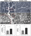
Figure 1
Figure 1
Semithin and ultrathin sections and quantitative analysis. Semithin section stained with Toluidine blue of saline + PLA group (A) and from inosine + PLA group (C), and ultrathin section of saline + PLA (B) and inosine + PLA (D). Note that saline + PLA has regenerative islets (red dot circles), different from inosine + PLA, which are surrounded by concentric perineurium layers (white arrow). In addition, the following graphics demonstrate the total number of myelinated fibers (E) and total number of blood vessels (F). In both groups, blue asterisks represent Schwann cells. Scale bar = 10 μm (A,C) and 2 μm (B,D); saline + PLA (n = 5) and inosine + PLA (n = 5); Values represent mean ± SEM (**p < 0.01).
Figure 2
Figure 2
Morphometric analysis of myelinated nerve fibers in semithin sections. Quantitative analysis of axon area (A), myelin area (B), fiber area (C), and g-ratio (D) from saline + PLA and inosine + PLA groups. Saline + PLA (n = 5) and inosine + PLA (n = 5); Values represent mean ± SEM (*p < 0.05).
Figure 3
Figure 3
Relative immunolabeled area for NFH. Cross-section of sciatic nerve labeled for NFH (green), DAPI (blue), and merge of saline + PLA (A–C) and inosine + PLA (D–F), and the graphic of relative immunolabeled area (G). Scale bar = 25 μm; Saline + PLA (n = 3) and inosine + PLA (n = 3); Values represent mean ± SEM (**p < 0.01).
Figure 4
Figure 4
Relative immunolabeled area for A2A and MBP. Cross-section of sciatic nerve labeled for A2A, MBP, and merge, from saline + PLA (A,D,G) and inosine + PLA animals (B,E,H), and graphics for the immunolabeled area of A2A (red) (C) and MBP (green) (F). In addition, the quantification of the colocalization between A2AR and MBP (G,H, white arrows), are demonstrated by Pearson’s coefficient in graph (I). Scale bar = 25 μm; Saline + PLA (n = 3) and inosne + PLA (n = 3); Values represent mean ± SEM (*p < 0.05 and **p < 0.01).
Figure 5
Figure 5
Muscle weight and NMJ assessment. Dissected gastrocnemius muscle from contralateral and saline + PLA (A) and contralateral and inosine + PLA (B). The quantification of dry weight is demonstrated in graph (E). The αbungarotoxin (red) labeling for saline + PLA (C) and inosine + PLA (D). The graphic demonstrates the quantification of NMJ (F). Moreover, note the presence of “pretzel” shape NMJ (white arrows), in both groups. Scale bar = 25 μm; Saline + PLA (n = 5) and inosine + PLA (n = 5); Values represent mean ± SEM (*p < 0.05 and **p < 0.01).
Figure 6
Figure 6
Functional analysis for motor and sensitive functions. To assess the motor function recovery, we performed the SFI. Footprints before (week 0) and after the injury (1 and 8 weeks) (A). Note that inosine + PLA presented a better recovery in the footprints, reflecting in better SFI, as observed in graph (B). To evaluate the recovery of sensitive functions, we performed analgesimeter (C) and pinprick (D). Note that inosine + PLA group presented a complete recovery after 8 weeks, compared to saline + PLA. Saline + PLA (n = 5) and inosine + PLA (n = 5); Values represent mean ± SEM (*p < 0.05, **p < 0.01, and ***p < 0.001).
Figure 7
Figure 7
Electroneuromyography evaluation of amplitude and latency of CMAP and NCV. Traces of normal (A), saline + PLA (B), and inosine + PLA (C), 8 weeks after nerve repair and treatment. To analyze the regeneration, we assessed the amplitude of CMAP (D) and to analyze the myelination, we assessed the latency of CMAP (E) and NCV (F). Normal (n = 3); saline + PLA (n = 5) and inosine + PLA (n = 5); Values represent mean ± SEM (** and ## p < 0.01; *** and ### p < 0.001. # represents normal vs saline; * represents inosine vs saline).

References

    1. Allodi I., Udina E., Navarro X. (2012). Specificity of peripheral nerve regeneration: interactions at the axon level. Prog. Neurobiol. 98, 16–37. doi: 10.1016/j.pneurobio.2012.05.005, PMID: - DOI - PubMed
    1. Arthur-Farraj P. J., Latouche M., Wilton D. K., Quintes S., Chabrol E., Banerjee A., et al. . (2012). C-Jun reprograms Schwann cells of injured nerves to generate a repair cell essential for regeneration. Neuron 75, 633–647. doi: 10.1016/j.neuron.2012.06.021, PMID: - DOI - PMC - PubMed
    1. Benowitz L. I., Goldberg D. E., Madsen J. R., Soni D., Irwin N. (1999). Inosine stimulates extensive axon collateral growth in the rat corticospinal tract after injury. Proc. Natl. Acad. Sci. USA. 96, 13486–13490. doi: 10.1073/pnas.96.23.13486, PMID: - DOI - PMC - PubMed
    1. Boerboom A., Dion V., Chariot A., Franzen R. (2017). Molecular mechanisms involved in Schwann cell plasticity. Front. Mol. Neurosci. 10:38. doi: 10.3389/fnmol.2017.00038, PMID: - DOI - PMC - PubMed
    1. Borea P. A., Gessi S., Merighi S., Vincenzi F., Varani K. (2018). Pharmacology of adenosine receptors: the state of the art. Physiol. Rev. 98, 1591–1625. doi: 10.1152/physrev.00049.2017, PMID: - DOI - PubMed

LinkOut - more resources