. 2025 Apr;64(4):756-768.
doi: 10.1002/mc.23885. Epub 2025 Jan 21.

LINC01305 and LAD1 Co-Regulate CTTN and N-WASP Phosphorylation, Mediating Cytoskeletal Reorganization to Promote ESCC Metastasis

Affiliations

LINC01305 and LAD1 Co-Regulate CTTN and N-WASP Phosphorylation, Mediating Cytoskeletal Reorganization to Promote ESCC Metastasis

Hang Yang et al. Mol Carcinog. 2025 Apr.

Abstract

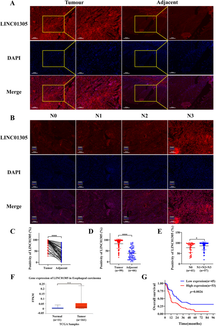
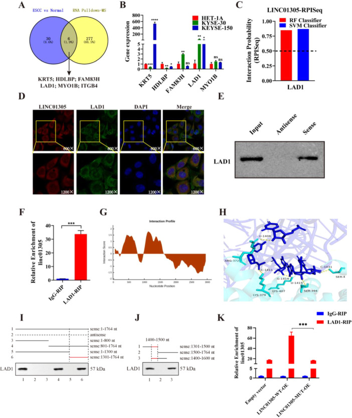
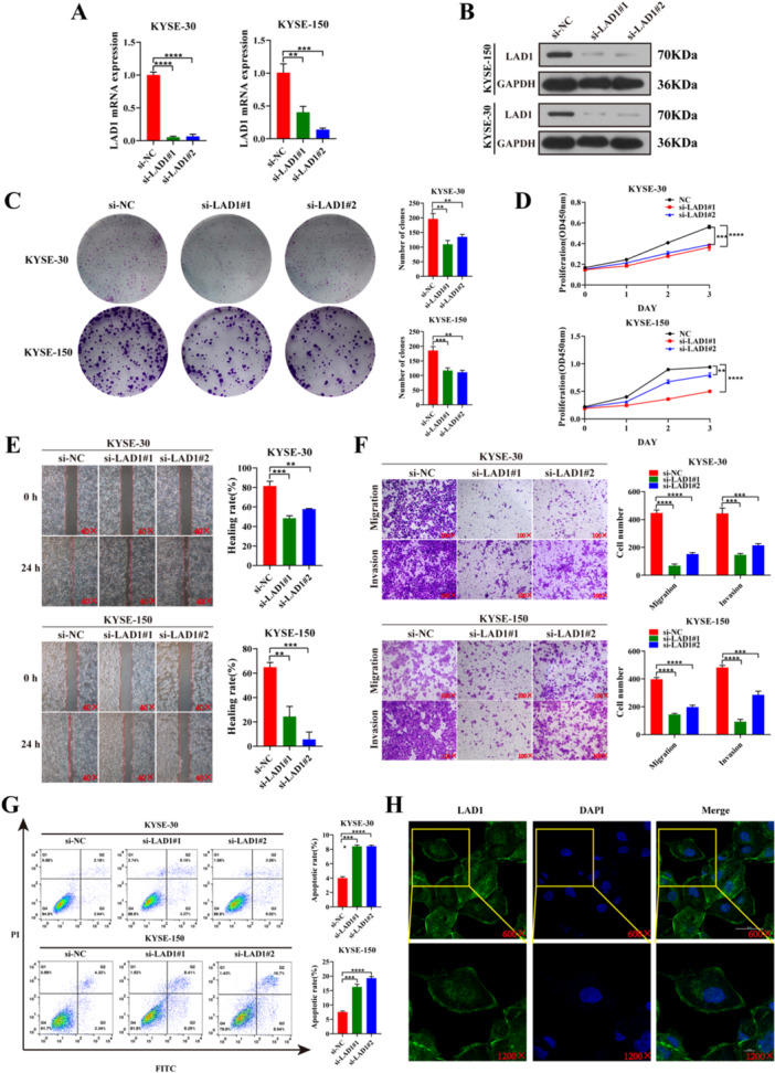
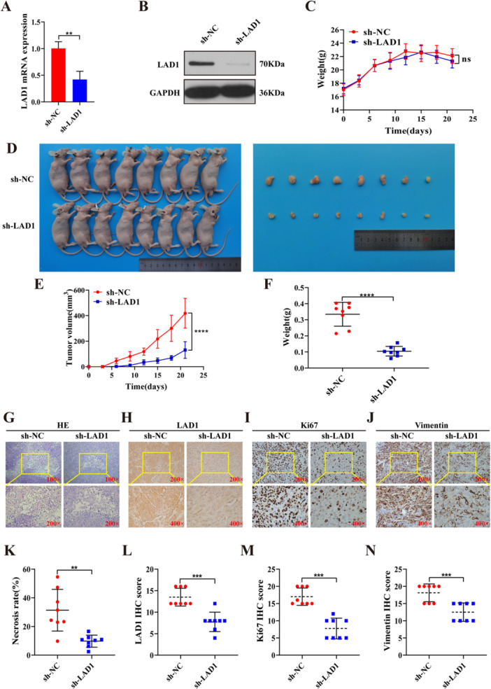
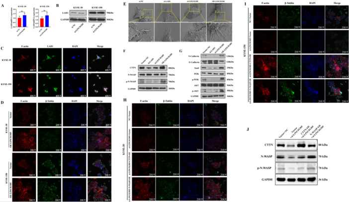
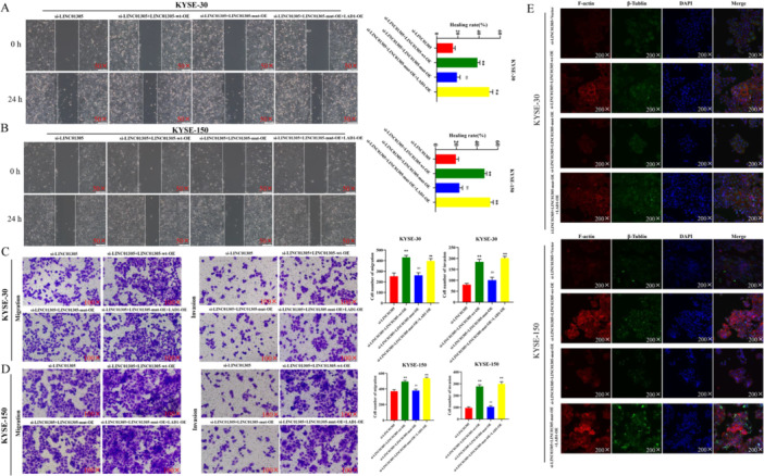
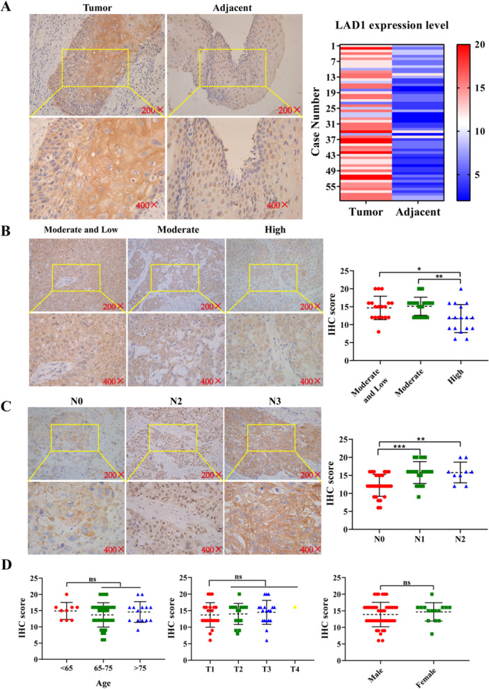
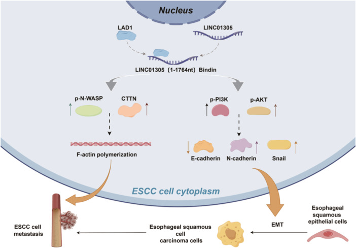
Esophageal squamous cell carcinoma (ESCC) is prone to metastasis and is a leading cause of mortality. The cytoskeleton is closely related to cell morphology and movement; however, little research has been conducted on ESCC metastasis. In this study, we found that the anchoring filament protein ladinin 1 (LAD1) specifically binds to LINC01305 for co-regulating the level of modulating cortactin proteins (CTTN) and neuronal Wiskott-Aldrich syndrome protein (N-WASP) phosphorylation, which mediates cytoskeletal reorganization and affects the metastasis of ESCC cells. Additionally, LINC01305 and LAD1 jointly promoted the epithelial-mesenchymal transition (EMT) process by activating the phosphoinositide-3-kinase-protein kinase B (PI3K/AKT) signaling pathway. Moreover, LINC01305 and LAD1 were related to the late clinical stage and lymph node metastasis of ESCC. Our study demonstrated that LINC01305 and LAD1 are major determinants of ESCC dissemination and revealed a novel molecular mechanism of cytoskeletal reorganization that controls ESCC metastasis. Trial Registration: N/A.

Keywords: ESCC; LAD1; LINC01305; cytoskeleton; metastasis.


Conflict of interest statement

The authors declare no conflicts of interest.

Figures

Figure 1
Figure 1
LINC01305 is highly expressed in ESCC tissues and cells and exerts pro‐cancer effects. (A) Representative Immunofluorescence maps of ESCC tissue and adjacent normal esophageal epithelial tissue. (B) Representative images of LINC01305 expression in ESCC tissues with different N stages. (C) Statistical plot of the results of the paired t‐test for 65 cases of ESCC tissue and corresponding adjacent normal tissue. (D) Statistical plot of unpaired t‐test for 99 cases of cancerous tissue and 66 cases of adjacent normal tissue. (E) Statistical plot of LINC01305 expression in relation to N‐staging of ESCC. (F) Differential expression of LINC01305 in esophageal cancer and normal esophageal tissues predicted by UALCAN online site. (G) Correlation of LINC01305 expression with poor prognosis of ESCC patients. *p < 0.05, **p < 0.01, ***p < 0.001, ****p < 0.0001.
Figure 2
Figure 2
LINC01305 specifically binds LAD1 at 1400–1500 nt. (A) Venn diagram of LINC01305 RNA pull‐down‐MS results with sequencing results of ESCC tissues. (B) RT‐PCR was performed to detect the differential expression of six candidate genes. (C) RIPSeq website online predicted the binding of LAD1 to LINC01305, and the RF and SVM scores were both greater than 0.5, suggesting the possibility of binding. (D) Cellular immunofluorescence probed the subcellular co‐localization of LINC01305 with LAD1. (E, F) LINC01305 RNA pull‐down assay and RIP assay verified the specific binding to LAD1. (G) CATRAPID online website predicted the binding site of LINC01305 with LAD1. (H) AlphaFold3 predicted the interaction between proteins and RNA. (I, J) The interaction between LINC01305 RNA and LAD1 protein was verified by RNA pull‐down and western blot in KYSE30 cells. (K) RIP‐qPCR illustrated that LINC01305 binds specifically to LAD1. *p < 0.05, **p < 0.01, ***p < 0.001, ****p < 0.0001.
Figure 3
Figure 3
LAD1 promotes malignant progression of ESCC cells in vitro. (A, B) RT‐PCR assay and WB assay to verify the efficiency of siRNA sequence knockdown. (C, D) CCK‐8 assay and colony formation assay to probe the effect of knockdown of LAD1 on the proliferation of ESCC cells in vitro. (E) Scratch assay to detect the effect of inhibition of LAD1 on the migratory ability of ESCC cells. (F) Transwell assay to probe the effect of knockdown of LAD1 on ESCC cell migration and invasion. (G) Flow cytometry to detect the effect of LAD1 on ESCC cell apoptosis. (H) Cellular immunofluorescence localization of LAD1. *p < 0.05, **p < 0.01, ***p < 0.001, ****p < 0.0001.
Figure 4
Figure 4
LAD1 promotes malignant progression of ESCC cells in vivo. (A, B) RT‐PCR assay and WB assay to detect the silencing efficiency of lentivirus. (C) Dynamic monitoring of the weight of nude mice in the sh‐NC group and the sh‐LAD1 group. (D) The tumor formation of nude mice in sh‐NC and sh‐LAD1 groups after subcutaneous injection of ESCC cells. (E) Changes in the volume of tumors of nude mice of the two groups at 21 days. (F) Two groups of difference in tumor volume weight of nude mice. (G) HE staining results of subcutaneous tumor sections of nude mice in the two groups, with the enlarged portion being necrotic areas. (H–J) Immunohistochemically staining detected the expression of LAD1, Ki‐67, and vimentin in the tumor of the two groups. (K) Statistics of tumor necrosis area in sh‐NC and sh‐LAD1 groups. (L–N) The Staining scores of LAD1, Ki‐67, and vimentin. *p < 0.05, **p < 0.01, ***p < 0.001, ****p < 0.0001.
Figure 5
Figure 5
LINC01305 co‐regulates ESCC cytoskeletal reorganization through activating CTTN and N‐WASP phosphorylation, cooperating to enhance ESCC metastasis. (A, B) The mRNA and protein expression levels of LAD1 after knockdown of LINC01305. (C) Co‐localization of LAD1 with F‐actin cytoskeleton fractions. (D) Effects of LINC01305 binding to LAD1 on the reorganization of the F‐actin cytoskeleton. (E) Scanning electron microscopic observation of the effects of LINC01305 and LAD1 on pseudopod growth in ESCC cells. (F) WB assay to detect the regulatory effect of LINC01305 with LAD1 on CTTN protein level and N‐WASP phosphorylation. (G) Effect of LINC01305 with LAD1 on EMT process in ESCC cells through PI3K/AKT signaling pathway. (H) Effects of CTTN and N‐WASP cooperate on the reorganization of the F‐actin cytoskeleton in KYSE30 cells. (I) Effects of CTTN and N‐WASP cooperate on the reorganization of the F‐actin cytoskeleton in KYSE150 cells. (J) WB assay to detect the regulatory effect of CTTN and N‐WASP overexpression in the context of LINC01305 knockdown. *p < 0.05, **p < 0.01, ***p < 0.001, ****p < 0.0001.
Figure 6
Figure 6
The suppressive effect of knockdown of LINC01305 can be reversed by overexpression of LAD1. (A, B) Scratch healing assay detects the salvage effect of overexpression of LAD1 on the migratory capacity of ESCC cells. (C, D) Transwell assay was performed to detect the salvage effect of overexpression of LAD1 on the inhibitory effect of knockdown of LINC01305. (E) Cellular immunofluorescence assay to detect the effect of overexpression of LAD1 on the cytoskeletal reorganization effect caused by knockdown of LINC01305. *p < 0.05, **p < 0.01, ***p < 0.001, ****p < 0.0001.
Figure 7
Figure 7
Clinical expression of LAD1. (A) Representative pictures of immunohistochemically staining of LAD1 expression in ESCC tissues and corresponding adjacent normal tissues (n = 60). (B) Relationship between LAD1 expression and clinic pathological differentiation of ESCC patients. (C) Relationship between LAD1 expression and lymph node metastasis staging of ESCC patients. (D) The relationship between LAD1 expression and ESCC patients' age, T‐stage, and gender. *p < 0.05, **p < 0.01, ***p < 0.001, ****p < 0.0001.
Figure 8
Figure 8
Model for the role of LINC01305 and LAD1 in ESCC. LINC01305 and LAD1 interaction promotes ESCC cytoskeletal reorganization and activates the EMT process through PI3K/AKT signaling pathway.

Similar articles

References

    1. Liu Z., Yang L. Y., Hao J. J., et al., “Nuclear‐Cytoplasmic Translocation of SQSTM1/p62 Protein Enhances ESCC Cell Migration and Invasion by Stabilizing EPLIN Expression,” Experimental Cell Research 435, no. 1 (2024): 113910, 10.1016/j.yexcr.2023.113910. - DOI - PubMed
    1. Sheikh M., Roshandel G., McCormack V., and Malekzadeh R., “Current Status and Future Prospects for Esophageal Cancer,” Cancers 15, no. 3 (2023): 765, 10.3390/cancers15030765. - DOI - PMC - PubMed
    1. Zhang Y. and Chen Y., “Stratification From Heterogeneity of the Cell‐Death Signal Enables Prognosis Prediction and Immune Microenvironment Characterization in Esophageal Squamous Cell Carcinoma,” Frontiers in Cell and Developmental Biology 10 (2022): 855404, 10.3389/fcell.2022.855404. - DOI - PMC - PubMed
    1. Zhang Y., Chen P., Guo Y., and Zhang Y., “Clinical Value of SIRT1 as a Prognostic Biomarker in Esophageal Squamous Cell Carcinoma, a Systematic Meta‐Analysis,” Open Medicine 17, no. 1 (2022): 527–535, 10.1515/med-2022-0454. - DOI - PMC - PubMed
    1. Yao J., Zhang Y., Li M., et al., “Single‐Cell RNA‐Seq Reveals the Promoting Role of Ferroptosis Tendency During Lung Adenocarcinoma EMT Progression,” Frontiers in Cell and Developmental Biology 9 (2022): 822315, 10.3389/fcell.2021.822315. - DOI - PMC - PubMed

MeSH terms