. 2025 Feb 25;99(2):e0192324.
doi: 10.1128/jvi.01923-24. Epub 2025 Jan 21.

Generation of chimeric forms of rhesus macaque rhadinovirus expressing KSHV envelope glycoproteins gH and gL for utilization in an NHP model of infection

Affiliations

Generation of chimeric forms of rhesus macaque rhadinovirus expressing KSHV envelope glycoproteins gH and gL for utilization in an NHP model of infection

Ryan D Estep et al. J Virol. .

Abstract

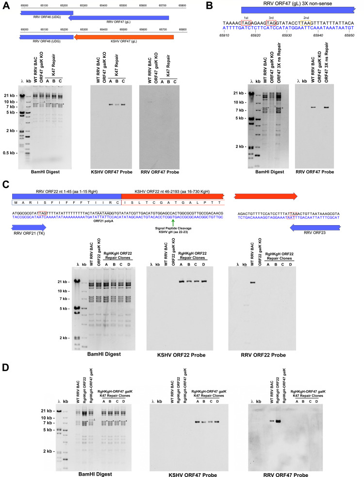
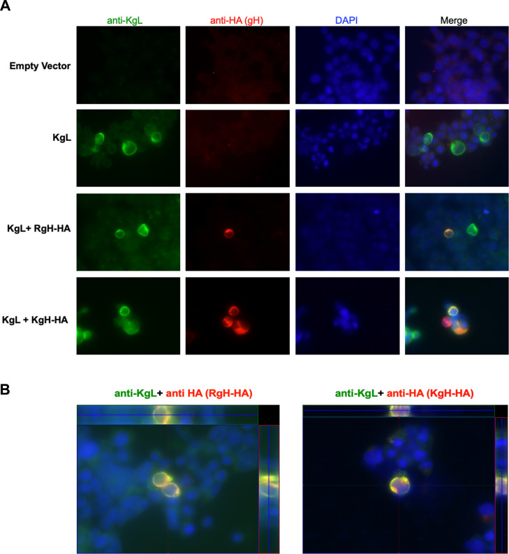
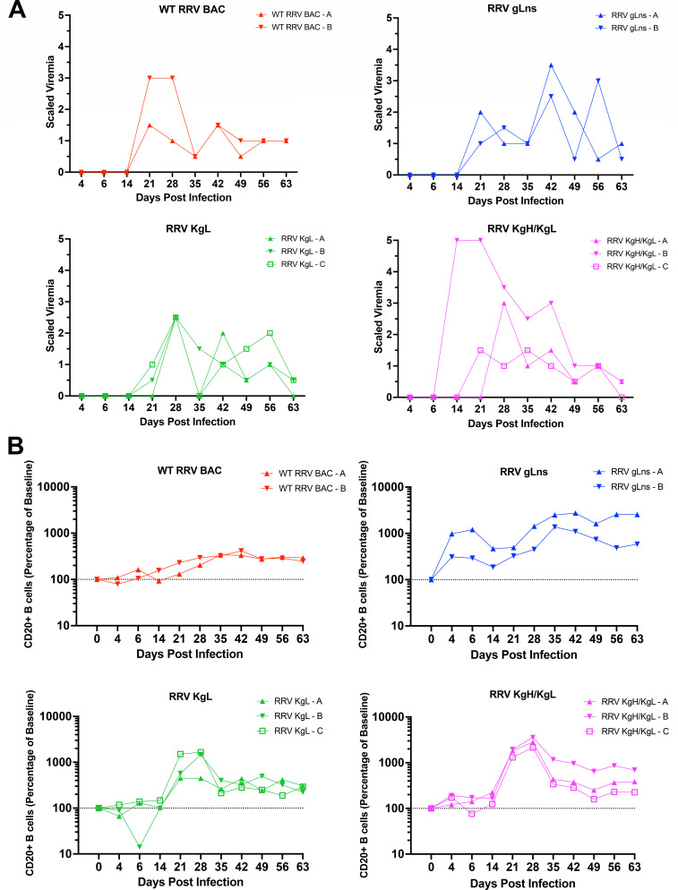
Kaposi's sarcoma-associated herpesvirus (KSHV) is a human gammaherpesvirus associated with Kaposi's sarcoma and B cell malignancies. Like all herpesviruses, KSHV contains conserved envelope glycoproteins (gps) involved in virus binding, entry, assembly, and release from infected cells, which are also targets of the immune response. Due to the lack of a reproducible animal model of KSHV infection, the precise functions of the KSHV gps during infection in vivo are not completely known. Fortunately, a nonhuman primate (NHP) model of KSHV infection and disease has been established utilizing closely related rhesus macaque rhadinovirus (RRV) that naturally infects rhesus macaques (RM) and possesses analogous gps to KSHV. To address the roles conserved envelope gps gH and gL play during KSHV infection in vivo, we utilized the pathogenic RRV17577 BAC to generate chimeric forms of RRV expressing KSHV gL or KSHV gH/gL, as well as an RRV mutant lacking gL expression. These viruses incorporate KSHV gH and gL into infectious virions, and although they display variable replication and differing plaque phenotypes in primary rhesus fibroblasts, they retain the ability to infect human B cells in vitro. Importantly, we also demonstrate that RRV gp chimeras can infect RM and induce the development of antibodies against KSHV. Overall, this work demonstrates that RRV gp chimeras can serve as important tools to assess the role of KSHV gH/gL in infection and disease while also providing an NHP model for testing the efficacy of KSHV gH and gL neutralizing antibodies and vaccine strategies to prevent and treat KSHV infection.IMPORTANCERhesus macaque rhadinovirus (RRV) is a rhesus macaque homolog of KSHV and serves as a model system for examining Kaposi's sarcoma-associated herpesvirus (KSHV) infection and pathogenesis in vivo. KSHV and RRV both encode conserved herpesvirus envelope glycoproteins, including gH and gL, that are important for regulating entry into host cells. In this study, we utilized the RRV BAC system to generate chimeric forms of RRV expressing KSHV gH and gL, as well as a mutant form of RRV lacking gL expression. Although these mutant and chimeric viruses can replicate in vitro, they do display growth properties different from wild-type RRV. Importantly, we demonstrate that RRV gp chimeras are capable of infecting rhesus macaques in vivo, inducing B cell hyperplasia, and promoting the development of anti-viral antibody responses that can also recognize KSHV antigens. RRV gp chimeras provide a novel system that allows for the examination of the role of KSHV gH and gL during infection in vivo.

Keywords: Kaposi's sarcoma-associated herpesvirus; RRV BAC; chimeric virus; glycoproteins; rhesus rhadinovirus; vaccines.


Conflict of interest statement

The authors declare no conflict of interest.

Figures

Fig 1
Fig 1
Generation of KSHV ORF47 chimeric, RRV ORF47 non-sense mutant, KSHV ORF22 hybrid chimeric, and KSHV ORF22 hybrid/ORF47 double chimeric RRV BAC clones. (A) Schematic depicting the insertion of KSHV ORF47 sequence in place of RRV ORF47 in the RRV genome, and BamHI restriction digestion and Southern blot analysis of RRV BAC clones. A 7.7 kb BamHI fragment in WT RRV BAC (*) shifts to 8.4 kb (Δ) upon replacement of RRV ORF47 with a galK cassette and reverts to 7.7 kb after repair with KSHV ORF47 sequence (+). Southern blot analysis for KSHV ORF47 was performed on digested DNA transferred to nitrocellulose, and the membrane was then stripped and reprobed for RRV ORF47. (B) Diagram of mutations inserted into the 5′ region of RRV ORF47 to generate an ORF47 3× non-sense repair fragment. TAG stop codons were introduced into the first and third reading frames (red dashed boxes), while a TAA stop codon in the second reading frame is naturally occurring (yellow dashed box). BamHI restriction digestion of RRV BAC clones followed by Southern blot analysis with an RRV ORF47 probe confirms the correct insertion of the ORF47 3× non-sense sequence. A 7.7 kb BamHI fragment in the wild-type RRV BAC (*) shifts to 8.4 kb (Δ) upon replacement of RRV ORF47 with a galK cassette and reverts to 7.7 kb after repair with ORF47 3× non-sense sequence (+). (C) Design of a hybrid RgHKgH ORF22 sequence encoding the first 15 amino acids of RRV gH fused to amino acids 16–730 of KSHV gH for insertion into the RRV genome. Stop codons of upstream ORF21 (TK), KSHV ORF22, and downstream ORF23 are noted by red dashed boxes, and the predicted poly A signal of ORF21 is noted by a gray dashed box. The signal peptide cleavage signal of KSHV gH is noted by a green arrow, with cleavage of the RgHKgH protein at this site allowing for the production of a processed form of KSHV gH protein lacking any RRV gH sequence. The 3′ end of KSHV ORF22 sequence was also inserted into RRV flanking arm sequence such that the stop codon of opposing ORF23 was not disrupted. BamHI restriction digestion and Southern blot analysis of RRV BAC clones confirm the correct insertion of RgHKgH hybrid ORF22 sequence in place of RRV ORF22. A 15.6 kb BamHI fragment in the wild-type RRV BAC shifts to 14.7 kb upon replacement of RRV ORF22 with a galK cassette and reverts to 15.7 kb after repair with RgHKgH ORF22 sequence to generate a chimeric BAC clone. Southern blot analysis for KSHV ORF22 was performed on digested DNA transferred to a nitrocellulose membrane, and the membrane was then stripped and reprobed for RRV ORF22. (D) Generation of a double chimeric RgHKgH hybrid ORF22/KSHV ORF47 RRV BAC clone. The same repair fragment from panel A was utilized to repair an RgHKgH ORF22 clone in which ORF47 was replaced with galK. BamHI restriction digestion and Southern blot analysis of RRV BAC clones confirm the correct insertion of KSHV ORF47 sequence in place of RRV ORF47 in the RRV BAC. A 7.7 kb BamHI fragment in the wild-type RRV BAC (*) shifts to 8.4 kb (Δ) upon replacement of RRV ORF47 with a galK cassette and reverts to 7.7 kb after repair with KSHV ORF47 sequence (+) to generate a chimeric BAC clone. Southern blot analysis for KSHV ORF47 was performed on digested DNA transferred to a nitrocellulose membrane, and the membrane was then stripped and reprobed for RRV ORF47.
Fig 2
Fig 2
Western blot analysis of purified RRV glycoprotein chimeric virions for the incorporation of KSHV gH or KSHV gL. (A) Equivalent PFUs of purified WT RRV BAC, RRV KgL, and RRV KgH/KgL were loaded and run on a 10% acrylamide gel, transferred to a nitrocellulose membrane, and probed with antibody specific for KSHV gH. The membrane was then stripped and re-probed for RRV major capsid protein (MCP) as a loading control. Signal for KSHV gH is only detected in RRV KgH/KgL virions. (B) Equivalent PFUs of purified WT RRV BAC, RRV gLns, RRV KgL, and RRV KgH/KgL were loaded and run on a 10% acrylamide gel, transferred to a nitrocellulose membrane, and probed with antibody specific for KSHV gL. The membrane was then stripped and re-probed for RRV MCP as a control. A volume of 5 and 20 mL of KSHV was also loaded as a control. Common bands of ~27 and ~15 kDa are detected in all viruses expressing KSHV gL, while an ~22 kDa band is detected only in RRV KgH/KgL and KSHV virions, and an ~18 kDa band is detected specifically in RRV KgH/KgL virions. The ~15 kDa band directly correlates with the predicted size of an unmodified form of KSHV gL lacking the signal peptide sequence (cleaved SP), while the larger ~22 and ~27 kDa bands represent putative modified forms of KSHV gL (denoted by *). No signal for KSHV gL was detected in WT RRV BAC or RRV gLns virions.
Fig 3
Fig 3
Immunofluorescence analysis to assess KSHV gL localization with RRV gH and KSHV gH in cells. HEK293T/17 cells were transfected with a vector expressing KSHV gL alone or in conjunction with vectors expressing C-terminal HA-tagged versions of RRV gH (RgH-HA) or KSHV gH (KgH-HA). Empty vector served as a negative control. Cells were fixed and stained with rabbit anti-KSHV gL antibody UK170 and mouse anti-HA antibody, followed by anti-rabbit-FITC and anti-mouse Texas Red secondary antibodies. DAPI staining was performed to visualize nuclei. (A) KSHV gL displays expression throughout the cytoplasm and at the plasma membrane and co-localizes with signal specific for RRV gH-HA and KSHV gH-HA, as indicated by yellow/orange staining patterns in merged images. (B) Z-stack images of stained cells further demonstrate the co-localization of KSHV gL with both RRV gH-HA and KSHV gH-HA in multiple planes in cells expressing these proteins. Side panels indicate the horizontal (top) and vertical (right side) cross-section of each image.
Fig 4
Fig 4
Plaque morphology and growth analysis of glycoprotein mutant and chimeric viruses. (A) 1oRF in 6-well dishes was infected with diluted stocks of WT RRV BAC, RRV gLns, RRV KgL, and RRV KgH/KgL incubated until the development of visible plaques and then fixed and stained with neutral red to visualize plaque morphology. Images depict a representative microscopic image of a single plaque from each well taken at 100× magnification. (B) Growth curve analysis of mutant and chimeric viruses in 1oRF. Cells were infected at an MOI of 2.5 (single step) or MOI of 0.1 (multi-step), and samples were harvested at the indicated time points. Plaque assay analysis was performed on 1oRF to determine viral titers in each sample. Error bars indicate standard deviation.
Fig 5
Fig 5
B cell infection and binding assays of RRV glycoprotein mutant and chimeric viruses. (A) Measurement of RRV genome copy levels during primary infection and establishment of latently infected BJAB cultures. BJAB cells were infected with WT RRV BAC, RRV gLns, RRV KgL, or RRV KgH/KgL at an MOI of 2, and DNA was isolated from cell samples at the indicated time points post-infection. qPCR was then performed using a primer and probe set specific for RRV viral macrophage inflammatory protein and normalized to GAPDH copy numbers to determine the relative amount of RRV genome copies in each sample (RRV vMIP/GAPDH ratio). Error bars indicate standard deviation. (B) BJAB cells were mixed with each virus at an MOI of 2 on ice, then incubated at 4°C for 2 hours to measure surface-bound virus, incubated at 37°C for 2 hours to measure internalized and any remaining surface-bound virus, or incubated at 37°C for 2 hours followed by treatment with trypsin to measure internalized virus only. After incubations, cells were washed with cold PBS, DNA was purified, and RRV vMIP/GAPDH ratios were determined by qPCR. Data represent the average of three independent experiments, and error bars indicate standard deviation. Relative to WT RRV BAC, RRV KgL and RRV KgH/KgL both display significantly higher levels of surface-bound virus (**P = 0.0011 and **P = 0.0097, respectively), internalized + surface-bound virus (**P = 0.0013 and **P = 0.0089, respectively), and internalized virus only (*P = 0.0176 and *P = 0.0138, respectively), as measured by unpaired t test. (C) Samples of latently infected BJAB cultures (from panel A) were taken at an early (day 54) and late (day 125) time point post-infection, treated with anti-human IgM to induce viral reactivation, or left untreated, and DNA was isolated for qPCR to determine RRV vMIP/GAPDH ratios in each sample. Error bars indicate standard deviation.
Fig 6
Fig 6
Development of viremia and B cell hyperplasia in infected rhesus macaques. (A) To assess the development of viremia during the acute phase of infection, PBMC samples were isolated from infected RM at the indicated time points, serially diluted 1:3, and co-cultured with primary rhesus fibroblasts. Viremia is defined as the limiting dilution factor where RRV CPE was detected, with a viremia scale of 1 representing the starting concentration of PBMCs (2 × 10⁵ PBMCs) utilized for co-culture. A viremia scale of 2 represents the first dilution where RRV CPE was detected in duplicate, and a maximum viremia scale of 5 indicates that all duplicate dilutions were positive for RRV CPE. A half scale indicates that RRV CPE was detected in one of the duplicates of the higher dilution. (B) The levels of total CD20 + B cells in infected RM were assessed by flow cytometry analysis of whole blood samples. All viruses examined induce an expansion of B cells in infected RM, indicating that RRV gLns, RRV KgL, and RRV KgH/KgL retain the ability to induce B cell hyperplasia, despite variances in gH and gL expression patterns from WT RRV BAC. Data are presented as percent change from baseline levels, which are set to 100% (dashed line).
Fig 7
Fig 7
Development of antibody responses in infected rhesus macaques. (A) Plasma from infected RM was analyzed for the presence of RRV-specific antibodies via ELISA, demonstrating the development of measurable antibody responses against RRV in all infected animals. Measurements for some time points in specific animals are absent due to a lack of sufficient samples for use in this analysis. (B) To assess the presence of KSHV-specific antibodies in infected RM, day 49 pi, plasma samples from each animal were utilized in western blot analysis against lysates from induced iSLK.219 cells or purified KSHV. Each plasma sample was diluted 1:500, with the exception of samples from RRV KgL-C, RRV KgH/KgL-B, and RRV KgH/KgL-C, which were utilized at a dilution of 1:100. Signals that are specific to plasma samples from animals infected with RRV KgL or RRV KgH/KgL are noted with arrows.
Fig 8
Fig 8
DNA viral loads in tissue samples of infected RM at the time of necropsy. Tissue samples, saliva, urine, and whole blood were collected from infected RM at the time of necropsy (WT RRV BAC-A d189 pi, WT RRV BAC-B d189 pi; RRV gLns-A d175 pi, RRV gLns-B d182 pi; RRV KgL-A d159 pi, RRV KgL-B d149 pi, RRV KgL-C d170 pi; RRV KgH/KgL-A d170 pi, RRV KgH/KgL-B d191 pi, RRV KgH/KgL-C d191 pi). DNA was isolated from each sample, and 100 ng was subjected to qPCR to assess the levels of RRV genomic DNA using a primer and probe set specific for RRV vMIP. Tissues analyzed include axillary lymph node (AX LN), bone marrow (BM), buccal tissue, cervical lymph node (CV LN), gum tissue, iliosacral lymph node (IL LN), inguinal lymph node (ING LN), kidney, mesenteric lymph node (MES LN), muscle, parotid gland, retropharyngeal lymph node (RP LN), submandibular lymph node (SM LN), superior mesenteric lymph node (SMES LN), spleen, saliva, tracheobronchial lymph node (TB LN), tonsil, urine, and whole blood (WB). Animals RRV KgL-B and RRV KgH/KgL-C displayed no detectable viral loads in any sample examined.

Similar articles

References

    1. Cesarman E, Chang Y, Moore PS, Said JW, Knowles DM. 1995. Kaposi's sarcoma-associated herpesvirus-like DNA sequences in AIDS-related body-cavity-based lymphomas. N Engl J Med 332:1186–1191. doi:10.1056/NEJM199505043321802 - DOI - PubMed
    1. Chang Y, Cesarman E, Pessin MS, Lee F, Culpepper J, Knowles DM, Moore PS. 1994. Identification of herpesvirus-like DNA sequences in AIDS-associated Kaposi’s sarcoma. Science 266:1865–1869. doi:10.1126/science.7997879 - DOI - PubMed
    1. Oksenhendler E, Boulanger E, Galicier L, Du MQ, Dupin N, Diss TC, Hamoudi R, Daniel MT, Agbalika F, Boshoff C, Clauvel JP, Isaacson PG, Meignin V. 2002. High incidence of Kaposi sarcoma-associated herpesvirus-related non-Hodgkin lymphoma in patients with HIV infection and multicentric Castleman disease. Blood 99:2331–2336. doi:10.1182/blood.v99.7.2331 - DOI - PubMed
    1. Soulier J, Grollet L, Oksenhendler E, Cacoub P, Cazals-Hatem D, Babinet P, d’Agay MF, Clauvel JP, Raphael M, Degos L. 1995. Kaposi’s sarcoma-associated herpesvirus-like DNA sequences in multicentric Castleman’s disease. Blood 86:1276–1280. doi:10.1182/blood.V86.4.1276.bloodjournal8641276 - DOI - PubMed
    1. Cesarman E, Damania B, Krown SE, Martin J, Bower M, Whitby D. 2019. Kaposi sarcoma. Nat Rev Dis Primers 5:9. doi:10.1038/s41572-019-0060-9 - DOI - PMC - PubMed

Substances

LinkOut - more resources