. 2025 Feb 25;99(2):e0176024.
doi: 10.1128/jvi.01760-24. Epub 2025 Jan 21.

RNASEK interacting with PEDV structural proteins facilitates virus entry via clathrin-mediated endocytosis

Affiliations

RNASEK interacting with PEDV structural proteins facilitates virus entry via clathrin-mediated endocytosis

Wenzhen Qin et al. J Virol. .

Abstract

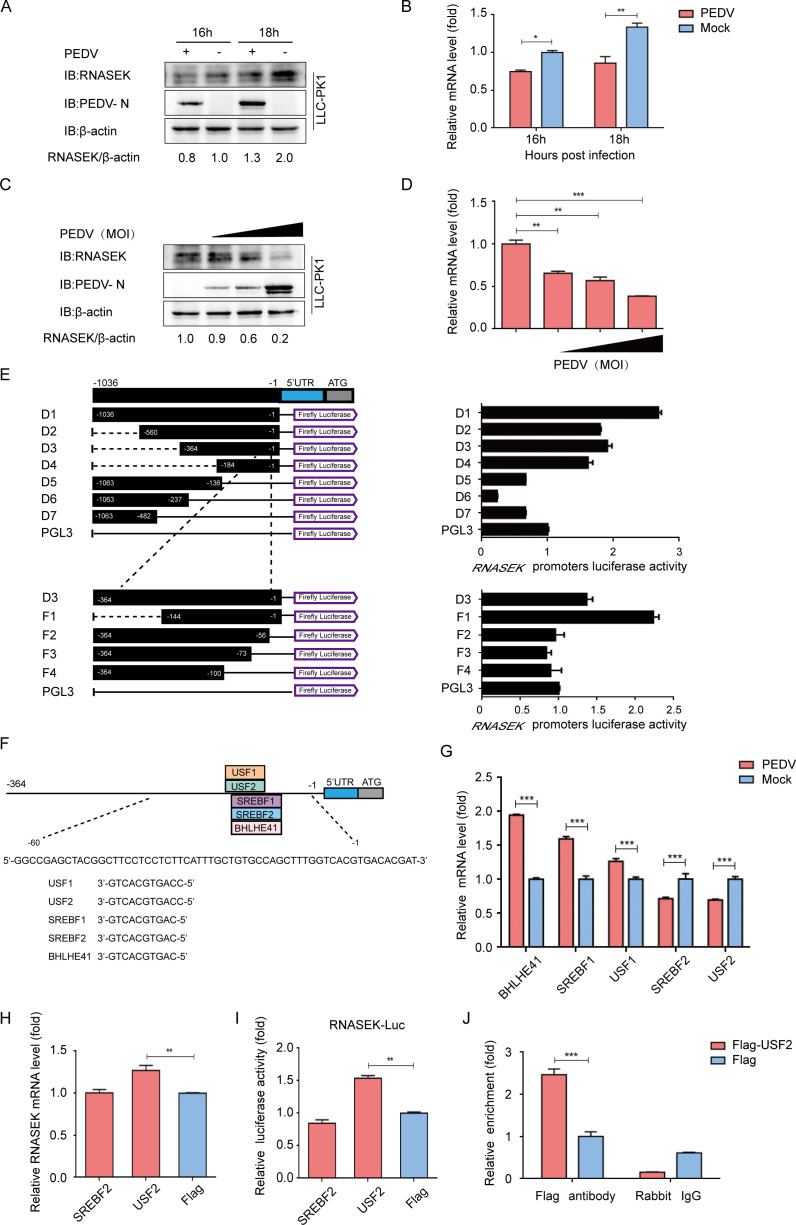
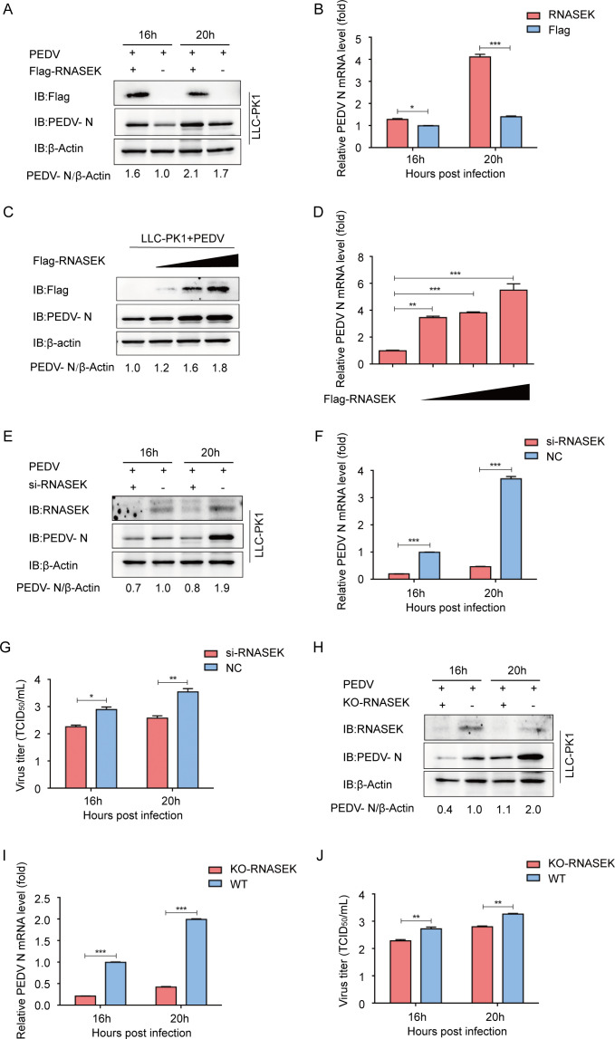
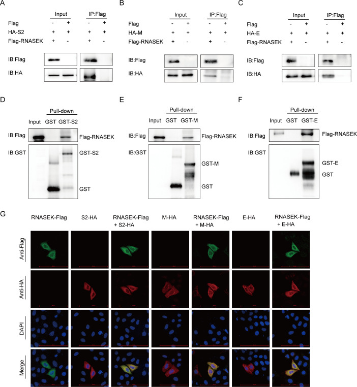
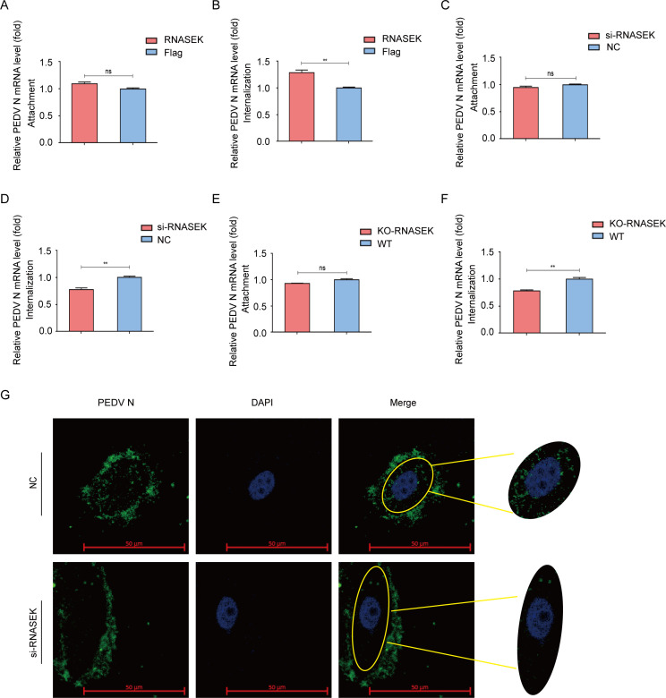
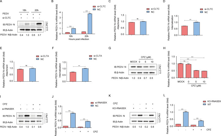
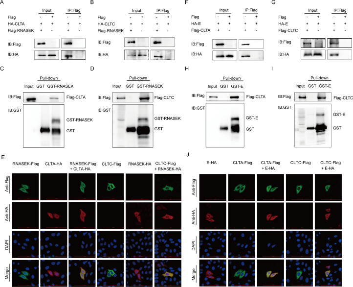
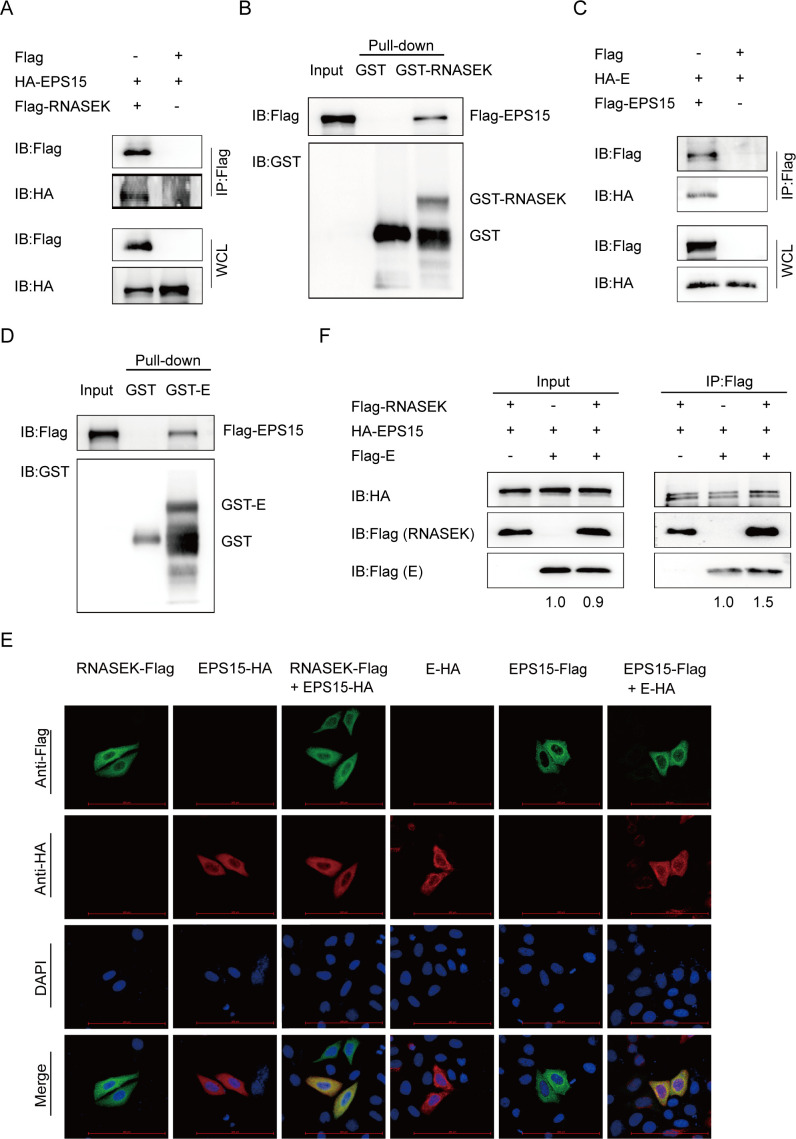
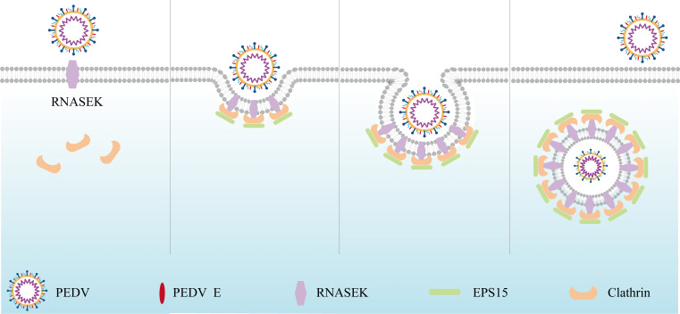
Porcine epidemic diarrhea virus (PEDV), as a type of Alphacoronavirus causing acute diarrhea and high death rate among sucking piglets, poses great financial damage to the swine industry. Nevertheless, the molecular mechanism whereby PEDV enters host cells is unclear, limiting the development of PED vaccines and anti-PEDV agents. The present study found that the host protein ribonuclease kappa (RNASEK) was regulated by USF2, a transcription factor, and facilitated the PEDV replication. RNASEK was identified as a novel binding partner of PEDV, which interacted with a spike (S), envelope (E), and membrane (M) proteins on PEDV virion surfaces to increase the uptake not for attachment of PEDV virions. PEDV enters cells through the endocytosis pathways. RNASEK knockdown or RNASEK knockout assay revealed that through clathrin-mediated endocytosis (CME), RNASEK promoted the internalization of PEDV virions. Clathrin and the adaptor protein EPS15 only interacted with PEDV E protein, demonstrating that the RNASEK could target more virions through interaction with PEDV S, E, and M proteins to clathrin and EPS15 proteins rather than merely interacting with PEDV E protein to mediate the PEDV entry through CME. Moreover, our findings suggest that RNASEK, a newly identified host-entry factor, facilitates PEDV internalization by increasing the interaction of PEDV virions and EPS15-clathrin complex and may also provide a potential target for anti-PEDV therapies.IMPORTANCEPEDV is the causative pathogen of porcine diarrhea, which is a highly infectious acute intestinal condition, that poses significant economic damage to the swine industry. However, the existing PED vaccines fail to provide adequate protection for piglets against PEDV infection. Although PEDV replication in cells has been widely described, the mechanisms beneath PEDV entry of the host cells are incompletely understood. In this study, we showed that RNASEK, regulated by the transcription factor USF2, is a new host factor increasing PEDV infection in LLC-PK1 cells. RNASEK can bind to multiple structural proteins of PEDV (S, E, and M proteins), therefore increasing the interaction between PEDV virions, clathrin, and EPS15 to promote PEDV virion entry. Apart from unraveling the entry mechanisms of PEDV, our findings also contributed to facilitating the development of anti-PEDV agents and PED vaccines.

Keywords: PEDV; RNASEK; endocytosis; virus entry.


Conflict of interest statement

The authors declare no conflict of interest.

Figures

Fig 1
Fig 1
PEDV infection causes USF2-mediated downregulation of RNASEK expression. (A and B) After PEDV infection (MOI = 1), the LLC-PK1 cells were gathered at 16 and 18 hpi. RNASEK expression was assessed by Western blotting in combination with qRT-PCR. β-actin was used as the sample loading control. (C and D) At 16 hpi, the LLC-PK1 cells challenged with varying MOIs of PEDV were harvested. Protein and mRNA levels of RNASEK were investigated through western blotting and qRT-PCR assays. (E) For the dual luciferase assay, truncated RNASEK promoter constructs comprising Renilla luciferase reporter vector (pRL-TK-luc) were transfected into HEK 293T cells. (F) Based on the JASPAR vertebrate database, the TFBS of the RNASEK promoter was predicted. (G) To assess the relative mRNA levels for predicted genes, qRT-PCR was performed on PEDV-infected LLC-PK1 cells. (H) The plasmid encoding Flag-SREBF2 or Flag-USF2 was transfected into LLC-PK1 cells, and the lysate was subjected to qRT-PCR analysis. (I) For the dual-luciferase activity evaluation, HEK 293T cells were transfected with RNASEK promoter-driven luciferase vector and SREBF2- or USF2-encoding plasmids. (J) For ChIP analysis, Flag-USF2 plasmid was transfected into LLC-PK1 cells. Data presented are indicated to be means ± SDs from triplicate samples. *P < 0.05; **P < 0.01; ***P < 0.001.
Fig 2
Fig 2
RNASEK promotes PEDV replication. (A and B) After transfection with the Flag-RNASEK encoding plasmid, the LLC-PK1 cells were challenged with PEDV (MOI = 1). Supernatants and cell lysates were performed by Western blotting and qRT-PCR. β-actin was used as the sample loading control. (C and D) After transfection with different concentrations of Flag-RNASEK plasmids, the LLC-PK1 cells were subjected to a PEDV challenge (MOI = 1). The samples were collected for western blotting and qRT-PCR. (E–G) After transfection with RNASEK siRNA, the LLC-PK1 cells were subjected to a PEDV challenge (MOI = 1). Sample analysis was performed through western blotting, qRT-PCR, and TCID50 assays. (H-J) PEDV replication in RNASEK KO LLC-PK1 cells was examined by Western blotting, qRT-PCR, and TCID50 assays. Data presented are suggested to be means ± SDs from triplicate samples. *P < 0.05; **P < 0.01; ***P < 0.001.
Fig 3
Fig 3
RNASEK interacted with the S2, M, E, and proteins of PEDV. (A–C) After co-transfection of HEK 293T cells with plasmids encoding Flag-RNASEK and HA-S2, HA-M, or HA-E, the associations between RNASEK and S2, M, and E proteins were assayed by Co-IP. (D–F) GST-S2, GST-M, GST-E, and RNASEK proteins were represented in bacterial strain BL21 (DE3). Meanwhile, the interactions of RNASEK with S2, M, and E proteins were investigated by GST pulldown assay. (G) After co-transfection of HeLa cells with plasmids encoding Flag-RNASEK and HA-S2, HA-M, or HA-E, the cells were exposed to incubation with designated primary antibody and fluorescently labeled secondary antibody, followed by results under confocal immunofluorescence microscopy. Scale bar, 100 µm.
Fig 4
Fig 4
RNASEK is involved in PEDV internalization. (A and C) After transfection with a plasmid coding RNASEK or RNASEK siRNA, the LLC-PK1 cells were subjected to a PEDV challenge (MOI = 1). After 60 min of viral attachment at 4°C, the virus RNA copies of attachment were assessed through qRT-PCR. (B and D) Following transfection with plasmid-coding RNASEK or RNASEK siRNA, the LLC-PK1 cells were subjected to a PEDV challenge (MOI = 1). Following 60 min of viral attachment at 4°C, the cells were washed three times in prechilled PBS and subsequently transferred to 37°C. At 90 min after infection, the cells were washed in pH 3.0 citric acid buffer and explored by qRT-PCR. (E and F) Attachment and internalization of PEDV in RNASEK KO LLC-PK1 cells were performed by qRT-PCR. (G) LLC-PK1 cells were subjected to RNASEK siRNA transfection, followed by a PEDV challenge. Immunofluorescence assay was employed to explore the internalization of PEDV. Scale bar, 50 µm.
Fig 5
Fig 5
RNASEK-mediated PEDV internalization is clathrin dependent. (A and B) Following 24 h of CLTC siRNA transfection, the LLC-PK1 cells were subjected to PEDV challenge and assayed by western blotting and qRT-PCR. (C-F) After 24 h of si-CLTC or si-CLTA transfection, the LLC-PK1 cells were subjected to a PEDV challenge (MOI = 1). Viral attachment and internalization assays were performed by qRT-PCR. (G and H) After 60 min of incubation with varying concentrations of CPZ, the LLC-PK1 cells were challenged with PEDV. Western blotting and qRT-PCR were conducted to assess the genomic DNA copies of PEDV in these cells. (I and J) After 1 h of transfection with si-RNASEK or pretreatment with 5 µM of CPZ at 37°C, the LLC-PK1 cells were incubated with PEDV, and subsequently assayed by qRT-PCR and western blotting. (K and L) After 60 min of pretreatment with 5 µM CPZ, the RNASEK KO LLC-PK1 cells were subjected to a PEDV challenge (MOI = 1). The viral genome level was examined by western blotting and qRT-PCR.
Fig 6
Fig 6
The interactions between RNASEK, clathrin, and PEDV structural proteins. (A and B) After transfection of HEK 293T cells with Flag-RNASEK and HA-CLTA or HA-CLTC encoding plasmids, a Co-IP assay was conducted by adopting anti-Flag binding beads, and the precipitated proteins were Western blotted. (C and D) The association of RNASEK with CLTA or CLTC was assessed by GST affinity–isolation assay. (E) After transfection of plasmids encoding RNASEK and CLTC or CLTA, the HeLa cells were labeled with designated primary and secondary antibodies. Nuclear staining was performed using DAPI, and fluorescence signals were identified by confocal immunofluorescent microscopy. Scale bars: 100 µm. (F and G) Plasmids encoding HA-E, Flag-CLTC, or Flag-CLTA were transfected into the HEK 293T cells. Co-IP assay was performed with anti-Flag-binding beads. (H and I) The interaction of PEDV E with CLTA or CLTC was examined through GST affinity–isolation assay. (J) After transfection with HA-E and Flag-CLTC or Flag-CLTA plasmids, the Hela cells were labeled with antibodies and assessed by confocal immunofluorescence microscopy. Scale bars: 100 µm.
Fig 7
Fig 7
RNASEK interacts with EPS15 and is implicated in PEDV internalization via CME. (A) HEK 293T cells transfected with HA-EPS15 and Flag-RNASEK encoding plasmids were exposed to Co-IP assay. (B) The association of RNASEK with EPS15 was assessed through GST affinity–isolation assay. (C) After transfecting HA-E and Flag-EPS15 vectors into HEK 293T cells, the interaction of EPS15 with E was analyzed through Co-IP. (D) After indicating EPS15 and GST-E proteins in strain BL21 (DE3), they were subjected to GST pulldown assay. (E) After transfecting plasmids encoding EPS15 and RNASEK or E into HeLa cells, confocal immunofluorescence microscopy was used for the fluorescence signal observation. Scale bars: 100 µm. (F) Lysates of HEK 293T cells overexpressing RNASEK, EPS15, and E were immunoprecipitated with anti-Flag antibodies.
Fig 8
Fig 8
Schematic depiction of RNASEK in boosting PEDV endocytosis as a host factor. RNASEK interacted with PEDV S2, M, and E proteins. Subsequently, the signal was transmitted to clathrin and EPS15 to form the virions-RNASEK-EPS15 axis. This process promoted the EPS15-clathrin complex formation and aggregation, causing the endocytosis of PEDV virions in the infection process.

Similar articles

References

    1. Wang D, Fang LR, Xiao SB. 2016. Porcine epidemic diarrhea in China. Virus Res 226:7–13. doi:10.1016/j.virusres.2016.05.026 - DOI - PMC - PubMed
    1. Li Y, Wu QX, Huang LL, Yuan C, Wang JL, Yang Q. 2018. An alternative pathway of enteric PEDV dissemination from nasal cavity to intestinal mucosa in swine. Nat Commun 9:3811. doi:10.1038/s41467-018-06056-w - DOI - PMC - PubMed
    1. Alonso C, Goede DP, Morrison RB, Davies PR, Rovira A, Marthaler DG, Torremorell M. 2014. Evidence of infectivity of airborne porcine epidemic diarrhea virus and detection of airborne viral RNA at long distances from infected herds. Vet Res 45:73. doi:10.1186/s13567-014-0073-z - DOI - PMC - PubMed
    1. Chen JF, Sun DB, Wang CB, Shi HY, Cui XC, Liu SW, Qiu HJ, Feng L. 2008. Molecular characterization and phylogenetic analysis of membrane protein genes of porcine epidemic diarrhea virus isolates in China. Virus Genes 36:355–364. doi:10.1007/s11262-007-0196-7 - DOI - PMC - PubMed
    1. Antas M, Woźniakowski G. 2019. Current status of porcine epidemic diarrhoea (PED) in European pigs. J Vet Res 63:465–470. doi:10.2478/jvetres-2019-0064 - DOI - PMC - PubMed

LinkOut - more resources