. 2025 Jan 28;122(4):e2417417122.
doi: 10.1073/pnas.2417417122. Epub 2025 Jan 21.

Cortical reorganization following dorsal spinal injuries in newborn monkeys reveals a critical period in the development of the somatosensory cortex

Affiliations

Cortical reorganization following dorsal spinal injuries in newborn monkeys reveals a critical period in the development of the somatosensory cortex

Neeraj Jain et al. Proc Natl Acad Sci U S A. .

Abstract

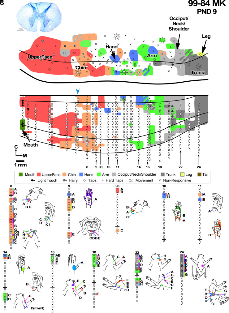
Lesions of the dorsal columns of the spinal cord in adult macaque monkeys lead to the loss of hand inputs and large-scale expansion of the face inputs in the hand region of the somatosensory cortex. Inputs from alternate spinal pathways do not reactivate the deafferented regions of area 3b. Here, we determined how transections of the dorsal columns done within a few days after birth affect the developing somatosensory cortex. Dorsal columns were transected between the 3rd and 12th postnatal day (PND), and the somatosensory cortex was mapped when the macaques were over 3 y old. There were two distinct outcomes depending on the age at the time of the lesion. In monkeys lesioned between the 3rd and 5th PND, neurons in the entire hand region of area 3b and the adjacent somatosensory cortex responded to touch on the hand. An alternate spinal pathway must have replaced the lost pathway. In monkeys lesioned between the 9th and 12th PND, neurons in the deafferented hand region did not respond to touch on the hand. There was medialward expansion of the face representation into the deafferented cortex and a lateral expansion of the arm representation as in lesioned adults. Thus, different mechanisms underlie the reorganization of area 3b and the adjacent somatosensory cortex following identical spinal cord injuries sustained as early or late newborns. The results suggest that alternate spinal cord pathways can develop within a critical period before the 9th PND, but not later.

Keywords: Macaca; area 3b; brain plasticity.


Conflict of interest statement

Competing interests statement:The authors declare no competing interest.

Figures

Fig. 1.
Fig. 1.
Somatotopy and receptive fields of neurons in area 3b and the adjacent caudal somatosensory areas 1 and 2 for monkey 97-63MK that received dorsal column lesion on PND 3. All of the body parts are represented in the expected somatotopic order from face lateral-most to foot medial-most. The Inset on Top Right shows the reconstruction of the spinal cord lesion in a coronal plane. The lesion is shown in black, gray matter is gray, and the white matter is white. (A) A reconstruction of the surface of the anterior postcentral gyrus showing somatotopy in area 1 and area 2. The thick line shows the lip of the CS, and the thin line is the cytoarchitectonic boundary between areas 1 and 2. The body parts are color-coded as per the key in the box. Stripes of two colors mark regions where stimulation of two corresponding body parts evoked a response. Nature of the neuronal responses is denoted by a symbol at each recording site. Neurons responding to light touch, black dots; taps, open dots; movement of hairs, dots with stippling; weak response to hard taps, triangle; response to movement of a body part, dot with arcs on Top and Bottom. Size of the dots depicts the nature of response, with larger dots depicting recording sites with a better response and smaller dots a weaker response; “x” denotes sites where no neuronal response was evoked by touch on any part of the body (see key). (B) Somatotopy of area 3b and adjacent area 1 in the posterior bank of the CS. The thick horizontal line at the top marks a depth of 1,200 µm from the cortical surface, the depth from which recordings were started in each electrode penetration. The border between area 1 and area 3b as well as approximate ventral border of area 3b is shown. The Bottom line delineates gray matter and white matter, and the line above it marks the border of area 3b and area 3a. The expected location of the hand–face border, as estimated to be near the tip of the intraparietal sulcus, is indicated by the blue arrowhead on the Top. The selected electrode penetrations for which the receptive fields are shown in “C” are marked by numbers. (C) Receptive fields for selected penetrations. Receptive fields of neurons at each recording site marked by letters are shown by the corresponding letter on the figurines on the Left. Dark horizontal lines mark borders of area 3b. Conventions, as for “A” and “B.” R, rostral; M, medial. The scale bar in “B” also applies to “A”; mvmt, movement. PND on which the lesion was made is shown on top.
Fig. 2.
Fig. 2.
Somatotopic organization of the digits representation in the hand region of area 3b and the adjacent somatosensory areas 1 and 2 of area 3b in monkey 97-63MK with transection of the dorsal column on PND 3. The data are represented as for Fig. 1 A and B. (A) Reconstruction of the digits representation on the surface of the postcentral gyrus and (B) the postcentral gyrus in the depth of the CS. Somatotopy of the digits is shown as per the key at the Bottom. Sites with multidigit receptive fields are marked with corresponding receptive field locations. Multidigit receptive fields in area 2 are as found in normal animals. A clear somatotopy from D1 to D5 is obvious. Conventions as for Fig. 1.
Fig. 3.
Fig. 3.
Somatotopy and receptive fields of neurons in area 3b and the adjacent caudal somatosensory areas 1 and 2 for monkey 97-58MK that received dorsal column lesion on PND 5. All of the body parts are represented in the expected somatotopic order from face lateral-most to foot medial-most. Note the lateromedial row of recording sites with dual receptive fields on the hand and chin. The Inset on Top Right shows the reconstruction of the spinal cord lesion in a coronal plane. (A) A reconstruction of the surface of the anterior postcentral gyrus showing somatotopy in area 1 and area 2. (B) Somatotopy of area 3b and adjacent area 1 in the posterior bank of the CS. (C) Receptive fields for selected penetrations. All conventions as in Fig. 1.
Fig. 4.
Fig. 4.
Somatotopy and receptive fields of neurons in area 3b and the adjacent caudal somatosensory areas 1 and 2 for monkey 99-84MK that received dorsal column lesion on PND 9. The neurons largely do not respond to touch on the hand. Note the expansion of the chin-responsive region medial to the expected hand–face border (blue arrowhead). The Inset on the Top Left shows a Nissl-stained section of the spinal cord rostral to the lesion site showing the asymmetrical dorsal columns with a large reduction of the dorsal columns ipsilateral to the lesion. (A) A reconstruction of the surface of the anterior postcentral gyrus showing somatotopy in area 1 and area 2. (B) Somatotopy of area 3b and adjacent area 1 in the posterior bank of the CS. (C) Receptive fields for selected penetrations. All conventions as in Fig. 1.
Fig. 5.
Fig. 5.
Sprouting of afferents from the trigeminal nucleus into the cuneate nucleus in monkey 99-84MK shown in representative coronal sections of the medulla. In “A” to “D” pairs of photomicrographs show part of the section processed to visualize CTB (Left) or CO activity (Right). Label in the trigeminal nucleus (Trig N; arrowhead) and in sprouted afferents in the cuneate nucleus (Cu N; arrow) is clearly visible. In “A” processes can be visualized as they extend from the trigeminal nucleus to the cuneate nucleus. CO-stained matching sections are shown to help visualize boundaries of the nuclei. Sections shown in “A” and “B,” and “C” and “D” are 108 µm apart, whereas “B” and “C” are 504 µm apart. “E” shows a complete section of the medulla corresponding to “C” stained for CO. The major nuclei are marked on the left half of the section and major tracts on the right half. The box marks the approximate region shown in “A” to “D.” Note the shrinkage of the cuneate and gracile nucleus on the left side, which is ipsilateral to the lesion. ALF, anterolateral fasciculus; ArF, arcuate fibers; CuF, cuneate fasciculus; DMV, dorsal motor nucleus of vagus nerve (X); DSCTr, dorsal spinocerebellar tract; ExCu, external cuneate nucleus; Gr, nucleus gracilis; GrF, fasciculus gracilis; Hy, Hypoglossal nucleus (XII); IO, inferior olivary nucleus; LRet, lateral reticular nucleus; MedL, medial lemniscus; Py, pyramidal tract; Ret, reticular formation; TgTr, trigeminal tract. The scale bar shown in “D” is also valid for “A” “B,” and “C.”

Similar articles

References

    1. Hubel D. H., Wiesel T. N., LeVay S., Plasticity of ocular dominance columns in monkey striate cortex. Philos. Trans. R. Soc. Lond. B Biol. Sci. 278, 377–409 (1977). - PubMed
    1. Takahata T., Patel N. B., Balaram P., Chino Y. M., Kaas J. H., Long-term histological changes in the macaque primary visual cortex and the lateral geniculate nucleus after monocular deprivation produced by early restricted retinal lesions and diffuser induced form deprivation. J. Comp. Neurol. 526, 2955–2972 (2018). - PMC - PubMed
    1. Warner C. E., et al. , Preservation of vision by the pulvinar following early-life primary visual cortex lesions. Curr. Biol. 25, 424–434 (2015). - PubMed
    1. Chino Y. M., Kaas J. H., Smith E. L. III, Langston A. L., Cheng H., Rapid reorganization of cortical maps in adult cats following restricted deafferentation in retina. Vision Res. 32, 789–796 (1992). - PubMed
    1. Schwaber M. K., Garraghty P. E., Kaas J. H., Neuroplasticity of the adult primate auditory cortex following cochlear hearing loss. Am. J. Otol. 14, 252–258 (1993). - PubMed

MeSH terms

LinkOut - more resources