. 2025 Jan 21;10(5):e187315.
doi: 10.1172/jci.insight.187315.

Map2k6 is a potent genetic modifier of arterial rupture in vascular Ehlers-Danlos syndrome mice

Affiliations

Map2k6 is a potent genetic modifier of arterial rupture in vascular Ehlers-Danlos syndrome mice

Caitlin J Bowen et al. JCI Insight. .

Abstract

Aortic dissection or rupture is a major cause of mortality in vascular Ehlers-Danlos syndrome (vEDS), a connective tissue disorder caused by heterozygous mutations in the collagen type III alpha 1 chain (COL3A1) gene. C57BL6/J (BL6) mice carrying the Col3a1G938D/+ mutation recapitulate the vEDS vascular phenotype and die suddenly of aortic rupture/dissection. However, 129S6/SvEvTac (referred to here as 129) mice expressing the same Col3a1G938D/+ mutation show near-complete lifelong protection from vascular rupture. To identify genetic modifiers of vascular risk in vEDS, we performed genome-wide genotyping of intercrossed BL6/129 vEDS mice stratified by survival and identified a significant protective locus encompassing a variant in Map2k6, encoding mitogen-activated protein kinase kinase 6 (M2K6), a p38-activating kinase. Genetic ablation of Map2k6 rendered previously protected 129 vEDS mice susceptible to aortic rupture, in association with reduced protein phosphatase 1 activity and increased PKC and ERK phosphorylation. Accelerated vascular rupture in vEDS mice treated with a pharmacological inhibitor of p38 was rescued by concomitant ERK antagonism, supporting an opposing role for ERK and p38 in the modification of aortic rupture risk in vEDS. These results suggest that pharmacologic strategies aimed at mimicking the effect of this natural protective pathway may attenuate aortic rupture risk in vEDS.

Keywords: Cardiology; Collagens; Genetic diseases; Genetic variation; Genetics; Vascular biology.


Conflict of interest statement

Conflict of interest: CJB, JFCG, and HCD have submitted patent applications for pharmacological treatment of vascular Ehlers-Danlos syndrome (US Patent 11938135, US Patent Application 18/024,662).

Figures

Figure 1
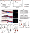
Figure 1. The 129Sve background protects vEDS mice from premature death.
(A) Kaplan-Meier survival curve comparing control (Col3a1+/+) (n = 199) and vEDS (Col3a1G938D/+) mice (n = 191) on a BL6 background with control (n = 18) and vEDS mice (n = 31) on a 129 background. Significant differences were calculated using log-rank (Mantel-Cox) analysis (*** P < 0.001). (B) Kaplan-Meier survival curve comparing male BL6 vEDS male (n = 82) and female (n = 109) mice with 129 vEDS male (n = 15) and female (n = 16) mice. Significant differences were calculated using log-rank (Mantel-Cox) analysis (*** P < 0.001). (C) Verhoeff–Van Gieson (VVG), Masson’s trichrome, and Picrosirius red (PSR) staining of control and vEDS aortic cross sections at 2 months of age. White arrows indicate elastin fiber breaks. Scale bar is 50 microns. (D) Quantification of aortic wall thickness in aortic cross sections. P value refers to 2-way ANOVA with Holm-Šídák post hoc test (*** P < 0.001). (E) Quantification of elastin fiber breaks in VVG-stained aortic cross sections. Asterisks identify discovery (q < Q) by Kruskal-Wallis test with post hoc false discovery rate correction. (F) Quantification of collagen content in aortic cross sections, as measured by normalized PSR intensity. P value refers to 2-way ANOVA with Holm-Šídák post hoc test (*** P < 0.001). (G) Immunoblot of aortic lysates obtained from the proximal descending aortas of control and vEDS mice in the BL6 and 129 backgrounds at 2 months of age, probed with antibodies directed against phosphorylated PKCβ at residue Ser660 (pPKC) and phosphorylated ERK (pERK1/2). (H) Quantification for immunoblot shown in G. P value refers to 2-way ANOVA with Holm-Šídák post hoc test for p-PKC and Kruskal-Wallis test for p-ERK (* P < 0.05, *** P < 0.001). For DF and H, each symbol represents an independent biological replicate, with unfilled symbols representing male samples. Error bars show mean ± SEM. Black circles represent control mice and red squares represent vEDS mice.
Figure 2
Figure 2. A single locus on distal chromosome 11 associates with protection from death by rupture in vEDS mice.
(A) Breeding scheme for the generation of vEDS mice of mixed background used for GWAS. (B) Survival and stratification strategy of vEDS mice of mixed background. Mice that died from vascular rupture before 12 weeks of age were coded as cases (BL6-like, shown in black), while those surviving more than 24 weeks are coded as controls (129-like, shown in red). Mice that did not die from vascular rupture (i.e., malocclusion) or died between 12 and 24 weeks of age were not included in the analysis, as defined prospectively. (C) Manhattan plot of mixed-background vEDS mice (n = 91 controls, n = 96 cases). The red line indicates the genome-wide significance threshold. (D) Filtering strategy used to identify candidate modifier genes within the locus of interest. (E) Graphical representation of the region of interest on chromosome 11 showing annotated transcripts color-coded based on the type of variant existing in the BL6 compared with the 129 genetic background. Black and gray shapes represent BL6-like mice, and red shapes represent 129-like mice. Bptf, bromodomain PHD finger transcription factor; Helz, helicase with zinc finger.
Figure 3
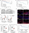
Figure 3. Map2k6 exerts a protective effect against death by aortic rupture in vEDS mice.
Kaplan-Meier survival curve comparing (A) male 129 vEDS Map2k6+/+ (n = 17), vEDS Map2k6+/– (n = 20), and vEDS Map2k6–/– mice (n = 10) and (B) female 129 vEDS Map2k6+/+ (n = 19), vEDS Map2k6+/– (n = 15), and vEDS Map2k6–/– mice (n = 12). Significant differences were calculated using log-rank (Mantel-Cox) analysis (* P < 0.05; ** P < 0.01). (C) Immunoblot analysis of phosphorylated PKC at residue Ser660 (pPKC) and phosphorylated ERK (pERK1/2) comparing aortic lysates obtained from the proximal descending aortas of mice at 2 months of age. (D) Quantification of p-PKC and p-ERK normalized to β-actin of control Map2k6+/+ (n = 3), vEDS Map2k6+/+ (n = 4), control Map2k6–/– (n = 7), and vEDS Map2k6–/– (n = 5) mice. P value refers to 2-way ANOVA with Holm-Šídák post hoc test (* P < 0.05, *** P < 0.001). (E) Immunofluorescence of sections from the proximal descending thoracic aorta of vEDS Map2k6+/+ and vEDS Map2k6–/– mice. The dashed line marks the approximate boundaries of the aortic wall. Scale bar is 50 microns. (F) Mean and total protein phosphatase 1 (PP1) dephosphorylation activity in aortic protein lysates from 129 vEDS Map2k6+/+ mice (n = 6) and 129 vEDS Map2k6–/– (n = 7). P value refers to unpaired t test with Welch’s correction (* P < 0.05, ** P < 0.01).DiFMU, 6,8-difluoro-7-hydroxy-4-methylcoumarin. In D and F, each symbol represents an independent biological replicate, with unfilled symbols representing male samples. Error bars show mean ± SEM. (G) Kaplan-Meier survival curve comparing control 129 vEDS Map2k6–/– mice (n = 33, 17 females and 16 males) with 129 vEDS Map2k6–/– (n = 11, 8 females and 3 males) mice receiving ruboxistaurin (PKC inhibitor) starting at postnatal day 21. Significant differences were calculated using log-rank (Mantel-Cox) analysis (* P < 0.05).
Figure 4
Figure 4. Inhibition of p38 activation increases the risk of vascular rupture in BL6 vEDS mice.
(A) Kaplan-Meier survival curve comparing saline-injected BL6 vEDS mice (n = 19, 7 female and 12 male) with BL6 vEDS mice (n = 14, 6 females and 8 males) receiving SB203580 (p38 inhibitor) via intraperitoneal injection and SB203580-treated also receiving cobimetinib, a MEK inhibitor (n = 14, 6 females and 8 males), starting at postnatal day 21 and continuing for 40 days thereafter. Significant differences were calculated using log-rank (Mantel-Cox) analysis (** P < 0.01, *** P < 0.001). (B) Immunoblot of aortic lysates from the proximal descending thoracic aortas of BL6 vEDS mice treated with placebo or p38 inhibitor at 45 days of age, probed with antibodies directed for p-PKCβ and p-ERK and quantification of immunoblot. Each symbol represents an independent biological replicate, with unfilled symbols representing male samples. Error bars show mean ± SEM. P value refers to unpaired t test with Welch’s correction (** P < 0.01). Red circles represent placebo-treated mice, and blue squares represent p38 inhibitor–treated mice. (C) Summary figure outlining contributors to risk of vascular rupture in the aorta of vEDS mouse models.

References

    1. Shalhub S, et al. A multi-institutional experience in the aortic and arterial pathology in individuals with genetically confirmed vascular Ehlers-Danlos syndrome. J Vasc Surg. 2019;70(5):1543–1554. doi: 10.1016/j.jvs.2019.01.069. - DOI - PMC - PubMed
    1. Pepin M, et al. Clinical and genetic features of Ehlers-Danlos syndrome type IV, the vascular type. N Engl J Med. 2000;342(10):673–680. doi: 10.1056/NEJM200003093421001. - DOI - PubMed
    1. Pepin MG, et al. Survival is affected by mutation type and molecular mechanism in vascular Ehlers-Danlos syndrome (EDS type IV) Genet Med. 2014;16(12):881–888. doi: 10.1038/gim.2014.72. - DOI - PubMed
    1. Smith LB, et al. Haploinsufficiency of the murine Col3a1 locus causes aortic dissection: a novel model of the vascular type of Ehlers-Danlos syndrome. Cardiovasc Res. 2011;90(1):182–190. doi: 10.1093/cvr/cvq356. - DOI - PMC - PubMed
    1. Smith LT, et al. Mutations in the COL3A1 gene result in the Ehlers-Danlos syndrome type IV and alterations in the size and distribution of the major collagen fibrils of the dermis. J Invest Dermatol. 1997;108(3):241–247. doi: 10.1111/1523-1747.ep12286441. - DOI - PubMed