. 2025 Jan 21;10(5):e186133.
doi: 10.1172/jci.insight.186133.

Neutrophils initiate proinflammatory immune responses in early endometriosis lesion development

Affiliations

Neutrophils initiate proinflammatory immune responses in early endometriosis lesion development

Taylor R Wilson et al. JCI Insight. .

Abstract

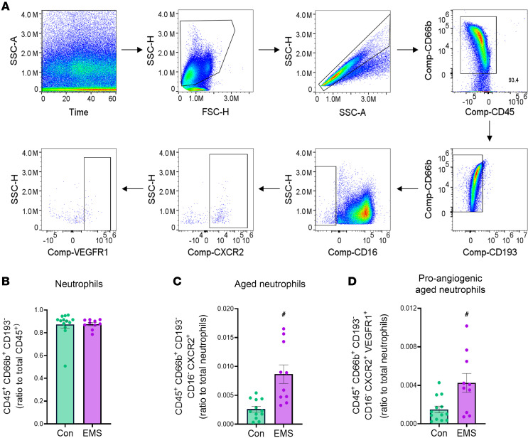
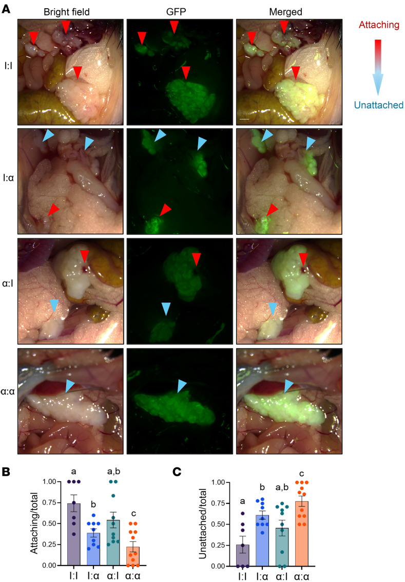
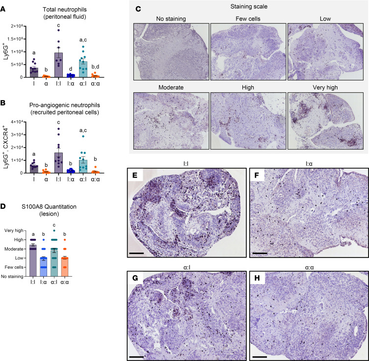
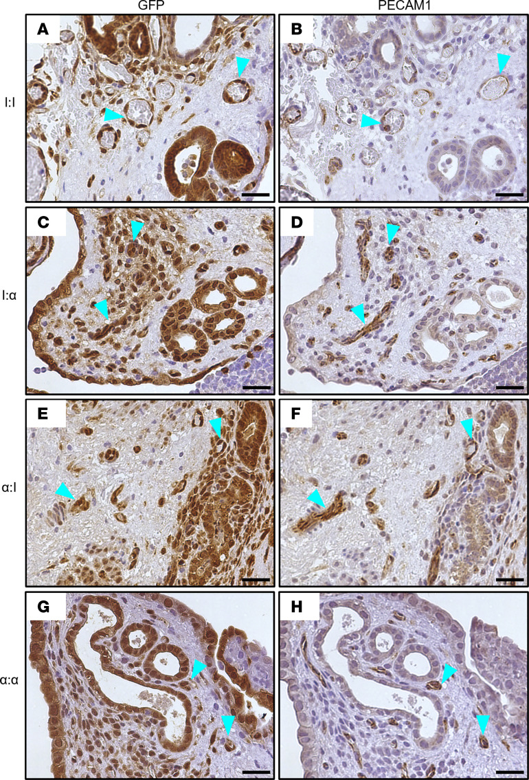
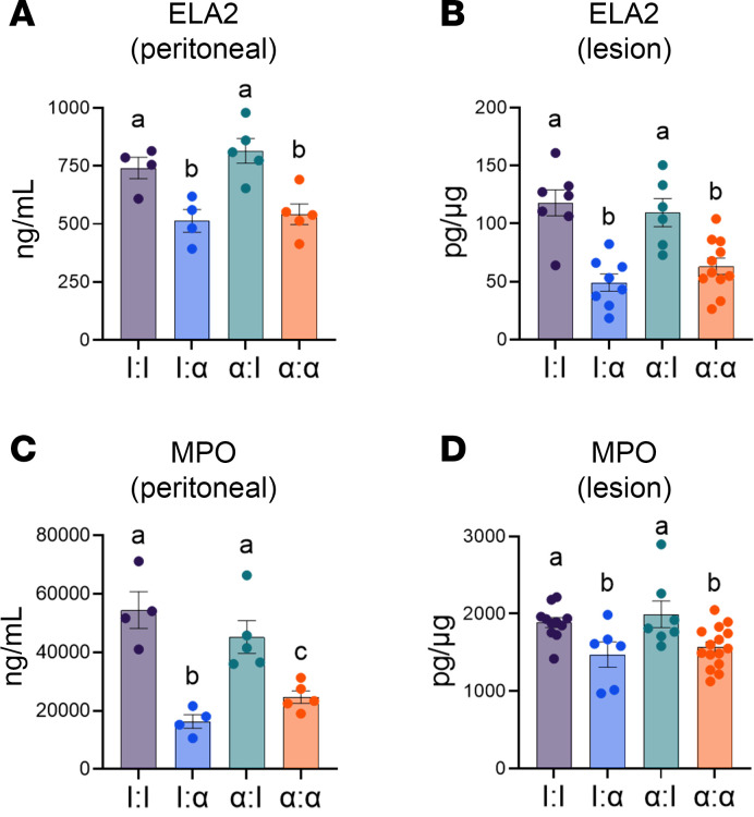
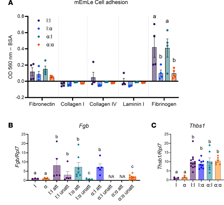
Endometriosis is a chronic gynecological disease that affects 1 in 10 reproductive-aged women. Most studies investigate established disease; however, the initiation and early events in endometriotic lesion development remain poorly understood. Our study used neutrophils from human menstrual effluent from patients with and without endometriosis for immunophenotyping, and it used a mouse model of endometriosis and a mouse endometriosis cell line to determine the role of neutrophils in the initiating events of endometriosis, including attachment and survival of minced endometrial pieces. In menstrual effluent from women with endometriosis, the ratios of aged and proangiogenic neutrophils increased compared with controls, indicating a potentially permissive proinflammatory microenvironment. In our endometriosis mouse model, knocking down neutrophil recruitment with α-CXCR2 into the peritoneum decreased endometrial tissue adhesion - supported by decreased levels of myeloperoxidase and neutrophil elastase in both developing lesions and peritoneal fluid. Fibrinogen was identified as the preferred substrate for endometrial cell adhesion in an in vitro adhesion assay and in developing lesions in vivo. Together, aged and proangiogenic neutrophils and their secretions likely promote attachment and formation of endometriotic lesions by releasing neutrophil extracellular traps and upregulating fibrinogen expression as a provisional matrix to establish attachment and survival in the development of endometriosis lesions.

Keywords: Immunology; Inflammation; Mouse models; Neutrophils; Reproductive biology.


Conflict of interest statement

Conflict of interest: KAB and TRW have a patent pending (no. 63/575,567) on menstrual effluent as a noninvasive diagnostic for endometriosis.

Figures

Figure 1
Figure 1. Women with endometriosis have higher levels of aged and proangiogenic neutrophils compared with healthy women.
(A) Gating strategy of total WBCs isolated from human menstrual effluent. Cells were gated on time, viable cells, single cells, CD45+, CD66b+, and CD193 to identify total neutrophils. Aged neutrophils were defined as CD45+CD66b+CD193CD16CXCR2+, and proangiogenic neutrophils are defined as CD45+CD66b+CD193CD16CXCR2+VEGFR1+. (BD) Quantitation of total neutrophils to total WBCs ratio, aged neutrophils to total neutrophil ratio, and proangiogenic aged neutrophils to total neutrophil ratio. Con, healthy controls (n = 13); EMS, endometriosis participants (n = 10). Data represent ± SEM. Statistical significance for each graph was determined by nonparametric, Mann-Whitney, 1-tailed U test. #P < 0.05.
Figure 2
Figure 2. Schematic representation of study treatment and experimental timeline.
Donor mice expressing GFP (green) begin treatment bidiurnally with α-CXCR2/IgG 5 days before collection of endometrial tissue (green diamonds). At 41 hours before endometrial collection, donor mice receive an i.p. injection of pregnant mare serum gonadotropin (PMSG) to synchronize uteri. Host mice (black) begin treatment bidiurnally with α-CXCR2/IgG 6 days before surgical induction of endometriosis (black diamonds) with a final dose administered the day of surgery. Mice are euthanized and lesions are collected 24 hours after surgical induction.
Figure 3
Figure 3. Knockdown of neutrophil recruitment in the SDME model decreased the attachment of minced endometrial pieces at 24 hours to form endometriosis lesions.
(A) Lesions (24 hours) were derived from GFP minced endometrial pieces and imaged at 488 nm and bright-field from I:I, I:α, α:I, and α:α groups. I, IgG; α, α-CXCR2. Lesions were defined as “attaching” or “unattached” based on color (pink, presence of a blood spot, or attachment for “attaching” versus white and no presence of attachment for “unattached”). Red arrowheads indicate “attaching.” Blue arrowheads indicate “unattached.” Original magnification, 7.5×. (B and C) Quantitation of attaching and unattached lesions. I:I (n = 7), I:α (n = 9), α:I (n = 10), and α:α (n = 11) biological replicates from 2 independent experiments. Data represent ± SEM. Statistical significance for each graph was determined by nonparametric, Kruskal-Wallis followed with 1-tailed Mann-Whitney U tests. Letters different from each other are statistically significant, P ≤ 0.05.
Figure 4
Figure 4. Knockdown of neutrophil recruitment in the SDME model decreased total and proangiogenic neutrophils in peritoneal fluid and lesions.
(A) Total neutrophils were gated as live cells, single cells, and Ly6G+ from I sham (n = 10), α sham (n = 9), I:I (n = 7), I:α (n = 9), α:I (n = 10), and α:α (n = 10) groups from biological replicates from 2 independent experiments. I, IgG; α, α-CXCR2. (B) Proangiogenic neutrophils were gated with CXCR4+ from the Ly6G+ population from I sham (n = 9), α sham (n = 10), I:I (n = 10), I:α (n = 10), α:I (n = 10), and α:α (n = 10) groups from biological replicates from 2 independent experiments. (C) Representative images of S100A8 staining scale. (D) Quantitation of S100A8 staining in lesions. I:I (n = 18), I:α (n = 20), α:I (n = 25), and α:α (n = 24) groups from biological replicates from 2 independent experiments from biological replicates from 2 independent experiments. (EH) Lesions stained with S100A8. Dark purple represents neutrophil infiltration into the lesion. Original magnification, 100×. Representative images from I:I, I:α, α:I, and α:α. Data for each graph represent ± SEM. Statistical significance for each graph was determined by nonparametric, Kruskal-Wallis followed with 1-tailed Mann-Whitney U tests. Letters different from each other are statistically significant. P ≤ 0.05. Scale bar: 100 μm. Original magnification, 100x (C, EH).
Figure 5
Figure 5. Knockdown of neutrophil recruitment in the SDME model decreased neutrophil infiltration into lesions and decreased further neutrophil recruitment.
(A) Neutrophil-associated gene expression targets from I uterus, α uterus, I:I, I:α, α:I, and α:α groups. Cxcr2: I uterus (n = 3), α uterus (n = 4), I:I (n = 16), I:α (n = 11), α:I (n = 10), and α:α (n = 15). S100a8: I uterus (n = 3), α uterus (n = 4), I:I (n = 16), I:α (n = 12), α:I (n = 11), and α:α (n = 16). S100a9: I uterus (n = 3), α uterus (n = 5), I:I (n = 15), I:α (n = 12), α:I (n = 11), and α:α (n = 16). Csf3r: I uterus (n = 3), α uterus (n = 5), I:I (n = 16), I:α (n = 11), α:I (n = 11), and α:α (n = 15) (B) Knockdown of neutrophil recruitment did not alter a subset of genes associated with endometriosis and inflammation, demonstrating uterine-associated processes in lesion development from I uterus, α uterus, I:I, I:α, α:I, and α:α groups. Il6: I uterus (n = 3), α uterus (n = 5), I:I (n = 7), I:α (n = 10), α:I (n = 12), and α:α (n = 11). Mmp3: I uterus (n = 3), α uterus (n = 5), I:I (n = 16), I:α (n = 12), α:I (n = 10), and α:α (n = 16). Itgb2: I uterus (n = 3), α uterus (n = 5), I:I (n = 16), I:α (n = 12), α:I (n = 11), and α:α (n = 16). Vegfa: I uterus (n = 3), α uterus (n = 5), I:I (n = 17), I:α (n = 12), α:I (n = 11), and α:α (n = 16). For each graph, biological replicates are shown from 2 independent experiments. Data for each graph represent ± SEM. Statistical significance for each graph was determined by nonparametric Kruskal-Wallis followed with 1-tailed Mann-Whitney U tests. Letters different from each other are statistically significant. P ≤ 0.05.
Figure 6
Figure 6. Endometriosis lesions include existing GFP+ donor-derived blood vessels and not de novo–derived blood vessels, regardless of neutrophil knockdown 24 hours after endometriosis induction.
(A, C, E, and G) Developing endometriosis lesions stained for GFP donor-derived tissue are GFP+ (brown stain) and contain endometrial glands from minced uterine tissue. (B, D, F, and H) Developing endometriosis lesions were stained to visualize endothelial cells lining blood vessels for PECAM1 (brown staining). Representative blood vessels (teal arrowheads) found in developing lesions from serial sections stained for GFP and PECAM1. GFP: I:I (n = 12), I:α (n = 16), α:I (n = 18), and α:α (n = 14). PECAM1: I:I (n = 12), I:α (n = 18), α:I (n = 18), and α:α (n = 14). Lesions per group represent biological replicates from 2 independent experiments. Scale bar: 25 μm. Original magnification, 400x.
Figure 7
Figure 7. Knockdown of neutrophil recruitment in the SDME model decreased NET formation in peritoneal fluid and developing lesions.
(AD) ELISA for ELA2 in peritoneal fluid from I:I (n = 4), I:α (n = 11), α:I (n = 5), and α:α (n = 5); ELA2 in lesions from I:I (n = 7), I:α (n = 8), α:I (n = 6), and α:α (n = 11); MPO in peritoneal fluid from I:I (n = 4), I:α (n = 4), α:I (n = 5), and α:α (n = 5); and MPO in lesions from I:I (n = 11), I:α (n = 6), α:I (n = 7), and α:α (n = 15). Each graph contains biological replicates from 2 independent experiments. Data for each graph represent ± SEM. Statistical significance for each graph was determined by nonparametric, Kruskal-Wallis followed with 1-tailed Mann-Whitney U tests. Letters different from each other are statistically significant. P ≤ 0.05.
Figure 8
Figure 8. mEmLe cells adhere to fibronectin in a neutrophil dependent manner, and the same fibrinogen response is observed in attaching lesion tissue.
(A) mEmLe cells were used in an adhesion array with fibronectin, collagen I, collagen IV, laminin I, and fibrinogen. Cells were allowed to adhere in the presence of peritoneal fluid from I:I, I:α, α:I, and α:α groups where I = IgG and α = α-CXCR2. n = 4 representative of 3 independent experiments. (B) Fibrinogen (Fgb) gene expression in minced endometrial pieces, attaching (att), and unattached (unatt) lesions from I uterus, α uterus I:I, I:α, α:I, and α:α groups. I uterus (n = 3), α uterus (n = 5), I:I att (n = 5), I:I unatt (n = 5), I:α att (n = 7), I:α unatt (n = 4), α:I att (n = 5), α:I unatt (n = NA), α:α att (n = NA), and α:α unatt (n = 10) for attaching and unattached lesions of biological replicates from 2 independent experiments. (C) Thrombospondin (Thbs1) gene expression in minced endometrial pieces and lesions from I uterus (n = 3), α uterus (n = 4), I:I (n = 11), I:α (n = 9), α:I (n = 7), and α:α (n = 9) groups. n = 3–5 for minced endometrial pieces, n = 11–17 for lesions of biological replicates from 2 independent experiments. NA = no gene expression. Data represent ± SEM. Statistical significance for each graph was determined nonparametric, Kruskal-Wallis followed with 1-tailed Mann-Whitney U tests. Letters different from each other are statistically significant. P ≤ 0.05.
Figure 9
Figure 9. Schematic representation of a proposed mechanism for neutrophil-mediated lesion adhesion and survival.
The early development of lesions consists of concurrent neutrophil-dependent and endometrial-dependent phases. Neutrophils recruited to the peritoneal cavity are polarized or activated toward an aged or proangiogenic phenotype (black arrows). Aged neutrophils release NETs (and their byproducts: MPO, ELA2), which respond to fibrinogen to initiate adhesion of minced endometrial pieces and subsequent survival to develop lesions. Proangiogenic neutrophils that may produce NETs and drive survival of minced endometrial pieces through angiogenesis are shown by the dotted pink line. Minced endometrial pieces express survival and adhesive factors to further establish lesion development through attachment, survival, and angiogenesis.

References

    1. Buck Louis GM, et al. Incidence of endometriosis by study population and diagnostic method: the ENDO study. Fertil Steril. 2011;96(2):360–365. doi: 10.1016/j.fertnstert.2011.05.087. - DOI - PMC - PubMed
    1. Eskenazi B, Warner ML. Epidemiology of endometriosis. Obstet Gynecol Clin North Am. 1997;24(2):235–258. doi: 10.1016/S0889-8545(05)70302-8. - DOI - PubMed
    1. Giudice LC, Kao LC. Endometriosis. Lancet. 2004;364(9447):1789–1799. doi: 10.1016/S0140-6736(04)17403-5. - DOI - PubMed
    1. Soliman AM, et al. Real-World Evaluation of Direct and Indirect Economic Burden Among Endometriosis Patients in the United States. Adv Ther. 2018;35(3):408–423. doi: 10.1007/s12325-018-0667-3. - DOI - PMC - PubMed
    1. Simoens S, et al. The burden of endometriosis: costs and quality of life of women with endometriosis and treated in referral centres. Hum Reprod. 2012;27(5):1292–1299. doi: 10.1093/humrep/des073. - DOI - PubMed