Review
. 2025 Feb;12(8):e2413426.
doi: 10.1002/advs.202413426. Epub 2025 Jan 21.

Ultrasensitive 129Xe Magnetic Resonance Imaging: From Clinical Monitoring to Molecular Sensing

Affiliations
Review

Ultrasensitive 129Xe Magnetic Resonance Imaging: From Clinical Monitoring to Molecular Sensing

Yuqi Yang et al. Adv Sci (Weinh). 2025 Feb.

Abstract

Magnetic resonance imaging (MRI) is a cornerstone technology in clinical diagnostics and in vivo research, offering unparalleled visualization capabilities. Despite significant advancements in the past century, traditional 1H MRI still faces sensitivity limitations that hinder its further development. To overcome this challenge, hyperpolarization methods have been introduced, disrupting the thermal equilibrium of nuclear spins and leading to an increased proportion of hyperpolarized spins, thereby enhancing sensitivity by hundreds to tens of thousands of times. Among these methods, hyperpolarized (HP) 129Xe MRI, also known as ultrasensitive 129Xe MRI, stands out for achieving the highest polarization enhancement and has recently received clinical approval. It effectively tackles the challenge of weak MRI signals from low proton density in the lungs. HP 129Xe MRI is valuable for assessing structural and functional changes in lung physiology during pulmonary disease progression, tracking cells, and detecting target molecules at pico-molar concentrations. This review summarizes recent developments in HP 129Xe MRI, including its physical principles, manufacturing methods, in vivo characteristics, and diverse applications in biomedical, chemical, and material sciences. In addition, it carefully discusses potential technical improvements and future prospects for enhancing its utility in these fields, further establishing HP 129Xe MRI's importance in advancing medical imaging and research.

Keywords: biosensors; hyperpolarization; magnetic resonance imaging; nanomaterials; the lungs.


Conflict of interest statement

The authors declare no conflict of interest.

Figures
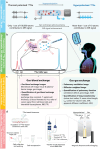
Figure 1
Figure 1
Hyperpolarized (HP) 129Xe is introduced to visualize the lungs and monitor target molecules in MRI. a) The process of generating HP 129Xe involves the transfer of angular momentum from photons to 129Xe atoms via an alkali metal intermediary, leading to an enhancement of the HP 129Xe magnetic resonance signal by over 10 000 times compared to that at thermal equilibrium. b) The produced HP 129Xe gas is inhaled, generating distinct magnetic resonance signals in pulmonary alveoli (0 ppm), lung tissues and plasma (197 ppm) and red blood cells (217 ppm), respectively. c) Using the diverse signals, ultrasensitive 129Xe MRI enables the quantitative assessment of gas–gas and gas–blood exchanges, as well as the visualization of lung structure and function. d) Typical procedures for identifying target molecules within living cells by introducing 129Xe‐based biosensors.
Figure 2
Figure 2
Quantification of lung microstructure and function by using noninvasive HP 129Xe MRI. a) The acinar lung airway model that describes the pulmonary microstructure parameters, and b) the diagram of the gas–blood exchange region of the alveoli. Reproduced with permission.[ 26 ] Copyright 2021, The American Association for the Advancement of Science.
Figure 3
Figure 3
Diffusion sensitive pulse gradient waveform introduced in simulations.
Figure 4
Figure 4
Images of human chest. a) The 1H MRI reveals a “black hole” in the lungs due to the lack of signal sources from protons. The HP 129Xe magnetic resonance spectrum and images show that 129Xe in the lung airspace registers a signal at 0 ppm, while dissolved 129Xe in TP and RBC shows signals around 197 and 217 ppm, respectively. b) A comparison of ventilation images between a healthy volunteer and a discharged COVID‐19 patient. Reproduced with permission.[ 44 ] Copyright 2024, Wiley. c) 129Xe MRI provides functional information of the lungs to guide radiotherapy planning. Reproduced with permission.[ 45 ] Copyright 2022, Wiley Periodicals, LLC on behalf of The American Association of Physicists in Medicine.
Figure 5
Figure 5
Endowing targeting ability to inert 129Xe. a) In a typical 129Xe probe, the host‐cage identifies targets by the guidance of the modified targeting molecule, and then captures 129Xe atoms to produce a specific MR signal. b) The exchange process between 129Xe atoms and host‐cages.
Figure 6
Figure 6
Different types of host cages for xenon atoms. There are three main categories, one for molecular‐hosts, another for nano‐hosts, and a combination of the two, which embeds the molecular hosts in a nanocarrier. Reproduced with permission.[ 116 ] Copyright 2006, American Chemical Society. Reproduced with permission.[ 117 ] Copyright 2015, Royal Society of Chemistry. Reproduced with permission.[ 118 ] Copyright 2008, Wiley. Reproduced with permission.[ 119 ] Copyright 2020, Springer Nature. Reproduced with permission.[ 120 ] Copyright 2020, American Chemical Society. Reproduced with permission.[ 121 ] Copyright 2013, American Chemical Society. Reproduced with permission.[ 122 ] Copyright 2014, Springer Nature. Reproduced with permission.[ 123 ] Copyright 2020, PNAS. Reproduced with permission.[ 124 ] Copyright 2024, American Chemical Society. Reproduced with permission.[ 125 ] Copyright 2020, Wiley‐VCH, Weinheim.
Figure 7
Figure 7
Hyperpolarized 129Xe chemical exchange saturation transfer (Hyper‐CSET): Amplifying the target 129Xe within “host‐cages” by indirectly detecting the reduction in signal of free HP 129Xe. a) An RF pulse is applied to target the HP 129Xe in host‐cages, resulting in depolarization of these 129Xe atoms and saturation of the corresponding MR signal. b) Subsequently, the 129Xe atoms within the host‐cages exchange with the free HP 129Xe atoms, c) leading to a reduction in MR signal of the free HP 129Xe due to the accompanying saturation transfe. d) A Hyper‐CSET spectrum is obtained by comparing the reduction in MR signal from free HP 129Xe atoms before and after the application of a saturation pulse, which reflects the specific MR signal generated by HP 129Xe within host‐cages.
Figure 8
Figure 8
Functionalization and applications of 129Xe hosts. a) CrypA was functionalized with a solubilizing group (purple) and a targeting group (yellow) via a rink amide resin linker. Adapted with permission.[ 149 ] Copyright 2001, PNAS. b) A “turn‐on” strategy for target analysis using a CB[6]‐based probe. Initially, the cavity of the CB[6] host was occupied by rotaxane molecules. Upon encountering the target molecule H2O2, the rotaxane was cleaved, freeing the host cavity for HP 129Xe exchange. As a result, the specific 129Xe MR signal for CB[6] was recovered with the introduction of the target molecules. Reproduced with permission.[ 158 ] Copyright 2019, Wiley. c) The ultrasensitive property of HP 129Xe allows for the differentiation of diastereoisomers of Fe‐MOP. Reproduced with permission.[ 137 ] Copyright 2022, Springer Nature. d) PFOB nano‐emulsions were functionalized with RGD peptides to target tumor cells, and resulting systems were imaged using ultrasensitive 129Xe MRI. Reproduced with permission.[ 159 ] Copyright 2019, American Chemical Society. e) GVs displayed a characteristic signal peak only when their structure remains intact; this peak vanished when the structure collapses. Reproduced with permission.[ 122 ] Copyright 2014, Springer Nature. f) Establishment of a 3D map to illustrate the relationships among the 129Xe CEST MRI contrast, MOL concentration, and saturation pulse, with its application in quantifying MOL‐hosts and the content of drug molecules within living cells. Reproduced with permission.[ 124 ] Copyright 2024, American Chemical Society.

Similar articles

References

    1. Castelvecchi D., Nat. Rev. Phys. 2023, 5, 554.
    1. Warren W. S., Richter W., Andreotti A. H., Farmer B. T., Science 1993, 262, 2005. - PubMed
    1. a) Siegal G., Selenko P., J. Magn. Reson. 2019, 306, 202; - PubMed
    2. b) Duong T. Q., Pardue M. T., Thule P. M., Olson D. E., Cheng H., Nair G., Li Y., Kim M., Zhang X., Shen Q., NMR Biomed. 2008, 21, 978. - PMC - PubMed
    1. a) Sereno M. I., Dale A. M., Reppas J. B., Kwong K. K., Belliveau J. W., Brady T. J., Rosen B. R., Tootell R. B. H., Science 1995, 268, 889; - PubMed
    2. b) Yu H., Shimakawa A., McKenzie C. A., Brodsky E., Brittain J. H., Reeder S. B., Magn. Reson. Med. 2008, 60, 1122. - PMC - PubMed
    1. Pekar J. J., IEEE Eng. Med. Biol. 2006, 25, 24. - PubMed

LinkOut - more resources