. 2025 Jan 21;21(1):e1012821.
doi: 10.1371/journal.ppat.1012821. eCollection 2025 Jan.

Gingipains protect Porphyromonas gingivalis from macrophage-mediated phagocytic clearance

Affiliations

Gingipains protect Porphyromonas gingivalis from macrophage-mediated phagocytic clearance

Magdalena Widziolek et al. PLoS Pathog. .

Abstract

Porphyromonas gingivalis (Pg) is a keystone pathogen in periodontitis, a highly prevalent disease manifested by chronic inflammation of the periodontium, alveolar bone resorption and tooth loss. During periodontitis pathobionts such as Pg can enter the bloodstream and growing evidence correlates periodontitis with increased risk of cardiovascular and neurodegenerative diseases. However, the mechanism by which immune cells respond to Pg challenge in vivo remains elusive. Pg produce aggressive proteolytic virulence factors termed gingipains which not only provide nutrients necessary for bacterial growth, but also subvert the host immune response, facilitating bacterial survival. Using transgenic zebrafish with fluorescently labelled macrophages and neutrophils, the role of gingipains in bacterial survival and interaction with phagocytes during systemic and local infection was examined. In contrast to the wild-type (W83) Pg, isogenic gingipain-null (ΔK/R-ab) or wild-type Pg treated with gingipain inhibitors caused less zebrafish mortality, bacteria were rapidly phagocytosed, acidified in phagosomes and eradicated when systemically injected, showing that gingipains are instrumental in preventing phagocytosis and intracellular killing of Pg by innate immune cells. Moreover, Pg were predominantly phagocytosed by macrophages, and gingipain depletion/inactivation increased bacterial phagocytosis when bacteria were injected either systemically or locally in the otic vesicle, with less bacteria internalised by neutrophils. This phenomenon was Pg-specific as Fusobacterium nucleatum caused neutrophil recruitment that then effectively phagocytosed these bacteria. These data demonstrate the important role of phagocytes, especially macrophages, in combating Pg infection and highlight the crucial protective role of gingipains in subverting the innate immune response. This study also emphasizes the advantages of using zebrafish to study interactions of Pg with phagocytes in vivo in real-time, providing a valuable experimental system for testing new therapeutic strategies aimed at reducing periodontal-associated systemic or neurodegenerative disease.


Conflict of interest statement

The authors have declared that no competing interests exist.

Figures

Fig 1
Fig 1. Wild-type Porphyromonas gingivalis (Pg) W83 strain displays greater levels of survival in systemically infected zebrafish larvae compared to gingipain-null ΔK/R-ab mutant.
Zebrafish embryos were infected systemically at 30 hpf with fluorescein-SE-labelled wild-type Pg W83 or the gingipain-null mutant ΔK/Rab. PBS was injected into control larvae. (A) Representative images of real-time light-sheet microscopy over 16 h showing the differences in the course of bacterial eradication. White arrowheads point to Pg localised in puncta. Scale bar = 100 μm. (B) Zebrafish larvae were infected with Pg W83 or ΔK/R-ab, and larvae were homogenized at indicated time post-infection (hpi), serially diluted and number of recovered bacteria were enumerated on ABA plates. CFU–colony forming unit. Data are presented as means ± SEM. Each dot represents the number of bacteria recovered from an individual larva. Statistical significance was analysed by Mann-Whitney test, *** p≤0.001, **** p ≤ 0.0001.
Fig 2
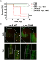
Fig 2. Myeloid cell-depleted zebrafish larvae are more susceptible to Porphyromonas gingivalis (Pg) infection.
(A) Kaplan-Meyer survival plot of pu.1 MO-treated or PBS-injected control zebrafish larvae upon systemic infection with the wild-type Pg W83 or ΔK/R-ab. Survival plots were analysed by log-rank test.*p≤0.05,***p≤0.001; hpi- hours post infection. (B) Representative images of the yolk and tail region of pu.1 MO-treated and untreated zebrafish larvae 2 hpi with fluorescein-SE (green) and pHrodo (red) dual-stained ΔK/R-ab. The untreated larvae display orange dots (white arrows) representing phagocytosed (phagosome acidified) ΔK/R-ab. Phagocytosed ΔK/R-ab were not detected in pu.1 MO due to removal of myeloid cells. White dashed lines indicate yolk or tail areas. Boxes show zoomed regions of the images. h-heart. Scale bar = 100 μm.
Fig 3
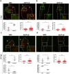
Fig 3. Gingipains prevent phagosome acidification of internalized Porphyromonas gingivalis (Pg).
Real-time in vivo examination of the acidification of the wild-type Pg W83 and ΔK/R-ab. Pg were stained with fluorescein-SE (green) and pHrodo (red) and zebrafish larvae (30 hpf) were infected into the Duct of Cuvier. Representative images along with quantification of the phagosome-acidified bacteria within selected area and size of acidified bacterial clusters in the yolk (A and C) or the tail regions (B and D) at 2 hpi (A and B) or 24 hpi (C and D). White dashed lines indicate yolk or tail areas. Boxes show zoomed regions of the images. y = yolk, h = heart. Scale bar = 100 μm. Data presented are means ± SD. Each dot represents signal quantified from a single zebrafish larva. Statistical significance was analysed by unpaired t-test with Welch’s correction or Mann-Whitney test where required. *≤0.05, **p≤0.01, ****p≤0.0001.
Fig 4
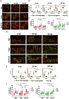
Fig 4. Inactivation of gingipains by KYT inhibitors increases larvae survival and increases phagocytosis of wild-type W83 Porphyromonas gingivalis (Pg).
(A) Kaplan-Meier survival plot of zebrafish larvae (30 hpf) infected with wild-type Pg W83 pre-treated or untreated with KYT inhibitors. Comparisons between survival curves were made using the log rank test. *p≤0.05,***p≤0.001; hpi- hours post infection. Real-time analysis of the acidification process during phagocytosis of the Pg W83 treated/untreated with KYT inhibitors in the yolk (B) and tail (C) regions 2 hpi. Pg were stained with fluorescein-SE (green) and pHrodo (red). White dashed lines indicate yolk or tail areas. Boxes show zoomed regions of the images. y-yolk, h-heart. Scale bar = 100 μm. Quantified data from the yolk (D) and the tail (E) regions. Data are presented as means ± SD. Each dot represents signal quantified from a single larva. Statistical significance was analysed by unpaired t-test with Welch’s correction. **p≤0.01.
Fig 5
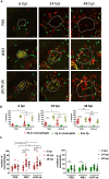
Fig 5. The role of macrophages and neutrophils in the eradication of Porphyromonas gingivalis (Pg) infection.
Zebrafish larvae (30 hpf) were infected systemically with AlexaFluor647–SE labelled wild-type Pg W83 or ΔK/R-ab and real-time examination of phagocytosis was performed using transgenic zebrafish larvae Tg(mpeg1:mCherry; red macrophages) and Tg(mpx:EGFP, green neutrophils). PBS was injected as a control. Representative images of the yolk (A) or the tail (D) regions at 2, 24 or 48 hours post-infection (hpi). Scale bar = 50 μm. Quantified percentage of bacteria phagocytosed by macrophages, neutrophils and non-phagocytosed (free) bacteria at 2, 24 and 48 hpi in the yolk (B) and the tail (E) regions. Number of macrophages and neutrophils in the yolk (C) and the tail (F) regions at 2, 24 or 48 hpi. White dashed boxes correspond to zoomed regions of the images. Each dot represents quantified data from a single larva obtained in at least 3 independent experiments. Graphs show means ± SD. Differences between groups were analysed by Two-Way ANOVA.*p≤0.05,**p≤0.01,***p≤0.001,****p≤0.0001.
Fig 6
Fig 6. Flow cytometry analysis of phagocytosis of Pg W83 and ΔK/R-ab mutant by macrophages and neutrophils in zebrafish larvae.
Transgenic zebrafish larvae Tg(mpeg1:mCherry) x Tg(mpx:EGFP) were infected with AF647-SE-stained Pg W83 or ΔK/R-ab and flow cytometry analysis was performer 24 hpi. Gating strategy used for flow cytometry analysis from zebrafish larval homogenate (A). Gating strategy used to enumerate population of EGFP+ neutrophils (B-E) or mCherry+ macrophages (F-I) 24 hpi. Percentage of neutrophils or macrophages associated with bacteria (J). Mean fluorescence intensity of neutrophils and macrophages associated with bacteria (K). Representative confocal microscopy image of cells sorted from infected larvae (L). Red–macrophages, green—neutrophils, yellow- Pg. hpi–hours post infection.
Fig 7
Fig 7. The role and migration of macrophages and neutrophils during local infection of Porphyromonas gingivalis (Pg) into the otic vesicle.
Zebrafish larvae (30 hpf) were infected locally into the otic vesicle with AlexaFluor647–Se-labelled wild-type Pg W83 or ΔK/R-ab and real-time examination of phagocytosis and cell migration was performed using transgenic zebrafish larvae Tg(mpeg1:mCherry) x Tg(mpx:EGFP). PBS was injected as a control. (A) Representative images of the otic vesicle at 6, 24 or 48 hours post-infection (hpi). Scale bar = 50 μm. White dashed lines indicate otic vesicle location. (B) Quantified percentage of bacteria phagocytosed by macrophages, neutrophils and non-phagocytosed (i.e., free Pg) at 2, 24 and 48 hpi in the otic vesicle and surrounding region. (C) Number of macrophages and neutrophils attracted toward the infection site. Each dot represents quantified data from a single larva obtained in at least 3 independent experiments. Graphs show means ± SD. Differences between groups were analysed by Two-Way ANOVA. *p≤0.05,**p≤0.01,***p≤0.001,****p≤0.0001.

References

    1. Abusleme L, Hoare A, Hong BY, Diaz PI. Microbial signatures of health, gingivitis, and periodontitis. Periodontol 2000. 2021;86(1):57–78. doi: 10.1111/prd.12362 - DOI - PubMed
    1. Hajishengallis G, Lamont RJ. Polymicrobial communities in periodontal disease: Their quasi-organismal nature and dialogue with the host. Periodontol 2000. 2021;86(1):210–30. doi: 10.1111/prd.12371 - DOI - PMC - PubMed
    1. Hajishengallis G, Darveau RP, Curtis MA. The keystone-pathogen hypothesis. Nat Rev Microbiol. 2012;10(10):717–25. doi: 10.1038/nrmicro2873 - DOI - PMC - PubMed
    1. Saglie FR, Pertuiset JH, Smith CT, Nestor MG, Carranza FA Jr., Newman MG, et al.. The presence of bacteria in the oral epithelium in periodontal disease. III. Correlation with Langerhans cells. J Periodontol. 1987;58(6):417–22. doi: 10.1902/jop.1987.58.6.417 - DOI - PubMed
    1. Vitkov L, Klappacher M, Hannig M, Krautgartner WD. Neutrophil fate in gingival crevicular fluid. Ultrastruct Pathol. 2010;34(1):25–30. doi: 10.3109/01913120903419989 - DOI - PubMed

MeSH terms

Substances

LinkOut - more resources